Click image to see more details
Product Info Summary
| SKU: | PB9319 |
|---|---|
| Size: | 100 μg/vial |
| Reactive Species: | Human, Mouse, Rat |
| Host: | Rabbit |
| Application: | Flow Cytometry, IP, WB |
Customers Who Bought This Also Bought

Product info
Product Name
Anti-PKC beta 1/PRKCB Antibody Picoband®
SKU/Catalog Number
PB9319
PB0355 is an alternative SKU for this antibody, used in previous lots.
Size
100 μg/vial
Form
Lyophilized
Description
Boster Bio Anti-PKC beta 1/PRKCB Antibody Picoband® catalog # PB9319. Tested in Flow Cytometry, IP, WB applications. This antibody reacts with Human, Mouse, Rat. The brand Picoband indicates this is a premium antibody that guarantees superior quality, high affinity, and strong signals with minimal background in Western blot applications. Only our best-performing antibodies are designated as Picoband, ensuring unmatched performance.
Storage & Handling
Store at -20˚C for one year from date of receipt. After reconstitution, at 4˚C for one month. It can also be aliquotted and stored frozen at -20˚C for six months. Avoid repeated freeze-thaw cycles.
Cite This Product
Anti-PKC beta 1/PRKCB Antibody Picoband® (Boster Biological Technology, Pleasanton CA, USA, Catalog # PB9319)
Host
Rabbit
Contents
Each vial contains 4 mg Trehalose, 0.9 mg NaCl and 0.2 mg Na2HPO4.
Clonality
Polyclonal
Isotype
Rabbit IgG
Immunogen
E.coli-derived human PKC beta 1 recombinant protein (Position: E542-V671). Human PKC beta 1 shares 100% amino acid (aa) sequence identity with both mouse and rat PKC beta 1.
Cross-reactivity
No cross-reactivity with other proteins
Reactive Species
PB9319 is reactive to PRKCB in Human, Mouse, Rat
Observed Molecular Weight
80 kDa
Calculated molecular weight
76.9 kDa
Background of PRKCB
Protein kinase C beta type is an enzyme that in humans is encoded by the PRKCB gene. It is a member of the protein kinase C (PKC) gene family. PKC family members phosphorylate a wide variety of protein targets and are known to be involved in diverse cellular signaling pathways. PKC family members also serve as major receptors for phorbol esters, a class of tumor promoters. This protein kinase has been reported to be involved in many different cellular functions, such as B cell activation, apoptosis induction, endothelial cell proliferation, and intestinal sugar absorption. It has been found that PRKCB activated by oxidative conditions in the cell, induces phosphorylation of p66 (SHC) and triggers mitochondrial accumulation of the protein after it is recognized by the prolyl isomerase PIN1.
Antibody Validation
Boster validates all antibodies on WB, IHC, ICC, Immunofluorescence, and ELISA with known positive control and negative samples to ensure specificity and high affinity, including thorough antibody incubations.
Application & Images
Applications
PB9319 is guaranteed for Flow Cytometry, IP, WB Boster Guarantee
Recommend Dilution
| Application | Dilution | Species |
|---|---|---|
| Western blot | 0.1-0.5μg/ml | Human, Mouse, Rat |
| Immunoprecipitation | 0.5-2 μg/ml | Human |
| Flow Cytometry(Fixed) | 1-3 μg/1x106 cells | Human |
Tested application
Suggested blocking solution with 5% non-fat milk or BSA; (*)Recommended protein loading: 20-40 µg per lane
Validation Images & Assay Conditions
Click image to see more details
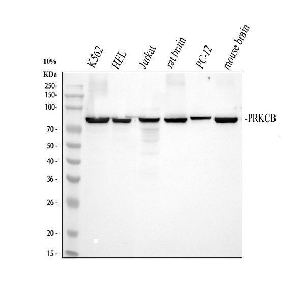
Western blot analysis of PKC beta 1 using anti-PKC beta 1 antibody (PB9319).
Electrophoresis was performed on a 5-20% SDS-PAGE gel at 80V (Stacking gel) / 120V (Resolving gel) for 2 hours. The sample well of each lane was loaded with 30 ug of sample under reducing conditions.
Lane 1: human K562 whole cell lysates,
Lane 2: human HEL whole cell lysates,
Lane 3: human Jurkat whole cell lysates,
Lane 4: rat brain tissue lysates,
Lane 5: rat PC-12 whole cell lysates,
Lane 6: mouse brain tissue lysates.
After electrophoresis, proteins were transferred to a nitrocellulose membrane at 150 mA for 50-90 minutes. Blocked the membrane with 5% non-fat milk/TBS for 1.5 hour at RT. The membrane was incubated with rabbit anti-PKC beta 1 antigen affinity purified polyclonal antibody (Catalog # PB9319) at 0.5 μg/mL overnight at 4°C, then washed with TBS-0.1%Tween 3 times with 5 minutes each and probed with a goat anti-rabbit IgG-HRP secondary antibody at a dilution of 1:5000 for 1.5 hour at RT. The signal is developed using an ECL Plus Western Blotting Substrate (Catalog # AR1196-200) with Tanon 5200 system. A specific band was detected for PKC beta 1 at approximately 80 kDa. The expected band size for PKC beta 1 is at 77 kDa.

Click image to see more details
Immunoprecipitating (IP) PKC beta 1 in Jurkat whole cell lysate.
Western blot analysis of PKC beta 1 using anti-PKC beta 1 antibody (PB9319);
Lane 1: Jurkat whole cell lysates (30ug);
Lane 2: Rabbit control IgG instead of anti-PKC beta 1 antibody in Jurkat whole cell lysate;
Lane 3: anti-PKC beta 1 antibody (2μg) + Jurkat whole cell lysate (500μg).
After electrophoresis, proteins were transferred to a membrane. Then the membrane was incubated with rabbit anti-PKC beta 1 antigen affinity purified polyclonal antibody (PB9319) at a dilution of 0.5 μg/mL and probed with a goat anti-rabbit IgG-HRP secondary antibody (Catalog # BA1054). The signal is developed using ECL Plus Western Blotting Substrate (Catalog # AR1196-200). A specific band was detected for PKC beta 1 at approximately 80 kDa. The expected band size for PKC beta 1 is at 77 kDa.

Click image to see more details
Flow Cytometry analysis of Jurkat cells using anti-PKC beta 1 antibody (PB9319).
Overlay histogram showing Jurkat cells stained with PB9319 (Blue line). To facilitate intracellular staining, cells were fixed with 4% paraformaldehyde and permeabilized with permeabilization buffer. The cells were blocked with 10% normal goat serum. And then incubated with rabbit anti-PKC beta 1 Antibody (PB9319, 1 μg/1x106 cells) for 30 min at 20°C. DyLight®488 conjugated goat anti-rabbit IgG (BA1127, 5-10 μg/1x106 cells) was used as secondary antibody for 30 minutes at 20°C. Isotype control antibody (Green line) was rabbit IgG (1 μg/1x106) used under the same conditions. Unlabelled sample without incubation with primary antibody and secondary antibody (Red line) was used as a blank control.
Specific Publications For Anti-PKC beta 1/PRKCB Antibody Picoband® (PB9319)
Loading publications
Recommended Resources
Here are featured tools and databases that you might find useful.
- Boster's Pathways Library
- Protein Databases
- Bioscience Research Protocol Resources
- Data Processing & Analysis Software
- Photo Editing Software
- Scientific Literature Resources
- Research Paper Management Tools
- Molecular Biology Software
- Primer Design Tools
- Bioinformatics Tools
- Phylogenetic Tree Analysis
Customer Reviews
Have you used Anti-PKC beta 1/PRKCB Antibody Picoband®?
Share your experimental results or join a short interview to earn up to $1,000 in product credits or other rewards.
0 Reviews For Anti-PKC beta 1/PRKCB Antibody Picoband®
Customer Q&As
Have a question?
Find answers in Q&As, reviews.
Can't find your answer?
Submit your question
1 Customer Q&As for Anti-PKC beta 1/PRKCB Antibody Picoband®
Question
We are currently using anti-PKC beta 1/PRKCB antibody PB9319 for human tissue, and we are satisfied with the WB results. The species of reactivity given in the datasheet says human, mouse, rat. Is it true that the antibody can work on zebrafish tissues as well?
Verified Customer
Verified customer
Asked: 2020-03-06
Answer
The anti-PKC beta 1/PRKCB antibody (PB9319) has not been validated for cross reactivity specifically with zebrafish tissues, though there is a good chance of cross reactivity. We have an innovator award program that if you test this antibody and show it works in zebrafish you can get your next antibody for free. Please contact me if I can help you with anything.
Boster Scientific Support
Answered: 2020-03-06
