Click image to see more details
Product Info Summary
| SKU: | RP1058 |
|---|---|
| Size: | 100 μg/vial |
| Reactive Species: | Human, Mouse, Rat |
| Host: | Rabbit |
| Application: | IHC, WB |
Customers Who Bought This Also Bought

Product info
Product Name
Anti-PKC delta/PRKCD Antibody Picoband®
SKU/Catalog Number
RP1058
PB0356 is an alternative SKU for this antibody, used in previous lots.
Size
100 μg/vial
Form
Lyophilized
Description
Boster Bio Anti-PKC delta/PRKCD Antibody catalog # RP1058. Tested in IHC, WB applications. This antibody reacts with Human, Mouse, Rat. The brand Picoband indicates this is a premium antibody that guarantees superior quality, high affinity, and strong signals with minimal background in Western blot applications. Only our best-performing antibodies are designated as Picoband, ensuring unmatched performance.
Storage & Handling
Store at -20˚C for one year from date of receipt. After reconstitution, at 4˚C for one month. It can also be aliquotted and stored frozen at -20˚C for six months. Avoid repeated freeze-thaw cycles.
Cite This Product
Anti-PKC delta/PRKCD Antibody Picoband® (Boster Biological Technology, Pleasanton CA, USA, Catalog # RP1058)
Host
Rabbit
Contents
Each vial contains 4 mg Trehalose, 0.9 mg NaCl and 0.2 mg Na2HPO4.
Clonality
Polyclonal
Isotype
Rabbit IgG
Immunogen
E.coli-derived human PKC delta recombinant protein (Position: M1-E160). Human PKC delta shares 95% and 93% amino acid (aa) sequence identity with mouse and rat PKC delta, respctively.
Cross-reactivity
No cross-reactivity with other proteins
Reactive Species
RP1058 is reactive to PRKCD in Human, Mouse, Rat
Observed Molecular Weight
78 kDa
Calculated molecular weight
77.5 kDa
Background of PRKCD
Protein kinase C delta type, also called PKCD, is an enzyme that in humans is encoded by the PRKCD gene. The PRKCD gene encodes a member of the protein kinase C family, members of which are critical for regulation of cell survival, proliferation, and apoptosis. This gene is mapped to 3p21.1. Studies both in human and mice demonstrate that this kinase is involved inB cell signaling and in the regulation of growth, apoptosis, and differentiation of a variety of cell types. PRKCD is also regulated by phosphorylation on various tyrosine residues including Y311 (by SRC). It has been showed that PRKCD phosphorylates NLRC4 and that this phosphorylation is critical for inflammasome assembly. What’s more, it is also a part of a noncanonical WNT signaling cascade.
Antibody Validation
Boster validates all antibodies on WB, IHC, ICC, Immunofluorescence, and ELISA with known positive control and negative samples to ensure specificity and high affinity, including thorough antibody incubations.
Application & Images
Applications
RP1058 is guaranteed for IHC, WB Boster Guarantee
Recommend Dilution
| Application | Dilution | Species |
|---|---|---|
| Western blot | 0.1-0.5μg/ml | Human, Mouse, Rat |
| Immunohistochemistry (Paraffin-embedded Section) | 0.5-1μg/ml | Human |
Tested application
Suggested blocking solution with 5% non-fat milk or BSA; (*)Recommended protein loading: 20-40 µg per lane
Use TE buffer pH 9.0 for antigen retrieval; (*) citrate buffer pH 6.0 is an alternative.
Validation Images & Assay Conditions
Click image to see more details
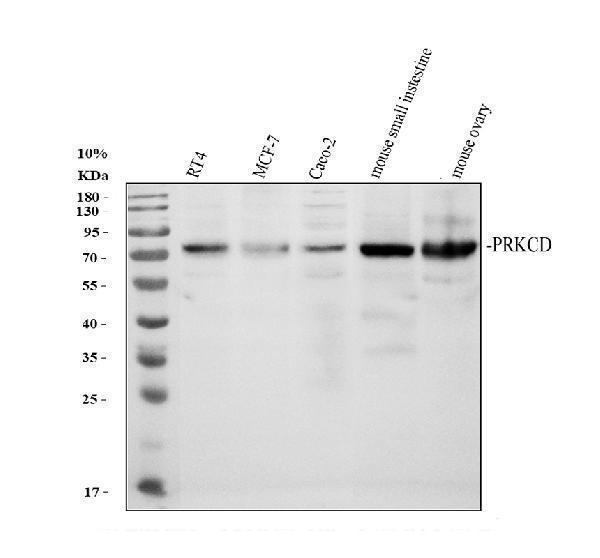
Western blot analysis of PRKCD using anti-PRKCD antibody (RP1058).
Electrophoresis was performed on a 10% SDS-PAGE gel at 80V (Stacking gel) / 120V (Resolving gel) for 2 hours. The sample well of each lane was loaded with 30 ug of sample under reducing conditions.
Lane 1: human RT4 whole cell lysates,
Lane 2: human MCF-7 whole cell lysates,
Lane 3: human CACO-2 whole cell lysates,
Lane 4: mouse small intestine tissue lysates.
Lane 5: mouse ovary tissue lysates.
After electrophoresis, proteins were transferred to a nitrocellulose membrane at 150 mA for 50-90 minutes. Blocked the membrane with 5% non-fat milk/TBS for 1.5 hour at RT. The membrane was incubated with rabbit anti-PRKCD antigen affinity purified polyclonal antibody (Catalog # RP1058) at 0.5 μg/mL overnight at 4°C, then washed with TBS-0.1%Tween 3 times with 5 minutes each and probed with a goat anti-rabbit IgG-HRP secondary antibody at a dilution of 1:5000 for 1.5 hour at RT. The signal is developed using an ECL Plus Western Blotting Substrate (Catalog # AR1196-200) with Tanon 5200 system. A specific band was detected for PRKCD at approximately 78 kDa. The expected band size for PRKCD is at 78 kDa.

Click image to see more details
Western blot analysis of PRKCD using anti-PRKCD antibody (RP1058).
Electrophoresis was performed on a 10% SDS-PAGE gel at 80V (Stacking gel) / 120V (Resolving gel) for 2 hours. The sample well of each lane was loaded with 30 ug of sample under reducing conditions.
Lane 1: rat brain tissue lysates,
Lane 2: rat lung tissue lysates,
Lane 3: mouse brain tissue lysates,
Lane 4: mouse lung tissue lysates.
After electrophoresis, proteins were transferred to a nitrocellulose membrane at 150 mA for 50-90 minutes. Blocked the membrane with 5% non-fat milk/TBS for 1.5 hour at RT. The membrane was incubated with rabbit anti-PRKCD antigen affinity purified polyclonal antibody (Catalog # RP1058) at 0.5 μg/mL overnight at 4°C, then washed with TBS-0.1%Tween 3 times with 5 minutes each and probed with a goat anti-rabbit IgG-HRP secondary antibody at a dilution of 1:5000 for 1.5 hour at RT. The signal is developed using an ECL Plus Western Blotting Substrate (Catalog # AR1196-200) with Tanon 5200 system. A specific band was detected for PRKCD at approximately 78 kDa. The expected band size for PRKCD is at 78 kDa.

Click image to see more details
IHC analysis of PRKCD using anti-PRKCD antibody (RP1058).
PRKCD was detected in a paraffin-embedded section of human tonsil tissue. Heat mediated antigen retrieval was performed in EDTA buffer (pH 8.0, epitope retrieval solution). The tissue section was blocked with 10% goat serum. The tissue section was then incubated with 2 μg/ml rabbit anti-PRKCD Antibody (RP1058) overnight at 4°C. Peroxidase Conjugated Goat Anti-rabbit IgG was used as secondary antibody and incubated for 30 minutes at 37°C. The tissue section was developed using HRP Conjugated Rabbit IgG Super Vision Assay Kit (Catalog # SV0002) with DAB as the chromogen.

Click image to see more details
IHC analysis of PRKCD using anti-PRKCD antibody (RP1058).
PRKCD was detected in a paraffin-embedded section of human prostatic cancer tissue. Heat mediated antigen retrieval was performed in EDTA buffer (pH 8.0, epitope retrieval solution). The tissue section was blocked with 10% goat serum. The tissue section was then incubated with 2 μg/ml rabbit anti-PRKCD Antibody (RP1058) overnight at 4°C. Peroxidase Conjugated Goat Anti-rabbit IgG was used as secondary antibody and incubated for 30 minutes at 37°C. The tissue section was developed using HRP Conjugated Rabbit IgG Super Vision Assay Kit (Catalog # SV0002) with DAB as the chromogen.
Specific Publications For Anti-PKC delta/PRKCD Antibody Picoband® (RP1058)
Loading publications
Recommended Resources
Here are featured tools and databases that you might find useful.
- Boster's Pathways Library
- Protein Databases
- Bioscience Research Protocol Resources
- Data Processing & Analysis Software
- Photo Editing Software
- Scientific Literature Resources
- Research Paper Management Tools
- Molecular Biology Software
- Primer Design Tools
- Bioinformatics Tools
- Phylogenetic Tree Analysis
Customer Reviews
Have you used Anti-PKC delta/PRKCD Antibody Picoband®?
Share your experimental results or join a short interview to earn up to $1,000 in product credits or other rewards.
0 Reviews For Anti-PKC delta/PRKCD Antibody Picoband®
Customer Q&As
Have a question?
Find answers in Q&As, reviews.
Can't find your answer?
Submit your question
3 Customer Q&As for Anti-PKC delta/PRKCD Antibody Picoband®
Question
I was wanting to use your anti-PKC delta/PRKCD antibody for WB for human platelet on frozen tissues, but I want to know if it has been tested for this particular application. Has this antibody been tested and is this antibody a good choice for human platelet identification?
Verified Customer
Verified customer
Asked: 2019-09-13
Answer
It shows on the product datasheet, RP1058 anti-PKC delta/PRKCD antibody has been tested for WB on human tissues. We have an innovator award program that if you test this antibody and show it works in human platelet in IHC-frozen, you can get your next antibody for free.
Boster Scientific Support
Answered: 2019-09-13
Question
Thank you for helping with my inquiry over the phone. Here are the WB image, lot number and protocol we used for platelet using anti-PKC delta/PRKCD antibody RP1058. Let me know if you need anything else.
Verified Customer
Verified customer
Asked: 2019-05-17
Answer
We appreciate the data. You have provided everything we needed. Our lab team are working to resolve your inquiry as quickly as possible, and we appreciate your patience and understanding! Please let me know if there is anything you need in the meantime.
Boster Scientific Support
Answered: 2019-05-17
Question
We are currently using anti-PKC delta/PRKCD antibody RP1058 for human tissue, and we are satisfied with the WB results. The species of reactivity given in the datasheet says human. Is it true that the antibody can work on primate tissues as well?
E. Huang
Verified customer
Asked: 2015-12-14
Answer
The anti-PKC delta/PRKCD antibody (RP1058) has not been tested for cross reactivity specifically with primate tissues, though there is a good chance of cross reactivity. We have an innovator award program that if you test this antibody and show it works in primate you can get your next antibody for free. Please contact me if I can help you with anything.
Boster Scientific Support
Answered: 2015-12-14
