Click image to see more details
-

-
-
-
-
+4
Product Info Summary
| SKU: | PB9721 |
|---|---|
| Size: | 100 μg/vial |
| Reactive Species: | Human, Mouse, Rat |
| Host: | Rabbit |
| Application: | Flow Cytometry, IP, IF, IHC, ICC, WB |
Customers Who Bought This Also Bought

Product info
Product Name
Anti-SAPK4/MAPK13 Antibody Picoband®
SKU/Catalog Number
PB9721
Size
100 μg/vial
Form
Lyophilized
Description
Boster Bio Anti-SAPK4/MAPK13 Antibody Picoband® catalog # PB9721. Tested in Flow Cytometry, IP, ICC/IF, IHC, WB applications. This antibody reacts with Human, Mouse, Rat. The brand Picoband indicates this is a premium antibody that guarantees superior quality, high affinity, and strong signals with minimal background in Western blot applications. Only our best-performing antibodies are designated as Picoband, ensuring unmatched performance.
Storage & Handling
Store at -20˚C for one year from date of receipt. After reconstitution, at 4˚C for one month. It can also be aliquotted and stored frozen at -20˚C for six months. Avoid repeated freeze-thaw cycles.
Cite This Product
Anti-SAPK4/MAPK13 Antibody Picoband® (Boster Biological Technology, Pleasanton CA, USA, Catalog # PB9721)
Host
Rabbit
Contents
Each vial contains 4 mg Trehalose, 0.9 mg NaCl and 0.2 mg Na2HPO4.
Clonality
Polyclonal
Isotype
Rabbit IgG
Immunogen
A synthetic peptide corresponding to a sequence at the C-terminus of human SAPK4 , different from the related mouse sequence by two amino acids, and from the related rat sequence by three amino acids.
Cross-reactivity
No cross-reactivity with other proteins.
Reactive Species
PB9721 is reactive to MAPK13 in Human, Mouse, Rat
Observed Molecular Weight
42 kDa
Calculated molecular weight
42.1 kDa
Background of MAPK13
MAPK13 (Mitogen-Activated Protein Kinase 13), also called p38-DELTA or Stress-Activated Protein Kinase 4 (SAPK4), is an enzyme that in humans is encoded by the MAPK13 gene. The protein encoded by this gene is a member of the MAP kinase family. MAP kinases act as an integration point for multiple biochemical signals, and are involved in a wide variety of cellular processes such as proliferation, differentiation, transcription regulation and development. This kinase is closely related to p38 MAP kinase, both of which can be activated by proinflammatory cytokines and cellular stress. MAP kinase kinases 3, and 6 can phosphorylate and activate this kinase. Transcription factor ATF2, and microtubule dynamics regulator stathmin have been shown to be the substrates of this kinase.
Antibody Validation
Boster validates all antibodies on WB, IHC, ICC, Immunofluorescence, and ELISA with known positive control and negative samples to ensure specificity and high affinity, including thorough antibody incubations.
Application & Images
Applications
PB9721 is guaranteed for Flow Cytometry, IP, IF, IHC, ICC, WB Boster Guarantee
Recommend Dilution
| Application | Dilution | Species |
|---|---|---|
| Western blot | 0.1-0.5μg/ml | Human, Mouse, Rat |
| Immunohistochemistry (Paraffin-embedded Section) | 2-5μg/ml | Human, Mouse, Rat |
| Immunocytochemistry/Immunofluorescence | 5 μg/ml | Human |
| Immunoprecipitation | 0.5-2 μg/ml | Human |
| Flow Cytometry(Fixed) | 1-3 μg/1x106 cells | Human |
Tested application
Suggested blocking solution with 5% non-fat milk or BSA; (*)Recommended protein loading: 20-40 µg per lane
Use TE buffer pH 9.0 for antigen retrieval; (*) citrate buffer pH 6.0 is an alternative.
Validation Images & Assay Conditions
Click image to see more details
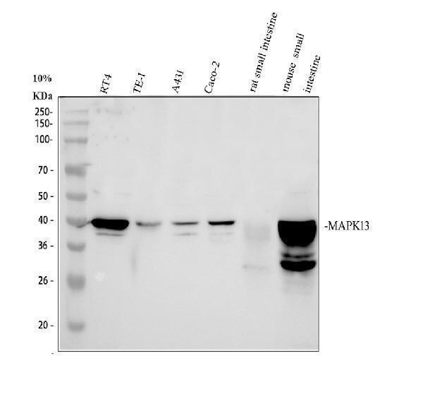
Western blot analysis of SAPK4 using anti-SAPK4 antibody (PB9721).
Electrophoresis was performed on a 10% SDS-PAGE gel at 80V (Stacking gel) / 120V (Resolving gel) for 2 hours. The sample well of each lane was loaded with 30 ug of sample under reducing conditions.
Lane 1: human RT4 whole cell lysates,
Lane 2: human TE-1 whole cell lysates,
Lane 3: human A431 whole cell lysates,
Lane 4: human Caco-2 whole cell lysates,
Lane 5: rat small intestine tissue lysates,
Lane 6: mouse small intestine tissue lysates.
After electrophoresis, proteins were transferred to a nitrocellulose membrane at 150 mA for 50-90 minutes. Blocked the membrane with 5% non-fat milk/TBS for 1.5 hour at RT. The membrane was incubated with rabbit anti-SAPK4 antigen affinity purified polyclonal antibody (PB9721) at 0.5 μg/mL overnight at 4°C, then washed with TBS-0.1%Tween 3 times with 5 minutes each and probed with a goat anti-rabbit IgG-HRP secondary antibody (Catalog # BA1054) at a dilution of 1:5000 for 1.5 hour at RT. The signal is developed using an ECL Plus Western Blotting Substrate (Catalog # AR1196-200) with Tanon 5200 system. A specific band was detected for SAPK4 at approximately 42 kDa. The expected band size for SAPK4 is at 42 kDa.

Click image to see more details
IHC analysis of SAPK4 using anti-SAPK4 antibody (PB9721).
SAPK4 was detected in a paraffin-embedded section of human breast cancer tissue. Heat mediated antigen retrieval was performed in EDTA buffer (pH 8.0, epitope retrieval solution). The tissue section was blocked with 10% goat serum. The tissue section was then incubated with 2 μg/ml rabbit anti-SAPK4 Antibody (PB9721) overnight at 4°C. Peroxidase Conjugated Goat Anti-rabbit IgG was used as secondary antibody and incubated for 30 minutes at 37°C. The tissue section was developed using HRP Conjugated Rabbit IgG Super Vision Assay Kit (Catalog # SV0002) with DAB as the chromogen.

Click image to see more details
IHC analysis of SAPK4 using anti-SAPK4 antibody (PB9721).
SAPK4 was detected in a paraffin-embedded section of human ovary cancer tissue. Heat mediated antigen retrieval was performed in EDTA buffer (pH 8.0, epitope retrieval solution). The tissue section was blocked with 10% goat serum. The tissue section was then incubated with 2 μg/ml rabbit anti-SAPK4 Antibody (PB9721) overnight at 4°C. Peroxidase Conjugated Goat Anti-rabbit IgG was used as secondary antibody and incubated for 30 minutes at 37°C. The tissue section was developed using HRP Conjugated Rabbit IgG Super Vision Assay Kit (Catalog # SV0002) with DAB as the chromogen.

Click image to see more details
IHC analysis of SAPK4 using anti-SAPK4 antibody (PB9721).
SAPK4 was detected in a paraffin-embedded section of mouse colon tissue. Heat mediated antigen retrieval was performed in EDTA buffer (pH 8.0, epitope retrieval solution). The tissue section was blocked with 10% goat serum. The tissue section was then incubated with 2 μg/ml rabbit anti-SAPK4 Antibody (PB9721) overnight at 4°C. Peroxidase Conjugated Goat Anti-rabbit IgG was used as secondary antibody and incubated for 30 minutes at 37°C. The tissue section was developed using HRP Conjugated Rabbit IgG Super Vision Assay Kit (Catalog # SV0002) with DAB as the chromogen.

Click image to see more details
IHC analysis of SAPK4 using anti-SAPK4 antibody (PB9721).
SAPK4 was detected in a paraffin-embedded section of rat colon tissue. Heat mediated antigen retrieval was performed in EDTA buffer (pH 8.0, epitope retrieval solution). The tissue section was blocked with 10% goat serum. The tissue section was then incubated with 2 μg/ml rabbit anti-SAPK4 Antibody (PB9721) overnight at 4°C. Peroxidase Conjugated Goat Anti-rabbit IgG was used as secondary antibody and incubated for 30 minutes at 37°C. The tissue section was developed using HRP Conjugated Rabbit IgG Super Vision Assay Kit (Catalog # SV0002) with DAB as the chromogen.

Click image to see more details
IF analysis of SAPK4 using anti-SAPK4 antibody (PB9721) and anti-Alpha Tubulin antibody (M03989-3).
SAPK4 was detected in an immunocytochemical section of U2OS cells. Enzyme antigen retrieval was performed using IHC enzyme antigen retrieval reagent (AR0022) for 15 mins. The cells were blocked with 10% goat serum. And then incubated with 5 μg/mL rabbit anti-SAPK4 Antibody (PB9721) and mouse anti-Alpha Tubulin antibody (M03989-3) overnight at 4°C. Fluoro488 Conjugated Goat Anti-Rabbit IgG (BA1127) and Cy3 Conjugated Goat Anti-Mouse IgG (BA1031) were used as secondary antibody at 1:500 dilution and incubated for 30 minutes at 37°C. Visualize using a fluorescence microscope and filter sets appropriate for the label used.

Click image to see more details
Immunoprecipitating (IP) SAPK4 in RT4 whole cell lysate.
Western blot analysis of SAPK4 using anti-SAPK4 antibody (PB9721);
Lane 1: RT4 whole cell lysates (30ug);
Lane 2: Rabbit control IgG instead of anti-SAPK4 antibody in RT4 whole cell lysate;
Lane 3: anti-SAPK4 antibody (2μg) + RT4 whole cell lysate (500μg).
After electrophoresis, proteins were transferred to a membrane. Then the membrane was incubated with rabbit anti-SAPK4 antigen affinity purified polyclonal antibody (PB9721) at a dilution of 0.5 μg/mL and probed with a goat anti-rabbit IgG-HRP secondary antibody (Light Chain). The signal is developed using ECL Plus Western Blotting Substrate (Catalog # AR1196-200). A specific band was detected for SAPK4 at approximately 42 kDa. The expected band size for SAPK4 is at 42 kDa.

Click image to see more details
Flow Cytometry analysis of CACO-2 cells using anti-SAPK4 antibody (PB9721).
Overlay histogram showing CACO-2 cells stained with PB9721 (Blue line). To facilitate intracellular staining, cells were fixed with 4% paraformaldehyde and permeabilized with permeabilization buffer. The cells were blocked with 10% normal goat serum. And then incubated with rabbit anti-SAPK4 Antibody (PB9721, 1 μg/1x106 cells) for 30 min at 20°C. Fluoro488 conjugated goat anti-rabbit IgG (BA1127, 5-10 μg/1x106 cells) was used as secondary antibody for 30 minutes at 20°C. Isotype control antibody (Green line) was rabbit IgG (1 μg/1x106) used under the same conditions. Unlabelled sample without incubation with primary antibody and secondary antibody (Red line) was used as a blank control.
Specific Publications For Anti-SAPK4/MAPK13 Antibody Picoband® (PB9721)
Loading publications
Recommended Resources
Here are featured tools and databases that you might find useful.
- Boster's Pathways Library
- Protein Databases
- Bioscience Research Protocol Resources
- Data Processing & Analysis Software
- Photo Editing Software
- Scientific Literature Resources
- Research Paper Management Tools
- Molecular Biology Software
- Primer Design Tools
- Bioinformatics Tools
- Phylogenetic Tree Analysis
Customer Reviews
Have you used Anti-SAPK4/MAPK13 Antibody Picoband®?
Share your experimental results or join a short interview to earn up to $1,000 in product credits or other rewards.
0 Reviews For Anti-SAPK4/MAPK13 Antibody Picoband®
Customer Q&As
Have a question?
Find answers in Q&As, reviews.
Can't find your answer?
Submit your question
6 Customer Q&As for Anti-SAPK4/MAPK13 Antibody Picoband®
Question
Does PB9721 anti-SAPK4/MAPK13 antibody work on parafin embedded sections? If so, which fixation method do you recommend we use (PFA, paraformaldehyde, other)?
Verified Customer
Verified customer
Asked: 2020-02-24
Answer
It shows on the product datasheet, PB9721 anti-SAPK4/MAPK13 antibody as been tested on WB. It is best to use PFA for fixation because it has better tissue penetration ability. PFA needs to be prepared fresh before use. Long term stored PFA turns into formalin, as the PFA molecules congregate and become formalin.
Boster Scientific Support
Answered: 2020-02-24
Question
We are currently using anti-SAPK4/MAPK13 antibody PB9721 for human tissue, and we are content with the WB results. The species of reactivity given in the datasheet says human, mouse, rat. Is it true that the antibody can work on goat tissues as well?
Verified Customer
Verified customer
Asked: 2019-10-24
Answer
The anti-SAPK4/MAPK13 antibody (PB9721) has not been validated for cross reactivity specifically with goat tissues, but there is a good chance of cross reactivity. We have an innovator award program that if you test this antibody and show it works in goat you can get your next antibody for free. Please contact me if I can help you with anything.
Boster Scientific Support
Answered: 2019-10-24
Question
I see that the anti-SAPK4/MAPK13 antibody PB9721 works with WB, what is the protocol used to produce the result images on the product page?
Verified Customer
Verified customer
Asked: 2019-09-25
Answer
You can find protocols for WB on the "support/technical resources" section of our navigation menu. If you have any further questions, please send an email to support@bosterbio.com
Boster Scientific Support
Answered: 2019-09-25
Question
Does anti-SAPK4/MAPK13 antibody PB9721 work for WB with pituitary?
D. Johnson
Verified customer
Asked: 2018-09-27
Answer
According to the expression profile of pituitary, MAPK13 is highly expressed in pituitary. So, it is likely that anti-SAPK4/MAPK13 antibody PB9721 will work for WB with pituitary.
Boster Scientific Support
Answered: 2018-09-27
Question
Can you help my question with product PB9721, anti-SAPK4/MAPK13 antibody. I was wondering if it would be possible to conjugate this antibody with biotin. I would need it to be without BSA or sodium azide. I am planning on using a buffer exchange of sodium azide with PBS only. Would there be problems for me to conjugate the antibody and store it in -20 degrees in small aliquots?
L. Rodriguez
Verified customer
Asked: 2015-11-25
Answer
We suggest not storing this antibody with PBS buffer only in -20 degrees. If you want to store it in -20 degrees it is best to add some cryoprotectant like glycerol. If you want carrier free PB9721 anti-SAPK4/MAPK13 antibody, we can provide it to you in a special formula with trehalose and/or glycerol. These molecules will not interfere with conjugation chemistry and provide a good level of protection for the antibody from degradation. Please be sure to specify this in your purchase order.
Boster Scientific Support
Answered: 2015-11-25
Question
I was wanting to use your anti-SAPK4/MAPK13 antibody for WB for rat pituitary on frozen tissues, but I want to know if it has been tested for this particular application. Has this antibody been tested and is this antibody a good choice for rat pituitary identification?
J. Gonzalez
Verified customer
Asked: 2014-07-16
Answer
It shows on the product datasheet, PB9721 anti-SAPK4/MAPK13 antibody has been tested for IHC, WB on human, mouse, rat tissues. We have an innovator award program that if you test this antibody and show it works in rat pituitary in IHC-frozen, you can get your next antibody for free.
Boster Scientific Support
Answered: 2014-07-16
