Click image to see more details
Product Info Summary
| SKU: | M00349-3 |
|---|---|
| Size: | 100 μg/vial |
| Reactive Species: | Human, Mouse |
| Host: | Mouse |
| Application: | IHC, WB |
Customers Who Bought This Also Bought

Product info
Product Name
Anti-SOD2 Antibody Picoband® (monoclonal, 2B12B1)
SKU/Catalog Number
M00349-3
Size
100 μg/vial
Form
Lyophilized
Description
Boster Bio Anti-SOD2 Antibody Picoband® (monoclonal, 2B12B1) catalog # M00349-3. Tested in IHC, WB applications. This antibody reacts with Human, Mouse. The brand Picoband indicates this is a premium antibody that guarantees superior quality, high affinity, and strong signals with minimal background in Western blot applications. Only our best-performing antibodies are designated as Picoband, ensuring unmatched performance.
Storage & Handling
At -20°C for one year from date of receipt. After reconstitution, at 4°C for one month. It can also be aliquotted and stored frozen at -20°C for six months. Avoid repeated freezing and thawing.
Cite This Product
Anti-SOD2 Antibody Picoband® (monoclonal, 2B12B1) (Boster Biological Technology, Pleasanton CA, USA, Catalog # M00349-3)
Host
Mouse
Contents
Each vial contains 4 mg Trehalose, 0.9 mg NaCl and 0.2 mg Na2HPO4.
Clonality
Monoclonal
Clone Number
2B12B1
Isotype
Mouse IgG2b
Immunogen
A synthetic peptide corresponding to a sequence at the C-terminus of human SOD2, different from the related mouse sequence by one amino acid, and from the related rat sequence by four amino acids.
Cross-reactivity
No cross-reactivity with other proteins.
Reactive Species
M00349-3 is reactive to SOD2 in Human, Mouse
Observed Molecular Weight
25 kDa
Calculated molecular weight
24.8 kDa
Background of SOD2
SOD2(Superoxide Dismutase 2), also called IPO-B or MNSOD, is a mitochondrial matrix enzyme that scavenges oxygen radicals produced by the extensive oxidation-reduction and electron transport reactions occurring in mitochondria. This gene is a member of the iron/manganese superoxide dismutase family. Using a somatic cell hybrid panel containing different segments of chromosome 6, they demonstrated that SOD2 is located in the region 6q25.3-qter which, together with the FISH analysis, indicated that SOD2 is in the distal portion of 6q25. The SOD2 gene encodes an intramitochondrial free radical scavenging enzyme that is the first line of defense against superoxide produced as a byproduct of oxidative phosphorylation. Adeno-associated viral delivery of the human SOD2 gene resulted in suppression of optic nerve degeneration and rescue of retinal ganglion cells. The findings suggested that reactive oxygen species contributed to retinal cell death and optic nerve damage in mice with complex I deficiency, and that expression of SOD2 attenuated the disease process.
Antibody Validation
Boster validates all antibodies on WB, IHC, ICC, Immunofluorescence, and ELISA with known positive control and negative samples to ensure specificity and high affinity, including thorough antibody incubations.
Application & Images
Applications
M00349-3 is guaranteed for IHC, WB Boster Guarantee
Recommend Dilution
| Application | Dilution | Species |
|---|---|---|
| Western blot | 0.25-0.5 μg/ml | Human, Mouse |
| Immunohistochemistry(Paraffin-embedded Section) | 2-5 μg/ml | Human |
Tested application
Suggested blocking solution with 5% non-fat milk or BSA; (*)Recommended protein loading: 20-40 µg per lane
Use TE buffer pH 9.0 for antigen retrieval; (*) citrate buffer pH 6.0 is an alternative.
Validation Images & Assay Conditions
Click image to see more details
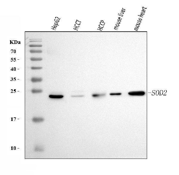
Western blot analysis of SOD2 using anti-SOD2 antibody (M00349-3).
Electrophoresis was performed on a 5-20% SDS-PAGE gel at 70V (Stacking gel) / 90V (Resolving gel) for 2-3 hours. The sample well of each lane was loaded with 30 ug of sample under reducing conditions.
Lane 1: human HepG2 whole cell lysates,
Lane 2: human HCCT tissue lysates,
Lane 3: human HCCP tissue lysates,
Lane 4: mouse liver tissue lysates,
Lane 5: mouse heart tissue lysates.
After electrophoresis, proteins were transferred to a nitrocellulose membrane at 150 mA for 50-90 minutes. Blocked the membrane with 5% non-fat milk/TBS for 1.5 hour at RT. The membrane was incubated with mouse anti-SOD2 antigen affinity purified monoclonal antibody (Catalog # M00349-3) at 0.5 μg/mL overnight at 4°C, then washed with TBS-0.1%Tween 3 times with 5 minutes each and probed with a goat anti-mouse IgG-HRP secondary antibody at a dilution of 1:10000 for 1.5 hour at RT. The signal is developed using an Enhanced Chemiluminescent detection (ECL) kit (Catalog # EK1001) with Tanon 5200 system. A specific band was detected for SOD2 at approximately 25 kDa. The expected band size for SOD2 is at 25 kDa.

Click image to see more details
IHC analysis of SOD2 using anti-SOD2 antibody (M00349-3).
SOD2 was detected in a paraffin-embedded section of human adenocarcinoma of the right colon tissue. Heat mediated antigen retrieval was performed in EDTA buffer (pH 8.0, epitope retrieval solution). The tissue section was blocked with 10% goat serum. The tissue section was then incubated with 2 μg/ml mouse anti-SOD2 Antibody (M00349-3) overnight at 4°C. Peroxidase Conjugated Goat Anti-mouse IgG was used as secondary antibody and incubated for 30 minutes at 37°C. The tissue section was developed using HRP Conjugated Mouse IgG Super Vision Assay Kit (Catalog # SV0001) with DAB as the chromogen.

Click image to see more details
IHC analysis of SOD2 using anti-SOD2 antibody (M00349-3).
SOD2 was detected in a paraffin-embedded section of human liver cancer tissue. Heat mediated antigen retrieval was performed in EDTA buffer (pH 8.0, epitope retrieval solution). The tissue section was blocked with 10% goat serum. The tissue section was then incubated with 2 μg/ml mouse anti-SOD2 Antibody (M00349-3) overnight at 4°C. Peroxidase Conjugated Goat Anti-mouse IgG was used as secondary antibody and incubated for 30 minutes at 37°C. The tissue section was developed using HRP Conjugated Mouse IgG Super Vision Assay Kit (Catalog # SV0001) with DAB as the chromogen.

Click image to see more details
IHC analysis of SOD2 using anti-SOD2 antibody (M00349-3).
SOD2 was detected in a paraffin-embedded section of human lymphoma tissue. Heat mediated antigen retrieval was performed in EDTA buffer (pH 8.0, epitope retrieval solution). The tissue section was blocked with 10% goat serum. The tissue section was then incubated with 2 μg/ml mouse anti-SOD2 Antibody (M00349-3) overnight at 4°C. Peroxidase Conjugated Goat Anti-mouse IgG was used as secondary antibody and incubated for 30 minutes at 37°C. The tissue section was developed using HRP Conjugated Mouse IgG Super Vision Assay Kit (Catalog # SV0001) with DAB as the chromogen.
Specific Publications For Anti-SOD2 Antibody Picoband® (monoclonal, 2B12B1) (M00349-3)
Loading publications
Recommended Resources
Here are featured tools and databases that you might find useful.
- Boster's Pathways Library
- Protein Databases
- Bioscience Research Protocol Resources
- Data Processing & Analysis Software
- Photo Editing Software
- Scientific Literature Resources
- Research Paper Management Tools
- Molecular Biology Software
- Primer Design Tools
- Bioinformatics Tools
- Phylogenetic Tree Analysis
Customer Reviews
Have you used Anti-SOD2 Antibody Picoband® (monoclonal, 2B12B1)?
Share your experimental results or join a short interview to earn up to $1,000 in product credits or other rewards.
0 Reviews For Anti-SOD2 Antibody Picoband® (monoclonal, 2B12B1)
Customer Q&As
Have a question?
Find answers in Q&As, reviews.
Can't find your answer?
Submit your question
