Click image to see more details
Product Info Summary
| SKU: | A00662-2 |
|---|---|
| Size: | 100 μg/vial |
| Reactive Species: | Human, Mouse, Rat |
| Host: | Rabbit |
| Application: | ELISA, WB |
Customers Who Bought This Also Bought

Product info
Product Name
Anti-TFEB Antibody Picoband®
SKU/Catalog Number
A00662-2
Size
100 μg/vial
Form
Lyophilized
Description
Boster Bio Anti-TFEB Antibody Picoband® catalog # A00662-2. Tested in ELISA, WB applications. This antibody reacts with Human, Mouse, Rat. The brand Picoband indicates this is a premium antibody that guarantees superior quality, high affinity, and strong signals with minimal background in Western blot applications. Only our best-performing antibodies are designated as Picoband, ensuring unmatched performance.
Storage & Handling
Store at -20˚C for one year from date of receipt. After reconstitution, at 4˚C for one month. It can also be aliquotted and stored frozen at -20˚C for six months. Avoid repeated freeze-thaw cycles.
Cite This Product
Anti-TFEB Antibody Picoband® (Boster Biological Technology, Pleasanton CA, USA, Catalog # A00662-2)
Host
Rabbit
Contents
Each vial contains 4 mg Trehalose, 0.9 mg NaCl and 0.2 mg Na2HPO4.
Clonality
Polyclonal
Isotype
Rabbit IgG
Immunogen
E.coli-derived human TFEB recombinant protein (Position: E68-L350).
Cross-reactivity
No cross-reactivity with other proteins.
Reactive Species
A00662-2 is reactive to TFEB in Human, Mouse, Rat
Observed Molecular Weight
65 kDa
Calculated molecular weight
52.9 kDa
Background of TFEB
Transcription factor EB is a protein that in humans is encoded by the TFEB gene. TFEB is a master gene for lysosomal biogenesis. It encodes a transcription factor that coordinates expression of lysosomal hydrolases, membrane proteins and genes involved in autophagy. Upon nutrient depletion and under aberrant lysosomal storage conditions such as in lysosomal storage diseases, TFEB translocates from the cytoplasm to the nucleus, resulting in the activation of its target genes. TFEB overexpression in cultured cells induces lysosomal biogenesis, exocytosis and autophagy. Viral-mediated TFEB overexpression in cellular and mouse models of lysosomal storage disorders and in common neurodegenerative diseases such as Huntington, Parkinson and Alzheimer diseases, resulted in intracellular clearance of accumulating molecules and rescue of disease phenotypes. TFEB is activated by PGC1-alpha and promotes reduction of htt aggregation and neurotoxicity in a mouse model of Huntington disease. TFEB overexpression has been found in patients with renal cell carcinoma and pancreatic cancer and was shown to promote tumorogenesis via induction of varius oncogenic signals.
Antibody Validation
Boster validates all antibodies on WB, IHC, ICC, Immunofluorescence, and ELISA with known positive control and negative samples to ensure specificity and high affinity, including thorough antibody incubations.
Application & Images
Applications
A00662-2 is guaranteed for ELISA, WB Boster Guarantee
Recommend Dilution
| Application | Dilution | Species |
|---|---|---|
| Western blot | 0.25-0.5μg/ml | Human, Mouse, Rat |
| ELISA | 0.1-0.5μg/ml | - |
Tested application
Suggested blocking solution with 5% non-fat milk or BSA; (*)Recommended protein loading: 20-40 µg per lane
Validation Images & Assay Conditions
Click image to see more details
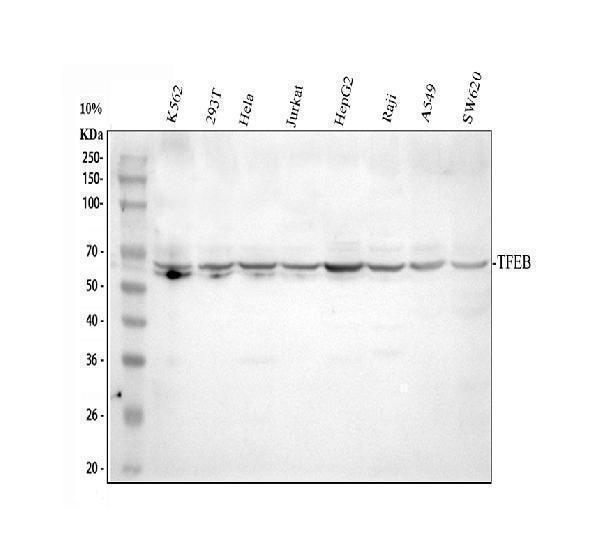
Western blot analysis of TFEB using anti-TFEB antibody (A00662-2).
Electrophoresis was performed on a 10% SDS-PAGE gel at 80V (Stacking gel) / 120V (Resolving gel) for 2 hours. The sample well of each lane was loaded with 30 ug of sample under reducing conditions.
Lane 1: human K562 whole cell lysates,
Lane 2: human 293T whole cell lysates,
Lane 3: human Hela whole cell lysates,
Lane 4: human Jurkat whole cell lysates,
Lane 5: human HepG2 whole cell lysates,
Lane 6: human Raji whole cell lysates,
Lane 7: human A549 whole cell lysates,
Lane 8: human SW620 whole cell lysates.
After Electrophoresis, proteins were transferred to a Nitrocellulose membrane at 150mA for 50-90 minutes. Blocked the membrane with 5% Non-fat Milk/ TBS for 1.5 hour at RT. The membrane was incubated with rabbit anti-TFEB antigen affinity purified polyclonal antibody (Catalog # A00662-2) at 0.5 μg/mL overnight at 4°C, then washed with TBS-0.1%Tween 3 times with 5 minutes each and probed with a goat anti-rabbit IgG-HRP secondary antibody at a dilution of 1:5000 for 1.5 hour at RT. The signal is developed using an ECL Plus Western Blotting Substrate (Catalog # AR1196-200) with Tanon 5200 system. A specific band was detected for TFEB at approximately 65 kDa. The expected band size for TFEB is at 53 kDa.
Specific Publications For Anti-TFEB Antibody Picoband® (A00662-2)
Loading publications
Recommended Resources
Here are featured tools and databases that you might find useful.
- Boster's Pathways Library
- Protein Databases
- Bioscience Research Protocol Resources
- Data Processing & Analysis Software
- Photo Editing Software
- Scientific Literature Resources
- Research Paper Management Tools
- Molecular Biology Software
- Primer Design Tools
- Bioinformatics Tools
- Phylogenetic Tree Analysis
Customer Reviews
Have you used Anti-TFEB Antibody Picoband®?
Share your experimental results or join a short interview to earn up to $1,000 in product credits or other rewards.
0 Reviews For Anti-TFEB Antibody Picoband®
Customer Q&As
Have a question?
Find answers in Q&As, reviews.
Can't find your answer?
Submit your question
