
Click image to see more details
-

-
-
-
-
+7
Product Info Summary
| SKU: | PB9234 |
|---|---|
| Size: | 100 μg/vial |
| Reactive Species: | Human, Mouse, Rat |
| Host: | Rabbit |
| Application: | Flow Cytometry, IF, IHC, ICC, WB |
Customers Who Bought This Also Bought

Product info
Product Name
Anti-ZO1 tight junction protein/TJP1 Antibody Picoband®
SKU/Catalog Number
PB9234
Size
100 μg/vial
Form
Lyophilized
Description
Boster Bio Anti-ZO1 tight junction protein/TJP1 Antibody Picoband® catalog # PB9234. Tested in Flow Cytometry, IF, IHC, ICC, WB applications. This antibody reacts with Human, Mouse, Rat. The brand Picoband indicates this is a premium antibody that guarantees superior quality, high affinity, and strong signals with minimal background in Western blot applications. Only our best-performing antibodies are designated as Picoband, ensuring unmatched performance.
Storage & Handling
Store at -20˚C for one year from date of receipt. After reconstitution, at 4˚C for one month. It can also be aliquotted and stored frozen at -20˚C for six months. Avoid repeated freeze-thaw cycles.
Cite This Product
Anti-ZO1 tight junction protein/TJP1 Antibody Picoband® (Boster Biological Technology, Pleasanton CA, USA, Catalog # PB9234)
Host
Rabbit
Contents
Each vial contains antibody formulated with stabilizing components, 0.9 mg NaCl, 0.2 mg Na2HPO4, and 0.05 mg NaN3.
*This antibody is supplied in a stabilized formulation.
Compatibility with conjugation reactions depends on the chemistry of the conjugation method used.
For conjugation methods that are not compatible with the stabilizing components present in this formulation, a carrier-free antibody format is required.
Clonality
Polyclonal
Isotype
Rabbit IgG
Immunogen
E.coli-derived human TJP1 recombinant protein (Position: H1178-F1527). Human TJP1 shares 82% amino acid (aa) sequence identity with mouse TJP1.
Cross-reactivity
No cross-reactivity with other proteins
Reactive Species
PB9234 is reactive to TJP1 in Human, Mouse, Rat
Observed Molecular Weight
220 kDa
Calculated molecular weight
195.5 kDa
Background of TJP1
Tight junction protein ZO-1 is a protein that in humans is encoded by the TJP1 gene. It is mapped to 15q13.1. This gene encodes a protein located on a cytoplasmic membrane surface of intercellular tight junctions. The encoded protein may be involved in signal transduction at cell–cell junctions. It has been found that injected CagA associates with the epithelial tight-junction scaffolding protein TJP1 and the transmembrane protein junctional adhesion molecule, causing an ectopic assembly of tight junction components at sites of bacterial attachment, and altering the composition and function of the apical-junctional complex.
Antibody Validation
Boster validates all antibodies on WB, IHC, ICC, Immunofluorescence, and ELISA with known positive control and negative samples to ensure specificity and high affinity, including thorough antibody incubations.
Application & Images
Applications
PB9234 is guaranteed for Flow Cytometry, IF, IHC, ICC, WB Boster Guarantee
Recommend Dilution
| Application | Dilution | Species |
|---|---|---|
| Western blot | 0.1-0.5μg/ml | Human, Mouse, Rat |
| Immunohistochemistry (Paraffin-embedded Section) | 0.5-1μg/ml | Human |
| Immunocytochemistry/Immunofluorescence | 2μg/ml | Human |
| Flow Cytometry (Fixed) | 1-3μg/1x106 cells | Human |
Tested application
Suggested blocking solution with 5% non-fat milk or BSA; (*)Recommended protein loading: 20-40 µg per lane
Use TE buffer pH 9.0 for antigen retrieval; (*) citrate buffer pH 6.0 is an alternative.
Validation Images & Assay Conditions
Click image to see more details
Western blot analysis of TJP1 using anti-TJP1 antibody (PB9234).
Electrophoresis was performed on a 5-20% SDS-PAGE gel at 70V (Stacking gel) / 90V (Resolving gel) for 2-3 hours. The sample well of each lane was loaded with 30 ug of sample under reducing conditions.
Lane 1: human PC-3 whole cell lysates,
Lane 2: human CACO-2 whole cell lysates,
Lane 3: human COLO320 whole cell lysates,
Lane 4: rat testis tissue lysates,
Lane 5: mouse testis tissue lysates.
After electrophoresis, proteins were transferred to a nitrocellulose membrane at 150 mA for 50-90 minutes. Blocked the membrane with 5% non-fat milk/TBS for 1.5 hour at RT. The membrane was incubated with rabbit anti-TJP1 antigen affinity purified polyclonal antibody (Catalog # PB9234) at 0.5 μg/mL overnight at 4°C, then washed with TBS-0.1%Tween 3 times with 5 minutes each and probed with a goat anti-rabbit IgG-HRP secondary antibody at a dilution of 1:5000 for 1.5 hour at RT. The signal is developed using an Enhanced Chemiluminescent detection (ECL) kit (Catalog # EK1002) with Tanon 5200 system. A specific band was detected for TJP1 at approximately 220 kDa. The expected band size for TJP1 is at 185 kDa.

Click image to see more details
IHC analysis of TJP1 using anti-TJP1 antibody (PB9234).
TJP1 was detected in paraffin-embedded section of human intestinal cancer tissue. Heat mediated antigen retrieval was performed in citrate buffer (pH6, epitope retrieval solution) for 20 mins. The tissue section was blocked with 10% goat serum. The tissue section was then incubated with 1μg/ml rabbit anti-TJP1 Antibody (PB9234) overnight at 4°C. Biotinylated goat anti-rabbit IgG was used as secondary antibody and incubated for 30 minutes at 37°C. The tissue section was developed using Strepavidin-Biotin-Complex (SABC)(Catalog # SA1022) with DAB as the chromogen.

Click image to see more details
IF analysis of TJP1 using anti-TJP1 antibody (PB9234).
TJP1 was detected in immunocytochemical section of A431 cells. Enzyme antigen retrieval was performed using IHC enzyme antigen retrieval reagent (AR0022) for 15 mins. The cells were blocked with 10% goat serum. And then incubated with 2μg/mL rabbit anti-TJP1 Antibody (PB9234) overnight at 4°C. DyLight®488 Conjugated Goat Anti-Rabbit IgG (BA1127) was used as secondary antibody at 1:100 dilution and incubated for 30 minutes at 37°C. The section was counterstained with DAPI. Visualize using a fluorescence microscope and filter sets appropriate for the label used.

Click image to see more details
IF analysis of TJP1 using anti-TJP1 antibody (PB9234).
TJP1 was detected in an immunocytochemical section of Endothelial cells. Enzyme antigen retrieval was performed using IHC enzyme antigen retrieval reagent (AR0022) for 15 mins. The cells were blocked with 10% goat serum. And then incubated with 1:200 rabbit anti-TJP1 Antibody (PB9234) overnight at 4°C. DyLight®488 Conjugated Goat Anti-Rabbit IgG (BA1127) was used as secondary antibody at 1:500 dilution and incubated for 30 minutes at 37°C. The section was counterstained with DAPI. Visualize using a fluorescence microscope and filter sets appropriate for the label used.

Click image to see more details
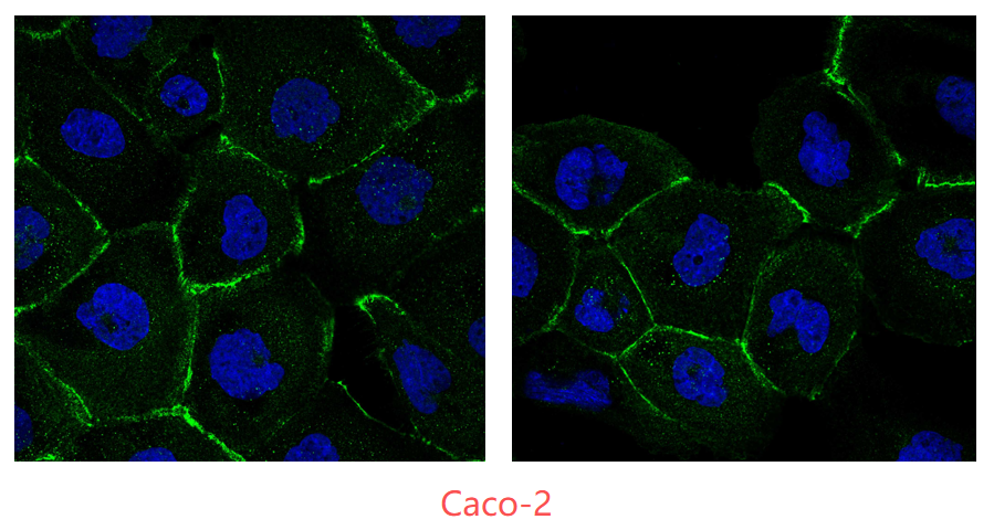
IF analysis of TJP1 using anti-TJP1 antibody (PB9234).
TJP1 was detected in an immunocytochemical section of human Caco-2 cells. Cells were normally cultured in 24-well plates using MEM supplemented with 20% fetal bovine serum. When the cell density reached approximately 60%, the culture was stopped. The medium was removed, cells were washed three times with PBS, fixed with 4% paraformaldehyde for 15 minutes, and then washed three times with PBS before further use. The cells were blocked with 10% goat serum. And then incubated with 1:200 rabbit anti-TJP1 Antibody (PB9234) overnight at 4°C. DyLight®488 Conjugated Goat Anti-Rabbit IgG (BA1127) was used as secondary antibody at 1:500 dilution and incubated for 45 minutes at 37°C. The section was counterstained with DAPI. Visualize using a fluorescence microscope and filter sets appropriate for the label used.

Click image to see more details
Flow Cytometry analysis of K562 cells using anti-TJP1 antibody (PB9234).
Overlay histogram showing K562 cells stained with PB9234 (Blue line). The cells were fixed with 4% paraformaldehyde and blocked with 10% normal goat serum. And then incubated with rabbit anti-TJP1 Antibody (PB9234,1μg/1x106 cells) for 30 min at 20°C. DyLight®488 conjugated goat anti-rabbit IgG (BA1127, 5-10μg/1x106 cells) was used as secondary antibody for 30 minutes at 20°C. Isotype control antibody (Green line) was rabbit IgG (1μg/1x106) used under the same conditions. Unlabelled sample without incubation with primary antibody and secondary antibody (Red line) was used as a blank control.

Click image to see more details
Effects of MT water extract on regulating macrophage polarization, maintaining intestinal barrier integrity, and alleviating oxidative stress in the colons of DSS-induced UC mice. (A) IHC staining of iNOS, Arg1, occludin, and ZO-1 expressions in mouse colon tissues. Quantitative analysis of the integrated optical density (IOD) of (B) iNOS, (C) Arg1, (D) occludin, and (E) ZO-1 in each group. The levels of (F) IL-6, (G) IL-10, (H) MDA, and (I) GSH were measured and analyzed. Data were presented as means ± SD for three independent trials. One-way ANOVA followed by Tukey's multiple comparison test was performed to compare the differences between groups. *p < 0.05 versus control and #p < 0.05 versus DSS alone group. Arg1, arginase 1; DSS, dextran sulfate sodium; IL, interleukin; iNOS, inducible nitric oxide synthase; MDA, malondialdehyde; MT, Medulla Tetrapanacis.
Index in Food Frontiers under a CC BY license. DOI: 10.1002/fft2.70164

Click image to see more details
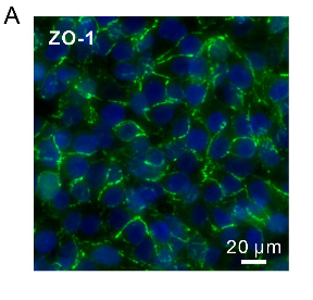
The subcellular localization and relative expression level detection of ZO-1 and villin in the intestinal mucosa of IBD at 7 days after termination of DSS intake. (A) The ZO-1 immunohistochemistry staining of the small intestinal epithelial TJP (brown dots): (A1) the normal group: the villi and crypts were arranged compactly, and the ZO-1-positive staining (representing the TJP) showed the dotted line (brown) along the surface of the villi and the crypts; (A2) the DSS group: ZO-1 distributed dispersively in the residual villi of the small intestinal mucosa; (A3) the DSS + B. subtilis -fermented milk group: the ZO-1 staining formed the dotted line (brown, representing the TJP) at the subsurface of the regenerative villi. (B) The ZO-1 immunohistochemistry staining of the colonic epithelial TJP (brown dots): (B1) the normal group: ZO-1-positive staining distributed on the inner side of the epithelial cell membrane (representing the TJP); (B2) the DSS group: there was no ZO-1-positive staining in the scar; (B3) the DSS + B. subtilis -fermented milk group: the ZO-1-positive staining distributed on the inner side of the regenerative epithelial cell membrane (representing the TJP). (C) The villin immunohistochemistry staining (brown strip) of the small intestinal microvilli: (C1) the normal group: villin-positive staining showed a strip-like distribution on the surface of the villi in the normal small intestinal mucosa; (C2) the DSS group: villin distributed at the surface of the residual villi; (C3) the DSS + B. subtilis -fermented milk group: villin-positive staining formed an integrative strip (brown) enclosing the surface of the regenerative villi. (D) The villin immunohistochemistry staining of the colonic epithelium: (D1) the normal group: villin-positive staining (brown) showed banded distribution on the surface of the epithelium; (D2) the DSS group: almost no villin-positive staining was observed in the scar due to damage of the epithelium; (D3) the DSS + B. subtilis -fermented milk group: the villin-positive staining (brown) showed banded distribution on the surface of the regenerated epithelium in the colonic mucosa. (E,F) The western blotting analysis for the relative expression level of ZO-1 and villin in the samples contained equivalent ileum and colon. The expression level of ZO-1 and villin in the DSS group was significantly lower than that of the normal (control) group. The expression level of ZO-1 and villin and in the DSS + B. subtilis -fermented milk (FM) group was significantly higher than that of the DSS group ( n = 5, * represents p < 0.05, ** represents p < 0.01).
Index in PubMed under a CC BY license. PMID: 33519783

Click image to see more details
Simvastatin reversed protein expression of high glucose-induced ZO-1 and VE-Cadherin. RAECs were incubated with high glucose for 24h, which was treated with simvastatin (SIM, 5μM) in the presence or absence of NLRP3 siRNA or MCC950 (15nM). Representative Western blot gel documents (A, B) and summarized data showing the protein expression of ZO-1 (C, D) and VE-Cadherin (E, F) . (G) Representative fluorescence images showing the cell membrane fluorescence of ZO-1 from at least three independent experiments. (H) Frequency histogram of ZO-1 in the membranes showing the protein expression of ZO-1 by flowcytometry. * P <0.05 vs. Scram Vehl or Ctrl Vehl; # P <0.05 vs. HG treated group (n=4).
Index in PubMed under a CC BY license. PMID: 29207644

Click image to see more details
Effect of Lacticaseibacillus rhamnosus E9 administration on tight junction proteins and SOX-10 in ileum of MPTP-induced model of PD compared to the control. ( A ) Immunohistochemical staining of ileal ZO-1 (100 ×), Occludin (100 ×) (staining intensity is evaluated in ideal mucosa), and SOX-10 (200 ×, inset-4000 ×) (Arrows are nuclear positivity in myenteric plexus). ( B ) Protein expression of ileal ZO-1, Occludin, and SOX-10 in PD and control mice. ( C ) Gene expression of ileal ZO-1 and Occludin in PD and control mice. MPTP mice were received (i.p.) 30 mg/kg MPTP-HCl daily for 5 consecutive days (MPTP and MPTP + P). Probiotic mice were administered 1 dose (10 8 CFU/mouse/day) daily of L. rhamnosus E9 for fifteen days and sacrificed after the last dose (P and MPTP + P). *p ≤ 0.05, control vs MPTP (n:4–5/group).
Index in PubMed under a CC BY license. PMID: 38965287

Click image to see more details
Tight junction formation by HBMVEC under flow conditions as indicated by immunofluorescence staining of ZO-1. PKCδ inhibition (PKCδ- i ) attenuates TNF-α-induced tight junction damage in vitro in B 3 C. When cultured with normal media, tight junctions were fully established between adjacent cells ( a ). Tight junction expression was disrupted after 4 h of TNF-α activation ( b ), while PKCδ inhibition (TNF-α + PKCδ- i ) restored tight junction expression ( c ). HBMVEC cultured for 72 h under flow (0.1 μl/min) were stained with ZO-1 (red) and Hoechst 33342 (blue). d Quantitative analysis to the total tight junction fluorescence intensity confirmed our observation. Data are presented as mean ± SEM ( n = 3). *** p < 0.001 compared to no treatment and TNF-α + PKCδ- i by ANOVA with Tukey-Kramer post hoc
Index in PubMed under a CC BY license. PMID: 30400800
Specific Publications For Anti-ZO1 tight junction protein/TJP1 Antibody Picoband® (PB9234)
Loading publications
Recommended Resources
Here are featured tools and databases that you might find useful.
- Boster's Pathways Library
- Protein Databases
- Bioscience Research Protocol Resources
- Data Processing & Analysis Software
- Photo Editing Software
- Scientific Literature Resources
- Research Paper Management Tools
- Molecular Biology Software
- Primer Design Tools
- Bioinformatics Tools
- Phylogenetic Tree Analysis
Customer Reviews
Have you used Anti-ZO1 tight junction protein/TJP1 Antibody Picoband®?
Share your experimental results or join a short interview to earn up to $1,000 in product credits or other rewards.
2 Reviews For Anti-ZO1 tight junction protein/TJP1 Antibody Picoband®
In an immunofluorescence experiment using normally cultured Caco-2 cells, the ZO-1 antibody (PB9234) showed clear and specific membrane staining with low background, demonstrating reliable performance.
Excellent
| SKU | PB9234 |
|---|---|
| Application | Immunofluorescence |
| Sample | human Caco-2 cell line |
| Sample Processing Description | Cells were normally cultured in 24-well plates using MEM supplemented with 20% fetal bovine serum. When the cell density reached approximately 60%, the culture was terminated. The medium was removed, cells were washed three times with PBS, fixed with 4% paraformaldehyde for 15 minutes, and then washed three times with PBS before further use. |
| Other Reagents | Goat serum, DAPI, and an anti-fade mounting medium. |
| Primary Antibody | ZO1 tight junction protein/TJP1 Antibody Picoband® |
| Primary Incubation | 1:200, overnight at 4 ℃ |
| Secondary Antibody | Goat Anti-Rabbit IgG (H+L) Secondary Antibody, Fluoro488 Conjugated |
| Secondary Incubation | 45 min at 37℃ |
| Detection | Imaging system:Leica DM2500 |
| Results Summary | Caco-2 cells are a “gold standard” in vitro model for studying intestinal epithelial function, and ZO-1 is a key marker of barrier integrity. The images show immunofluorescence staining of ZO-1 in normally cultured Caco-2 cells to evaluate antibody performance. Clear membrane localization and accurate staining indicate that this antibody is suitable for downstream research applications. |
Guangyu Mei, Tongji University
Verified customer
Submitted 2026-02-06
I was so thrilled that I took the photo below — it felt like a true treasure.
Excellent
| SKU | PB9234 |
|---|---|
| Application | ICC/IF |
| Sample | Endothelial cells |
| Sample Processing Description | Endothelial cells were seeded in collagen gel-treated chips for 12 hours. |
| Primary Antibody | Anti-ZO1 tight junction protein/TJP1 Antibody Picoband® |
| Primary Incubation | 1:200, overnight at 4 ℃ |
| Blocking Agent | Ready-to-use Goat Serum |
| Secondary Antibody | Goat Anti-Rabbit IgG (H+L) Secondary Antibody, DyLight®488 Conjugated(BA1127) |
| Secondary Incubation | Incubate at room temperature for 1 hour |
| Other reagents | DAPI (AR1176), Anti-fade mounting medium |
| Detection | Fluorescence microscope |
| Results Summary | At a critical stage of our manuscript preparation, we needed to stain tight junction proteins in endothelial cells. After trying primary antibodies from many companies without achieving satisfactory results, it was ultimately Boster’s product that solved the problem (Figure 5A). I was so excited that I took the photo below — treating it like a true treasure. |
Wenting Fan
Verified customer
Submitted 2025-10-27
Customer Q&As
Have a question?
Find answers in Q&As, reviews.
Can't find your answer?
Submit your question
5 Customer Q&As for Anti-ZO1 tight junction protein/TJP1 Antibody Picoband®
Question
Is a blocking peptide available for product anti-ZO1 tight junction protein/TJP1 antibody (PB9234)?
Verified Customer
Verified customer
Asked: 2019-11-21
Answer
We do provide the blocking peptide for product anti-ZO1 tight junction protein/TJP1 antibody (PB9234). If you would like to place an order for it please contact support@bosterbio.com and make a special request.
Boster Scientific Support
Answered: 2019-11-21
Question
We are currently using anti-ZO1 tight junction protein/TJP1 antibody PB9234 for rat tissue, and we are happy with the WB results. The species of reactivity given in the datasheet says human, mouse, rat. Is it likely that the antibody can work on primate tissues as well?
Verified Customer
Verified customer
Asked: 2019-09-23
Answer
The anti-ZO1 tight junction protein/TJP1 antibody (PB9234) has not been tested for cross reactivity specifically with primate tissues, though there is a good chance of cross reactivity. We have an innovator award program that if you test this antibody and show it works in primate you can get your next antibody for free. Please contact me if I can help you with anything.
Boster Scientific Support
Answered: 2019-09-23
Question
For PB9234's WB image, the customer would like to confirm that the antibody dilution as 0.5ug/ml. Also, the customer wanted to know the detailed transfer conditions for the QC data, is the information on the WB image sufficient or did we have more information?
Verified Customer
Verified customer
Asked: 2019-07-29
Answer
The antibody dilution is 0.5ug/mL. After Electrophoresis, proteins were wet transferred to a Nitrocellulose membrane at 150mA for 90 minutes.
Boster Scientific Support
Answered: 2019-07-29
Question
Our lab want to know about to test anti-ZO1 tight junction protein/TJP1 antibody PB9234 on rat liver for research purposes, then I may be interested in using anti-ZO1 tight junction protein/TJP1 antibody PB9234 for diagnostic purposes as well. Is the antibody suitable for diagnostic purposes?
Verified Customer
Verified customer
Asked: 2018-10-10
Answer
The products we sell, including anti-ZO1 tight junction protein/TJP1 antibody PB9234, are only intended for research use. They would not be suitable for use in diagnostic work. If you have the means to develop a product into diagnostic use, and are interested in collaborating with us and develop our product into an IVD product, please contact us for more discussions.
Boster Scientific Support
Answered: 2018-10-10
Question
Would anti-ZO1 tight junction protein/TJP1 antibody PB9234 work on feline WB with cervix carcinoma erythroleukemia?
Verified Customer
Verified customer
Asked: 2018-01-03
Answer
Our lab technicians have not validated anti-ZO1 tight junction protein/TJP1 antibody PB9234 on feline. You can run a BLAST between feline and the immunogen sequence of anti-ZO1 tight junction protein/TJP1 antibody PB9234 to see if they may cross-react. If the sequence homology is close, then you can perform a pilot test. Keep in mind that since we have not validated feline samples, this use of the antibody is not covered by our guarantee. However we have an innovator award program that if you test this antibody and show it works in feline cervix carcinoma erythroleukemia in WB, you can get your next antibody for free.
Boster Scientific Support
Answered: 2018-01-03
