Click image to see more details
Product Info Summary
| SKU: | M00625 |
|---|---|
| Size: | 100ug |
| Reactive Species: | Human, Mouse |
| Host: | Mouse |
| Application: | ELISA, IHC, WB |
Customers Who Bought This Also Bought

Product info
Product Name
Anti-TRPC6 Monoclonal Antibody
SKU/Catalog Number
M00625
Size
100ug
Form
Liquid (sterile filtered)
Description
Boster Bio Anti-TRPC6 Monoclonal Antibody (Catalog # M00625). Tested in ELISA, IHC, WB applications. This antibody reacts with Human, Mouse.
Storage & Handling
Store vial at -20°C prior to opening. Aliquot contents and freeze at -20°C or below for extended storage. Avoid cycles of freezing and thawing. Centrifuge product if not completely clear after standing at room temperature. This product is stable for several weeks at 4°C as an undiluted liquid. Dilute only prior to immediate use. Expiration date is one (1) year from date of opening. (Ship on dry ice.)
Cite This Product
Anti-TRPC6 Monoclonal Antibody (Boster Biological Technology, Pleasanton CA, USA, Catalog # M00625)
Host
Mouse
Contents
0.02 M Potassium Phosphate, 0.15 M Sodium Chloride, pH 7.2, 0.01% (w/v) Sodium Azide
Clonality
Monoclonal
Clone Number
Clone: 3F2.H10.F2 IgG1 kappa
Isotype
IgG1 kappa
Immunogen
This monoclonal antibody was produced by repeated immunizations with a synthetic peptide corresponding to a region near the carboxy terminus of human TRPC6 protein.
Cross-reactivity
No cross reactivity with other proteins.
Reactive Species
M00625 is reactive to TRPC6 in Human, Mouse
Calculated molecular weight
106.3 kDa
Background of TRPC6
TRPC6, also known as TRP6, short transient receptor potential channel 6 and transient receptor potential cation channel subfamily C member 6, is thought to form a receptor-activated non-selective calcium permeant cation channel. TRPC6 is probably operated by a phosphatidylinositol second messenger system activated by receptor tyrosine kinases or G-protein coupled receptors. It is activated by diacylglycerol (DAG) in a membrane-delimited fashion, independently of protein kinase C and may not to be activated by intracellular calcium store depletion. Defects in this gene are a cause of focal segmental glomerulosclerosis (FSGS). Expression of this protein has been reported in tissues such as placenta, lung, spleen, ovary, small intestine, and renal podocytes. Immunohistochemistry studies using polyclonal antibodies to this target have shown moderate to strong staining in cell types such as neurons, breast, respiratory, squamous and prostate epithelium, epidermis, placental trophoblasts, dendritic cells, and subsets of immune cells, and faint to moderate staining of adrenal, colon, ganglion cells, hepatocytes, heart, and testis.
Antibody Validation
Boster validates all antibodies on WB, IHC, ICC, Immunofluorescence, and ELISA with known positive control and negative samples to ensure specificity and high affinity, including thorough antibody incubations.
Application & Images
Applications
M00625 is guaranteed for ELISA, IHC, WB Boster Guarantee
Recommend Dilution
| Application | Dilution | Species |
|---|---|---|
| ELISA: 1:10 | 000 - 1:50 | 000 |
| WB: 1:500- 1:2 | 000 | |
| Anti-TRPC6 monoclonal antibody (200-301-B59) clone # 3F2.H10.F2 was developed by Rockland Immunochemicals Inc. against human TRPC6 using conventional hybridoma technology by fusing splenocytes of a host animal immunized with a synthetic peptide corresponding to the cytosolic domain of TRPC6 with myeloma cells. The screening of clones during the subcloning process was based on immunohistochemistry using human tissue microarrays. The pathologist analyzing the staining patterns of clones reported that the antibody shows strong to moderate staining consistent with the localization of human TRPC6 in adrenal cortex | neurons | Purkinje cells, colon epithelium, cardiac myocytes, renal tubules, hepatocytes, skeletal muscle, pancreatic exocrine and islet cells, germinal center lymphocytes, plasma cells, Sertoli cells of the testes as well as staining more faintly other tissues known to be positive for the target protein (e.g., respiratory epithelium). Prostate and placenta were negative for staining. The antibody produced minimal to no background staining and appeared very specific at 2.5 µg/mL. The pattern of reactivity observed for this clone was also similar to other antibodies used for benchmarking purposes. Specific conditions for reactivity should be optimized by the end user, however, we suggest the use of formalin-fixed paraffin-embedded sections for immunohistochemistry. No pre-treatment of sample is required. While immunohistochemistry was used as the primary screening and release validation immunoassay, clone #3F2.H10.F2 was also screened by western blotting against known positive and negative control lysates. A single band is detected by this antibody in TRPC6 positive cells and tissues; however, the molecular weight of the band (~30 kDa) is not consistent with full length human TRPC6 (181 kDa). The band detected by this antibody may be the cleaved cytosolic domain of TRPC6 as the immunogen used for antibody production corresponds to an amino acid sequence located within this domain. However, no additional data is available to elucidate the molecular composition of this band. |
Validation Images & Assay Conditions
Click image to see more details
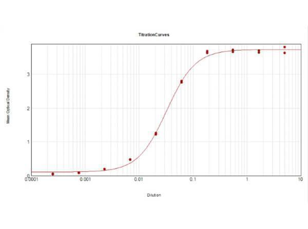
ELISA Results of Mouse Anti-TRPC6 Antibody. Each well was coated in duplicate with 0.1µg of conjugate. The working dilution is 1:31,000. The starting dilution of antibody was 5μg/ml and the X-axis represents the Log10 of a 3-fold dilution. This titration is a 4-parameter curve fit where the IC50 is defined as the titer of the antibody. Assay performed using HRP conjugation Stabilizer , Rabbit Anti-Mouse IgG HRP conjugated and TMB substrate .

Click image to see more details
Western Blot of Mouse Anti-TRPC6 Antibody. Lane 1: Opal Prestained Molecular Weight Marker . Lane 2: Mouse Pancreas Tissue Lysate [10µg]. Lane 3: MCF-7 Whole Cell Lysate [10µg]. Lane 4: A431 Whole Cell Lysate [10µg]. Lane 5: Jurkat Whole Cell Lysate [10µg]. Primary Antibody: Anti-TRPC6 at 1µg/mL overnight at 2-8°C. Secondary Antibody: Rabbit Anti-Mouse IgG Peroxidase 1:40000 for 30mins at RT. Blocking Buffer: BlockOut Buffer for 30mins at RT. Predicted MW: ~30kDa. Observed MW: ~40kDa. Exposure: 5sec.

Click image to see more details
Immunohistochemistry using Boster's anti-TRPC6 monoclonal antibody shows detection of TRPC6 in human adrenal (cortex) tissue (40X). The antibody was used a dilution to 2.5 µg/mL. The image shows strong staining with minimal background staining. Tissue was formalin fixed and paraffin embedded. No pre-treatment of sample was required. The image shows the localization of antibody as the precipitated red signal, with a hematoxylin purple nuclear counterstain. Personal communication, Andrew Elston, Lifespan Biosciences, Seattle, WA.

Click image to see more details
Western Blot of Mouse anti-TRPC6 Antibody. Lane 1: Mouse Kidney WCL . Load: 10 µg per lane. Primary antibody: TRPC6 Antibody at 1:1000 for overnight at 4°C. Secondary antibody: donkey anti-mouse DyLight™ 649 at 1:20,000 for 30 min at RT. Block: 30 min at RT.
Specific Publications For Anti-TRPC6 Monoclonal Antibody (M00625)
Loading publications
Recommended Resources
Here are featured tools and databases that you might find useful.
- Boster's Pathways Library
- Protein Databases
- Bioscience Research Protocol Resources
- Data Processing & Analysis Software
- Photo Editing Software
- Scientific Literature Resources
- Research Paper Management Tools
- Molecular Biology Software
- Primer Design Tools
- Bioinformatics Tools
- Phylogenetic Tree Analysis
Customer Reviews
Have you used Anti-TRPC6 Monoclonal Antibody?
Share your experimental results or join a short interview to earn up to $1,000 in product credits or other rewards.
0 Reviews For Anti-TRPC6 Monoclonal Antibody
Customer Q&As
Have a question?
Find answers in Q&As, reviews.
Can't find your answer?
Submit your question
4 Customer Q&As for Anti-TRPC6 Monoclonal Antibody
Question
I see that the anti-TRPC6 Monoclonal antibody M00625 works with IHC, what is the protocol used to produce the result images on the product page?
Verified Customer
Verified customer
Asked: 2020-04-28
Answer
You can find protocols for IHC on the "support/technical resources" section of our navigation menu. If you have any further questions, please send an email to support@bosterbio.com
Boster Scientific Support
Answered: 2020-04-28
Question
We are interested in to test anti-TRPC6 Monoclonal antibody M00625 on human placenta testis for research purposes, then I may be interested in using anti-TRPC6 Monoclonal antibody M00625 for diagnostic purposes as well. Is the antibody suitable for diagnostic purposes?
Verified Customer
Verified customer
Asked: 2019-06-24
Answer
The products we sell, including anti-TRPC6 Monoclonal antibody M00625, are only intended for research use. They would not be suitable for use in diagnostic work. If you have the means to develop a product into diagnostic use, and are interested in collaborating with us and develop our product into an IVD product, please contact us for more discussions.
Boster Scientific Support
Answered: 2019-06-24
Question
Here is the WB image, lot number and protocol we used for placenta testis using anti-TRPC6 Monoclonal antibody M00625. Please let me know if you require anything else.
Verified Customer
Verified customer
Asked: 2018-07-26
Answer
Thank you very much for the data. Our lab team are working to resolve this as quickly as possible, and we appreciate your patience and understanding! You have provided everything we needed. Please let me know if there is anything you need in the meantime.
Boster Scientific Support
Answered: 2018-07-26
Question
Is a blocking peptide available for product anti-TRPC6 Monoclonal antibody (M00625)?
N. Evans
Verified customer
Asked: 2016-06-16
Answer
We do provide the blocking peptide for product anti-TRPC6 Monoclonal antibody (M00625). If you would like to place an order for it please contact support@bosterbio.com and make a special request.
Boster Scientific Support
Answered: 2016-06-16
