Click image to see more details
-

-
-
-
-
+5
Product Info Summary
| SKU: | A01932-3 |
|---|---|
| Size: | 100 μg/vial |
| Reactive Species: | Human, Rat |
| Host: | Rabbit |
| Application: | ELISA, IF, IHC, ICC, WB |
Customers Who Bought This Also Bought

Product info
Product Name
Anti-TTC11/FIS1 Antibody Picoband®
SKU/Catalog Number
A01932-3
Size
100 μg/vial
Form
Lyophilized
Description
Boster Bio Anti-TTC11/FIS1 Antibody Picoband® catalog # A01932-3. Tested in ELISA, IF, IHC, ICC, WB applications. This antibody reacts with Human, Rat. The brand Picoband indicates this is a premium antibody that guarantees superior quality, high affinity, and strong signals with minimal background in Western blot applications. Only our best-performing antibodies are designated as Picoband, ensuring unmatched performance.
Storage & Handling
At -20°C for one year from date of receipt. After reconstitution, at 4°C for one month. It can also be aliquotted and stored frozen at -20°C for six months. Avoid repeated freezing and thawing.
Cite This Product
Anti-TTC11/FIS1 Antibody Picoband® (Boster Biological Technology, Pleasanton CA, USA, Catalog # A01932-3)
Host
Rabbit
Contents
Each vial contains 4 mg Trehalose, 0.9 mg NaCl, 0.2 mg Na2HPO4.
Clonality
Polyclonal
Isotype
Rabbit IgG
Immunogen
E.coli-derived human TTC11/FIS1 recombinant protein (Position: E18-S152).
Cross-reactivity
No cross-reactivity with other proteins.
Reactive Species
A01932-3 is reactive to FIS1 in Human, Rat
Observed Molecular Weight
17 kDa
Calculated molecular weight
16.9 kDa
Background of FIS1
Mitochondrial fission 1 protein (FIS1) is a protein that in humans is encoded by the FIS1 gene on chromosome 7. It is mapped to 7q22.1. The balance between fission and fusion regulates the morphology of mitochondria. TTC11 is a component of a mitochondrial complex that promotes mitochondrial fission. Its role in mitochondrial fission thus implicates it in the regulation of mitochondrial morphology, the cell cycle, and apoptosis. By extension, the protein is involved in associated diseases, including neurodegenerative diseases and cancers.
Antibody Validation
Boster validates all antibodies on WB, IHC, ICC, Immunofluorescence, and ELISA with known positive control and negative samples to ensure specificity and high affinity, including thorough antibody incubations.
Application & Images
Applications
A01932-3 is guaranteed for ELISA, IF, IHC, ICC, WB Boster Guarantee
Recommend Dilution
| Application | Dilution | Species |
|---|---|---|
| Western blot | 0.25-0.5 μg/ml | Human |
| Immunohistochemistry(Paraffin-embedded Section) | 2-5 μg/ml | Human, Rat |
| Immunocytochemistry/Immunofluorescence | 5 μg/ml | Human |
| ELISA | 0.1-0.5 μg/ml | - |
Tested application
Suggested blocking solution with 5% non-fat milk or BSA; (*)Recommended protein loading: 20-40 µg per lane
Use TE buffer pH 9.0 for antigen retrieval; (*) citrate buffer pH 6.0 is an alternative.
Validation Images & Assay Conditions
Click image to see more details
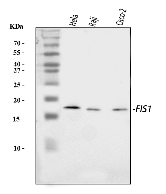
Western blot analysis of TTC11/FIS1 using anti-TTC11/FIS1 antibody (A01932-3).
Electrophoresis was performed on a 5-20% SDS-PAGE gel at 70V (Stacking gel) / 90V (Resolving gel) for 2-3 hours. The sample well of each lane was loaded with 30 ug of sample under reducing conditions.
Lane 1: human Hela whole cell lysates,
Lane 2: human Raji whole cell lysates,
Lane 3: human Caco-2 whole cell lysates.
After electrophoresis, proteins were transferred to a nitrocellulose membrane at 150 mA for 50-90 minutes. Blocked the membrane with 5% non-fat milk/TBS for 1.5 hour at RT. The membrane was incubated with rabbit anti-TTC11/FIS1 antigen affinity purified polyclonal antibody (Catalog # A01932-3) at 0.5 μg/mL overnight at 4°C, then washed with TBS-0.1%Tween 3 times with 5 minutes each and probed with a goat anti-rabbit IgG-HRP secondary antibody at a dilution of 1:5000 for 1.5 hour at RT. The signal is developed using an Enhanced Chemiluminescent detection (ECL) kit (Catalog # EK1002) with Tanon 5200 system. A specific band was detected for TTC11/FIS1 at approximately 17 kDa. The expected band size for TTC11/FIS1 is at 17 kDa.

Click image to see more details
DMOG treatment ameliorates mitochondrial dysfunction and enhances mitophagy and ATP production in aged MSCs. ( A ) Representative JC-1 fluorescence images showing mitochondrial membrane potential in different groups: CCCP (positive control for mitochondrial depolarization), P5 MSCs, P5 + H₂O₂-treated MSCs, P5 + H₂O₂+DMOG-treated MSCs, P15 MSCs, and P15 + DMOG-treated MSCs. JC-1 red fluorescence indicates high mitochondrial membrane potential, whereas green fluorescence indicates depolarized mitochondria. Scale bar = 100 μm. ( B ) Representative MitoSox Red fluorescence images showing mitochondrial ROS levels in different treatment groups. Scale bar = 10 μm. ( C , D ) Quantitative analysis of the mitochondrial membrane potential ( C ) and mitochondrial ROS levels ( D ). ( E ) Western blot analysis showing the expression levels of mitophagy-related proteins (MFN1, MFN2, and Fis1) in P5 and P15 MSCs with or without H₂O₂ and DMOG treatment. GAPDH was used as a loading control. ( F ) Representative confocal images of co-staining with LysoTracker Red (lysosomes) and MitoTracker Green (mitochondria) in MSCs under different conditions. Scale bar = 10 μm. ( G ) Quantitative analysis of the number of mitophagosomes per cell in the different groups. ( H ) Quantitative analysis of ATP production per cell in the different groups. Data are expressed as the mean ± SEM ( n = 3). * p < 0.05, ** p < 0.01. *** p < 0.001. Full-length blots are presented in Supplementary Materials - WB Raw Data Full size image
Index in PubMed under a CC BY license. PMID: 40457488

Click image to see more details
IHC analysis of TTC11/FIS1 using anti-TTC11/FIS1 antibody (A01932-3).
TTC11/FIS1 was detected in a paraffin-embedded section of human chronic tonsillitis tissue. Heat mediated antigen retrieval was performed in EDTA buffer (pH 8.0, epitope retrieval solution). The tissue section was blocked with 10% goat serum. The tissue section was then incubated with 2 μg/ml rabbit anti-TTC11/FIS1 Antibody (A01932-3) overnight at 4°C. Peroxidase Conjugated Goat Anti-rabbit IgG was used as secondary antibody and incubated for 30 minutes at 37°C. The tissue section was developed using HRP Conjugated Rabbit IgG Super Vision Assay Kit (Catalog # SV0002) with DAB as the chromogen.

Click image to see more details
IHC analysis of TTC11/FIS1 using anti-TTC11/FIS1 antibody (A01932-3).
TTC11/FIS1 was detected in a paraffin-embedded section of human laryngeal squamous cell carcinomas tissue. Heat mediated antigen retrieval was performed in EDTA buffer (pH 8.0, epitope retrieval solution). The tissue section was blocked with 10% goat serum. The tissue section was then incubated with 2 μg/ml rabbit anti-TTC11/FIS1 Antibody (A01932-3) overnight at 4°C. Peroxidase Conjugated Goat Anti-rabbit IgG was used as secondary antibody and incubated for 30 minutes at 37°C. The tissue section was developed using HRP Conjugated Rabbit IgG Super Vision Assay Kit (Catalog # SV0002) with DAB as the chromogen.

Click image to see more details
IHC analysis of TTC11/FIS1 using anti-TTC11/FIS1 antibody (A01932-3).
TTC11/FIS1 was detected in a paraffin-embedded section of human liver cancer tissue. Heat mediated antigen retrieval was performed in EDTA buffer (pH 8.0, epitope retrieval solution). The tissue section was blocked with 10% goat serum. The tissue section was then incubated with 2 μg/ml rabbit anti-TTC11/FIS1 Antibody (A01932-3) overnight at 4°C. Peroxidase Conjugated Goat Anti-rabbit IgG was used as secondary antibody and incubated for 30 minutes at 37°C. The tissue section was developed using HRP Conjugated Rabbit IgG Super Vision Assay Kit (Catalog # SV0002) with DAB as the chromogen.

Click image to see more details
IHC analysis of TTC11/FIS1 using anti-TTC11/FIS1 antibody (A01932-3).
TTC11/FIS1 was detected in a paraffin-embedded section of human placenta tissue. Heat mediated antigen retrieval was performed in EDTA buffer (pH 8.0, epitope retrieval solution). The tissue section was blocked with 10% goat serum. The tissue section was then incubated with 2 μg/ml rabbit anti-TTC11/FIS1 Antibody (A01932-3) overnight at 4°C. Peroxidase Conjugated Goat Anti-rabbit IgG was used as secondary antibody and incubated for 30 minutes at 37°C. The tissue section was developed using HRP Conjugated Rabbit IgG Super Vision Assay Kit (Catalog # SV0002) with DAB as the chromogen.

Click image to see more details
IHC analysis of TTC11/FIS1 using anti-TTC11/FIS1 antibody (A01932-3).
TTC11/FIS1 was detected in a paraffin-embedded section of rat gaster tissue. Heat mediated antigen retrieval was performed in EDTA buffer (pH 8.0, epitope retrieval solution). The tissue section was blocked with 10% goat serum. The tissue section was then incubated with 2 μg/ml rabbit anti-TTC11/FIS1 Antibody (A01932-3) overnight at 4°C. Peroxidase Conjugated Goat Anti-rabbit IgG was used as secondary antibody and incubated for 30 minutes at 37°C. The tissue section was developed using HRP Conjugated Rabbit IgG Super Vision Assay Kit (Catalog # SV0002) with DAB as the chromogen.

Click image to see more details
IHC analysis of TTC11/FIS1 using anti-TTC11/FIS1 antibody (A01932-3).
TTC11/FIS1 was detected in a paraffin-embedded section of human poison impregnated thyroid gland tissue. Heat mediated antigen retrieval was performed in EDTA buffer (pH 8.0, epitope retrieval solution). The tissue section was blocked with 10% goat serum. The tissue section was then incubated with 2 μg/ml rabbit anti-TTC11/FIS1 Antibody (A01932-3) overnight at 4°C. Peroxidase Conjugated Goat Anti-rabbit IgG was used as secondary antibody and incubated for 30 minutes at 37°C. The tissue section was developed using HRP Conjugated Rabbit IgG Super Vision Assay Kit (Catalog # SV0002) with DAB as the chromogen.

Click image to see more details
IF analysis of TTC11/FIS1 using anti-TTC11/FIS1 antibody (A01932-3).
TTC11/FIS1 was detected in an immunocytochemical section of MCF-7 cells. Enzyme antigen retrieval was performed using IHC enzyme antigen retrieval reagent (AR0022) for 15 mins. The cells were blocked with 10% goat serum. And then incubated with 5 μg/mL rabbit anti-TTC11/FIS1 Antibody (A01932-3) overnight at 4°C. DyLight®488 Conjugated Goat Anti-Rabbit IgG (BA1127) was used as secondary antibody at 1:100 dilution and incubated for 30 minutes at 37°C. The section was counterstained with DAPI. Visualize using a fluorescence microscope and filter sets appropriate for the label used.
Specific Publications For Anti-TTC11/FIS1 Antibody Picoband® (A01932-3)
Loading publications
Recommended Resources
Here are featured tools and databases that you might find useful.
- Boster's Pathways Library
- Protein Databases
- Bioscience Research Protocol Resources
- Data Processing & Analysis Software
- Photo Editing Software
- Scientific Literature Resources
- Research Paper Management Tools
- Molecular Biology Software
- Primer Design Tools
- Bioinformatics Tools
- Phylogenetic Tree Analysis
Customer Reviews
Have you used Anti-TTC11/FIS1 Antibody Picoband®?
Share your experimental results or join a short interview to earn up to $1,000 in product credits or other rewards.
0 Reviews For Anti-TTC11/FIS1 Antibody Picoband®
Customer Q&As
Have a question?
Find answers in Q&As, reviews.
Can't find your answer?
Submit your question
