Click image to see more details
-

-
-
-
-
+6
Product Info Summary
| SKU: | M06261 |
|---|---|
| Size: | 100 μl |
| Reactive Species: | Human, Mouse, Rat |
| Host: | Rabbit |
| Application: | IP, IHC, WB |
Customers Who Bought This Also Bought

Product info
Product Name
Anti-TWEAKR Rabbit Monoclonal Antibody
SKU/Catalog Number
M06261
BM4635 is an alternative SKU for this antibody, used in previous lots.
Size
100 μl
Form
Liquid
Description
Boster Bio Anti-TWEAKR Rabbit Monoclonal Antibody catalog # M06261. Tested in WB, IHC, IP applications. This antibody reacts with Human, Mouse, Rat.
Storage & Handling
Store at -20°C for one year. For short term storage and frequent use, store at 4°C for up to one month. Avoid repeated freeze-thaw cycles.
Cite This Product
Anti-TWEAKR Rabbit Monoclonal Antibody (Boster Biological Technology, Pleasanton CA, USA, Catalog # M06261)
Host
Rabbit
Contents
Rabbit IgG in stabilizing components, phosphate buffered saline, pH 7.4, 150mM NaCl, 0.02% sodium azide and 50% glycerol.
*This antibody is supplied in a stabilized formulation.
Compatibility with conjugation reactions depends on the chemistry of the conjugation method used.
For conjugation methods that are not compatible with the stabilizing components present in this formulation, a carrier-free antibody format is required.
Clonality
Monoclonal
Clone Number
GFF-20
Isotype
Rabbit IgG
Immunogen
A synthesized peptide derived from human TWEAKR
Reactive Species
M06261 is reactive to TNFRSF12A in Human, Mouse, Rat
Observed Molecular Weight
17,20 kDa
Calculated molecular weight
13.9 kDa
Antibody Validation
Boster validates all antibodies on WB, IHC, ICC, Immunofluorescence, and ELISA with known positive control and negative samples to ensure specificity and high affinity, including thorough antibody incubations.
Application & Images
Applications
M06261 is guaranteed for IP, IHC, WB Boster Guarantee
Recommend Dilution
WB 1:500-2000
IHC 1:50-200
IP 1:20
Tested application
Suggested blocking solution with 5% non-fat milk or BSA; (*)Recommended protein loading: 20-40 µg per lane
Validation Images & Assay Conditions
Click image to see more details
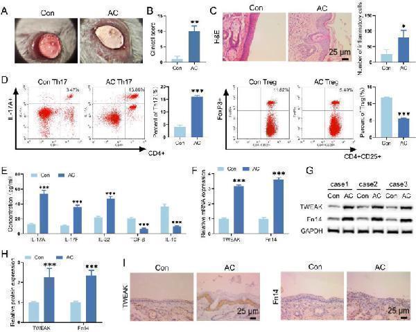
Th17/Treg cell differentiation ratio and TWEAK/Fn14 signaling level in AC mice. ( A ) The state of mice ocular surface was observed by slit-lamp. ( B ) The clinical scores of mice were performed according to the status of eyelid, conjunctiva and cornea. ( C ) HE staining was utilized to evaluate the pathological changes of conjunctival tissue in mice. ( D ) The proportion of Th17 or Treg cells in spleen of mice was assessed by flow cytometry. ( E ) The levels of Th17 and Treg cytokines in mice spleen were evaluated by ELISA. ( F-H ) TWEAK and Fn14 levels in ocular conjunctival tissue were measured by qRT-PCR and WB. ( I ) IHC was used to observe the protein level of TWEAK and Fn14 in ocular conjunctival tissue. * p < 0.05, ** p < 0.01, *** p < 0.001 vs. Control
Index in PubMed under a CC BY license. PMID: 39592944

Click image to see more details
TWEAK knockdown affected conjunctivitis in AC mice. ( A ) The effect of TWEAK knockdown on the state of mice ocular surface was observed under slit-lamp. ( B ) Clinical score of eyelid, conjunctiva and cornea to assess the effect of TWEAK knockdown in AC mice. ( C-D ) HE staining and TB staining were utilized to evaluate the effect of TWEAK knockdown on conjunctivitis in mice. ( E-G ) The levels of TWEAK and Fn14 in conjunctival tissue were detected by qRT-PCR, IHC and WB. * p < 0.05, ** p < 0.01, *** p < 0.001 vs. Con + AAV-shNC; # p < 0.05, ## p < 0.01, ### p < 0.001 vs. AC + AAV-shNC; ns, no significant difference
Index in PubMed under a CC BY license. PMID: 39592944

Click image to see more details
TWEAK regulated the Th17/Treg cell ratio in AC mice. ( A ) The effect of TWEAK knockdown on Th17 and Treg cell ratio in spleen of AC mice was observed by flow cytometry. ( B-C ) WB and IHC were employed to evaluate the expression levels of FoxP3 and RORγt in mice conjunctival tissue. ( D ) The effect of TWEAK knockdown on the levels of Th17 and Treg cytokines in mice spleen were evaluated by ELISA. * p < 0.05, ** p < 0.01, *** p < 0.001 vs. Con + AAV-shNC; # p < 0.05, ## p < 0.01, ### p < 0.001 vs. AC + AAV-shNC; ns, no significant difference
Index in PubMed under a CC BY license. PMID: 39592944

Click image to see more details
TWEAK knockdown promoted Nrf2/HO-1 signaling pathway in AC mice. ( A ) qRT-PCR was utilized to measure the effect of TWEAK knockdown on Nrf2 and HO-1 mRNA levels in conjunctival tissue of mice. ( B-C ) The effect of TWEAK knockdown on protein levels of Nrf2 and HO-1 in conjunctival tissue of mice were detected by WB and IHC. * p < 0.05, ** p < 0.01, *** p < 0.001 vs. Con + AAV-shNC; # p < 0.05, ## p < 0.01, ### p < 0.001 vs. AC + AAV-shNC; ns, no significant difference
Index in PubMed under a CC BY license. PMID: 39592944

Click image to see more details
Inhibition of Nrf2/HO-1 signaling pathway affected the improvement of conjunctivitis in AC mice from TWEAK knockdown. ( A ) The effect of Nrf2 inhibitor on ocular surface status was observed by slit-lamp in AC mice with TWEAK knockdown. ( B ) Clinical score of eyelid, conjunctiva and cornea to assess the effect of Nrf2 inhibitor in AC mice with TWEAK knockdown. ( C-D ) HE staining and TB staining were employed to evaluate the effect of Nrf2 inhibitor on conjunctivitis in AC mice with TWEAK knockdown. ( E-G ) The levels of Nrf2 and HO-1 in conjunctival tissue were detected by qRT-PCR, IHC and WB. * p < 0.05, ** p < 0.01, *** p < 0.001 vs. AC + AAV-shNC; # p < 0.05, ## p < 0.01, ### p < 0.001 vs. AC + AAV-shTWEAK
Index in PubMed under a CC BY license. PMID: 39592944

Click image to see more details
Inhibition of Nrf2/HO-1 signaling pathway affected Th17/Treg cell ratio in AC mice with TWEAK knockdown. ( A ) The effect of Nrf2 inhibitor on Th17/Treg cell ratio in AC mice with TWEAK knockdown was assessed by flow cytometry. ( B-C ) WB and IHC assays were employed to evaluate the expression of FoxP3 and RORγt in conjunctival tissue of AC mice. ( D ) The levels of Th17 and Treg cytokines in mice spleen were detected by ELISA. * p < 0.05, ** p < 0.01, *** p < 0.001 vs. AC + AAV-shNC; # p < 0.05, ## p < 0.01, ### p < 0.001 vs. AC + AAV-shTWEAK
Index in PubMed under a CC BY license. PMID: 39592944

Click image to see more details
Effects of ISO on the TWEAK/Fn14 signaling pathway in TGF-β1-induced HK-2 cells. (A, B) ISO directly interacts with Fn14, dose–response sensorgrams, and the affinity constant ( K d = 5.47 μM) of ISO with Fn14. (C–F) Western blot analysis of TWEAK, Fn14, and the phosphorylation levels of ERK1/2 in TGF-β1-induced HK-2 cells. β-actin was used as the loading control. Data are expressed as mean ± SD ( n = 3). # p < 0.05, ## p < 0.01 versus the sham group, * p < 0.05, ** p < 0.01 versus the UUO group.
Index in PubMed under a CC BY license. PMID: 40977693

Click image to see more details
Effect of ISO on the expression of TWEAK/Fn14 signaling pathway-related proteins in UUO rats. Representative images and quantification of Western blot results of the protein expression of TWEAK, Fn14, TRAF2, and BRAF (A–D) , and phosphorylation of MER1/2 and ERK1/2 (E–H) . β-actin was used as the loading control for total protein. (I–L) Relative optical density analysis of the nucleoprotein expression of p-SP1, Snail, and β-catenin. PCNA was used as the loading control for nucleoprotein. All data are expressed as the mean ± SD ( n = 5). ## p < 0.01 versus the sham group, * p < 0.05, ** p < 0.01 versus the UUO group.
Index in PubMed under a CC BY license. PMID: 40977693

Click image to see more details
Fn14 overexpression compromises the therapeutic effects of ISO and the regulation of ISO on the TWEAK/Fn14 pathway-related proteins on UUO model rats. (A, B) Serum Scr and BUN levels in different groups. (C, D) Representative images and the CVF of Masson staining (×400). (E–H) Representative and quantification analysis of Col III and FN immunohistochemical staining (×400). Nuclei were counterstained with DAPI (blue). (I–K) Western blot and the relative optical densities analysis of E-cadherin and α-SMA in kidney tissue. (L–O) Representative images and quantification of Western blot results of the protein expression of TWEAK, Fn14, and phosphorylation of ERK1/2. β-actin was used as the loading control. (P–R) Relative optical density analysis of the nucleoprotein expression of Snail and β-catenin. PCNA was used as the loading control for nucleoprotein. Data were shown as mean ± SD ( n = 3). ## p < 0.01 versus the sham group, * p < 0.05, ** p < 0.01 versus the UUO group, △ p < 0.05, △△ p < 0.01 versus the ISO group.
Index in PubMed under a CC BY license. PMID: 40977693

Click image to see more details
Western blot analysis of TWEAKR using anti-TWEAKR antibody (M06261).
Electrophoresis was performed on a 5-20% SDS-PAGE gel at 70V (Stacking gel) / 90V (Resolving gel) for 2-3 hours. The sample well of each lane was loaded with 30 ug of sample under reducing conditions.
Lane 1: human HUVEC whole cell lysates,
Lane 2: human A549 whole cell lysates,
Lane 3: rat PC-12 whole cell lysates,
Lane 4: rat C6 whole cell lysates,
Lane 5: mouse NIH/3T3 whole cell lysates,
Lane 6: mouse 4T1 whole cell lysates.
After electrophoresis, proteins were transferred to a nitrocellulose membrane at 150 mA for 50-90 minutes. Blocked the membrane with 5% non-fat milk/TBS for 1.5 hour at RT. The membrane was incubated with rabbit anti-TWEAKR antigen affinity purified monoclonal antibody (Catalog # M06261) at 1:1000 overnight at 4°C, then washed with TBS-0.1%Tween 3 times with 5 minutes each and probed with a goat anti-rabbit IgG-HRP secondary antibody at a dilution of 1:500 for 1.5 hour at RT. The signal is developed using an Enhanced Chemiluminescent detection (ECL) kit (Catalog # EK1002) with Tanon 5200 system. A specific band was detected for TWEAKR at approximately 17,20 kDa. The expected band size for TWEAKR is at 17,20 kDa.
Specific Publications For Anti-TWEAKR Rabbit Monoclonal Antibody (M06261)
Loading publications
Recommended Resources
Here are featured tools and databases that you might find useful.
- Boster's Pathways Library
- Protein Databases
- Bioscience Research Protocol Resources
- Data Processing & Analysis Software
- Photo Editing Software
- Scientific Literature Resources
- Research Paper Management Tools
- Molecular Biology Software
- Primer Design Tools
- Bioinformatics Tools
- Phylogenetic Tree Analysis
Customer Reviews
Have you used Anti-TWEAKR Rabbit Monoclonal Antibody?
Share your experimental results or join a short interview to earn up to $1,000 in product credits or other rewards.
0 Reviews For Anti-TWEAKR Rabbit Monoclonal Antibody
Customer Q&As
Have a question?
Find answers in Q&As, reviews.
Can't find your answer?
Submit your question
5 Customer Q&As for Anti-TWEAKR Rabbit Monoclonal Antibody
Question
We are currently using anti-TWEAKR Rabbit Monoclonal antibody M06261 for mouse tissue, and we are well pleased with the WB results. The species of reactivity given in the datasheet says human, mouse, rat. Is it true that the antibody can work on primate tissues as well?
Verified Customer
Verified customer
Asked: 2019-10-18
Answer
The anti-TWEAKR Rabbit Monoclonal antibody (M06261) has not been tested for cross reactivity specifically with primate tissues, though there is a good chance of cross reactivity. We have an innovator award program that if you test this antibody and show it works in primate you can get your next antibody for free. Please contact me if I can help you with anything.
Boster Scientific Support
Answered: 2019-10-18
Question
Does anti-TWEAKR Rabbit Monoclonal antibody M06261 work for WB with cortex of kidney?
Verified Customer
Verified customer
Asked: 2019-07-30
Answer
According to the expression profile of cortex of kidney, TNFRSF12A is highly expressed in cortex of kidney. So, it is likely that anti-TWEAKR Rabbit Monoclonal antibody M06261 will work for WB with cortex of kidney.
Boster Scientific Support
Answered: 2019-07-30
Question
Will M06261 anti-TWEAKR Rabbit Monoclonal antibody work on parafin embedded sections? If so, which fixation method do you recommend we use (PFA, paraformaldehyde, other)?
Verified Customer
Verified customer
Asked: 2019-05-16
Answer
It shows on the product datasheet, M06261 anti-TWEAKR Rabbit Monoclonal antibody as been tested on WB. It is best to use PFA for fixation because it has better tissue penetration ability. PFA needs to be prepared fresh before use. Long term stored PFA turns into formalin, as the PFA molecules congregate and become formalin.
Boster Scientific Support
Answered: 2019-05-16
Question
Would anti-TWEAKR Rabbit Monoclonal antibody M06261 work on dog WB with placenta?
J. Baker
Verified customer
Asked: 2014-08-28
Answer
Our lab technicians have not validated anti-TWEAKR Rabbit Monoclonal antibody M06261 on dog. You can run a BLAST between dog and the immunogen sequence of anti-TWEAKR Rabbit Monoclonal antibody M06261 to see if they may cross-react. If the sequence homology is close, then you can perform a pilot test. Keep in mind that since we have not validated dog samples, this use of the antibody is not covered by our guarantee. However we have an innovator award program that if you test this antibody and show it works in dog placenta in WB, you can get your next antibody for free.
Boster Scientific Support
Answered: 2014-08-28
Question
I was wanting to use your anti-TWEAKR Rabbit Monoclonal antibody for WB for rat cortex of kidney on frozen tissues, but I want to know if it has been tested for this particular application. Has this antibody been tested and is this antibody a good choice for rat cortex of kidney identification?
R. Carter
Verified customer
Asked: 2013-02-26
Answer
As indicated on the product datasheet, M06261 anti-TWEAKR Rabbit Monoclonal antibody has been tested for IP, WB on human, mouse, rat tissues. We have an innovator award program that if you test this antibody and show it works in rat cortex of kidney in IHC-frozen, you can get your next antibody for free.
Boster Scientific Support
Answered: 2013-02-26
