Click image to see more details
-

-
-
-
-
+5
Product Info Summary
| SKU: | M00584 |
|---|---|
| Size: | 100 μl |
| Reactive Species: | Human, Mouse, Rat |
| Host: | Rabbit |
| Application: | IF, IHC, ICC, WB |
Customers Who Bought This Also Bought

Product info
Product Name
Anti-ULK1/Atg1 Rabbit Monoclonal Antibody
SKU/Catalog Number
M00584
BM5170 is an alternative SKU for this antibody, used in previous lots.
Size
100 μl
Form
Liquid
Description
Boster Bio Anti-ULK1/Atg1 Rabbit Monoclonal Antibody catalog # M00584. Tested in WB, IHC, ICC/IF applications. This antibody reacts with Human, Mouse, Rat.
Storage & Handling
Store at -20°C for one year. For short term storage and frequent use, store at 4°C for up to one month. Avoid repeated freeze-thaw cycles.
Cite This Product
Anti-ULK1/Atg1 Rabbit Monoclonal Antibody (Boster Biological Technology, Pleasanton CA, USA, Catalog # M00584)
Host
Rabbit
Contents
Rabbit IgG in stabilizing components, phosphate buffered saline, pH 7.4, 150mM NaCl, 0.02% sodium azide and 50% glycerol.
*This antibody is supplied in a stabilized formulation.
Compatibility with conjugation reactions depends on the chemistry of the conjugation method used.
For conjugation methods that are not compatible with the stabilizing components present in this formulation, a carrier-free antibody format is required.
Clonality
Monoclonal
Clone Number
ABHH-21
Isotype
Rabbit IgG
Immunogen
A synthesized peptide derived from human ULK1
Reactive Species
M00584 is reactive to ULK1 in Human, Mouse, Rat
Observed Molecular Weight
130 kDa
Calculated molecular weight
112.6 kDa
Antibody Validation
Boster validates all antibodies on WB, IHC, ICC, Immunofluorescence, and ELISA with known positive control and negative samples to ensure specificity and high affinity, including thorough antibody incubations.
Application & Images
Applications
M00584 is guaranteed for IF, IHC, ICC, WB Boster Guarantee
Recommend Dilution
WB 1:500-2000
IHC 1:50-200
ICC/IF 1:50-200
Tested application
Suggested blocking solution with 5% non-fat milk or BSA; (*)Recommended protein loading: 20-40 µg per lane
Use TE buffer pH 9.0 for antigen retrieval; (*) citrate buffer pH 6.0 is an alternative.
Validation Images & Assay Conditions
Click image to see more details
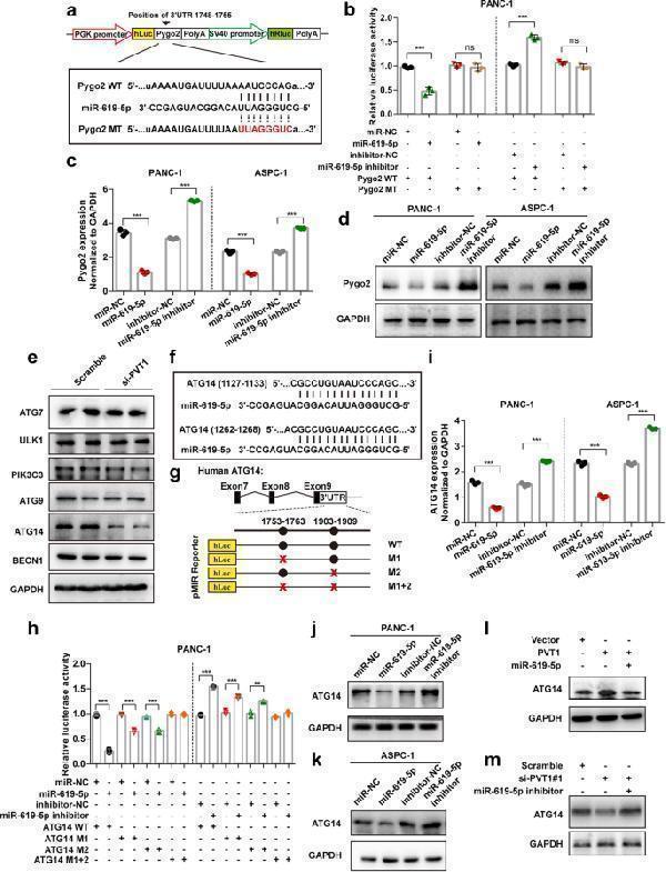
miR-619-5p negatively regulates Pygo2 and ATG14 expression. a The predicted miR-619-5p binding sequence in the Pygo2 3’UTR and the generation of dual-luciferase reporter plasmids of wild-type (WT) or mutant (MT) were shown. b Luciferase activity assays were performed in PANC-1 cells co-transfected with Pygo2 WT or Pygo2 MT and miR-619-5p mimic or miR-619-5p inhibitor. c and d The mRNA and protein levels of Pygo2 in PANC-1 and ASPC-1 cells after transfection with miR-619-5p mimics or miR-619-5p inhibitor. e Western blotting in PANC-1 cells transfected with PVT1 siRNA was carried out using the indicated antibodies. f and g The miR-619-5p binding sequence in the ATG14 3’UTR and the generation of dual-luciferase reporter plasmids of wild-type (WT) or mutant (MT) were shown. h Luciferase activity assays were performed in PANC-1 cells co-transfected with ATG14 WT or ATG14 MT and miR-619-5p mimic or miR-619-5p inhibitor. i-k The mRNA and protein levels of ATG14 in PANC-1 and ASPC-1 cells after transfection with miR-619-5p mimics or miR-619-5p inhibitor. l and m The expression of ATG14 after co-transfection with PVT1 and miR-619-5p mimics or PVT1 siRNA and miR-619-5p inhibitor. Data were represented as mean ± SD, * P < 0.05; ** P < 0.01; *** P < 0.001
Index in PubMed under a CC BY license. PMID: 32727463

Click image to see more details
PVT1/miR-619-5p axis promotes autophagic activity by regulating ATG14. a Western blotting analysis of PANC-1 and ASPC-1 cells after PVT1 knockdown with or without ATG14 overexpression was carried out with the indicated antibodies. b Western blotting analysis of PANC-1 and ASPC-1 cells after PVT1 overexpression with or without ATG14 knockdown was carried out with the indicated antibodies. c and d Representative confocal images of GFP-LC3 puncta in PANC-1 cells transfected with PVT1 siRNA with or without co-transfection of ATG14 overexpression plasmid with and without gemcitabine treatment. The number of GFP-LC3 puncta was quantified using ImageJ software. ( n = 10). Scale bars: 10 μm. e and f Representative confocal images of GFP-LC3 puncta in PANC-1 cells transfected with PVT1 overexpression plasmid with or without co-transfection with ATG14 siRNA with and without gemcitabine treatment. The number of GFP-LC3 puncta was quantified using ImageJ software. (n = 10). Scale bars: 10 μm. g and h Representative electronic micrographs of the autophagosomes or autolysosomes of PANC-1 cells co-transfected with PVT1 overexpression plasmid and/or ATG14 siRNA with or without gemcitabine treatment. Red arrows indicate autophagic structures. The number of autophagic structures per cell was quantified (n = 10). Scale bars: 2 μm. i and j Representative electronic micrographs of the autophagosomes or autolysosomes of PANC-1 cells co-transfected with miR-619-5p mimics and/or ATG14 siRNA with or without gemcitabine treatment. Red arrows indicate autophagic structures. The number of autophagic structures per cell was quantified (n = 10). Scale bars: 2 μm. k and l Western blotting analysis of PANC-1 and ASPC-1 cells after transfection with miR-619-5p mimics with or without ATG14 overexpression was carried out with the indicated antibodies. Data were represented as mean ± SD, * P < 0.05; ** P < 0.01; *** P < 0.001
Index in PubMed under a CC BY license. PMID: 32727463

Click image to see more details
PVT1 interacts with ATG14 and promotes PtdIns3K-C1 complex assembly. a The interaction between PVT1 and ATG14 in PANC-1 and ASPC-1 cells was confirmed by RNA pulldown followed by western blotting. b and c qRT-PCR analysis of PVT1 following RNA immunoprecipitation (RIP) assays in PANC-1 and ASPC-1 cells using anti-ATG14 antibody. RNA enrichment was determined relative to the IgG control. U6 was used as a non-specific control. d and e The interaction between PIK3C3 and ATG14 or BECN1 after PVT1 knockdown in PANC-1 cells. Immunoprecipitated endogenous PIK3C3 was quantified using Image Lab software and normalized against the amount of PIK3C3 in whole-cell lysates. f and g The interaction between PIK3C3 and ATG14 or BECN1 after PVT1 overexpression and/or miR-619-5p co-transfection in PANC-1 cells with or without gemcitabine (1 μM) treatment. Immunoprecipitated endogenous PIK3C3 was quantified using Image Lab software and normalized against the amount of PIK3C3 in whole-cell lysates. h The interaction between Bcl2 and BECN1 after PVT1 overexpression in PANC-1 cells with or without gemcitabine (1 μM) treatment. Immunoprecipitated endogenous Bcl2 was quantified using Image Lab software and normalized against the amount of PIK3C3 in whole-cell lysates. i The interaction between PIK3C3 and BECN1 after the overexpression of miR-619-5p mimics in PANC-1 cells with or without gemcitabine (1 μM) treatment. Immunoprecipitated endogenous PIK3C3 was quantified using Image Lab software and normalized against the amount of PIK3C3 in whole-cell lysates. j-l Different PtdIns3K-C1 complex components were immunoprecipitated from PANC-1 and ASPC-1 cells overexpressing PVT1 or miR-619-5p mimics with or without gemcitabine (1 μM) treatment with ATG14 antibody. PIK3C3 activity was measured by analyzing PtdIns3P production using ELISA as described in the Materials and Methods section. The fold change in PtdIns3P activity was calculated based on the concentration of PtdIns3P and normalized to the amount of ATG14 used in the assay. m and n Representative confocal images of GFP-ZFYVE1 puncta in control or PVT1- or miR-619-5p-transfected PANC-1 cells with or without gemcitabine induction. The numbers of GFP-ZFYVE1 puncta was quantified ( n = 10). Scale bars: 10 μm. Data were represented as mean ± SD, * P < 0.05; ** P < 0.01; *** P < 0.001
Index in PubMed under a CC BY license. PMID: 32727463

Click image to see more details
Western blot analysis of ULK1 using anti-ULK1 antibody (M00584).
Electrophoresis was performed on a 5-20% SDS-PAGE gel at 70V (Stacking gel) / 90V (Resolving gel) for 2-3 hours. The sample well of each lane was loaded with 30 ug of sample under reducing conditions.
Lane 1: human 293T whole cell lysates,
Lane 2: human A549 whole cell lysates,
Lane 3: human U-87MG whole cell lysates,
Lane 4: human Hela whole cell lysates,
Lane 5: rat liver tissue lysates,
Lane 6: rat PC-12 whole cell lysates,
Lane 7: mouse liver tissue lysates,
Lane 8: mouse Neuro-2a whole cell lysates.
After electrophoresis, proteins were transferred to a nitrocellulose membrane at 150 mA for 50-90 minutes. Blocked the membrane with 5% non-fat milk/TBS for 1.5 hour at RT. The membrane was incubated with rabbit anti-ULK1 antigen affinity purified monoclonal antibody (Catalog # M00584) at 1:500 overnight at 4°C, then washed with TBS-0.1%Tween 3 times with 5 minutes each and probed with a goat anti-rabbit IgG-HRP secondary antibody at a dilution of 1:1000 for 1.5 hour at RT. The signal is developed using an Enhanced Chemiluminescent detection (ECL) kit (Catalog # EK1002) with Tanon 5200 system. A specific band was detected for ULK1 at approximately 130 kDa. The expected band size for ULK1 is at 113 kDa.

Click image to see more details
IHC analysis of ULK1 using anti-ULK1 antibody (M00584).
ULK1 was detected in a paraffin-embedded section of human prostate cancer tissue. Heat mediated antigen retrieval was performed in EDTA buffer (pH 8.0, epitope retrieval solution). The tissue section was blocked with 10% goat serum. The tissue section was then incubated with 1:50 rabbit anti-ULK1 Antibody (M00584) overnight at 4°C. Peroxidase Conjugated Goat Anti-rabbit IgG was used as secondary antibody and incubated for 30 minutes at 37°C. The tissue section was developed using HRP Conjugated Rabbit IgG Super Vision Assay Kit (Catalog # SV0002) with DAB as the chromogen.

Click image to see more details
IHC analysis of ULK1 using anti-ULK1 antibody (M00584).
ULK1 was detected in a paraffin-embedded section of human spleen tissue. Heat mediated antigen retrieval was performed in EDTA buffer (pH 8.0, epitope retrieval solution). The tissue section was blocked with 10% goat serum. The tissue section was then incubated with 1:50 rabbit anti-ULK1 Antibody (M00584) overnight at 4°C. Peroxidase Conjugated Goat Anti-rabbit IgG was used as secondary antibody and incubated for 30 minutes at 37°C. The tissue section was developed using HRP Conjugated Rabbit IgG Super Vision Assay Kit (Catalog # SV0002) with DAB as the chromogen.

Click image to see more details
IHC analysis of ULK1 using anti-ULK1 antibody (M00584).
ULK1 was detected in a paraffin-embedded section of mouse brain tissue. Heat mediated antigen retrieval was performed in EDTA buffer (pH 8.0, epitope retrieval solution). The tissue section was blocked with 10% goat serum. The tissue section was then incubated with 1:50 rabbit anti-ULK1 Antibody (M00584) overnight at 4°C. Peroxidase Conjugated Goat Anti-rabbit IgG was used as secondary antibody and incubated for 30 minutes at 37°C. The tissue section was developed using HRP Conjugated Rabbit IgG Super Vision Assay Kit (Catalog # SV0002) with DAB as the chromogen.

Click image to see more details
IHC analysis of ULK1 using anti-ULK1 antibody (M00584).
ULK1 was detected in a paraffin-embedded section of rat brain tissue. Heat mediated antigen retrieval was performed in EDTA buffer (pH 8.0, epitope retrieval solution). The tissue section was blocked with 10% goat serum. The tissue section was then incubated with 1:50 rabbit anti-ULK1 Antibody (M00584) overnight at 4°C. Peroxidase Conjugated Goat Anti-rabbit IgG was used as secondary antibody and incubated for 30 minutes at 37°C. The tissue section was developed using HRP Conjugated Rabbit IgG Super Vision Assay Kit (Catalog # SV0002) with DAB as the chromogen.

Click image to see more details
Immunofluorescent analysis of 293 cells, using ULK1 Antibody.
Specific Publications For Anti-ULK1/Atg1 Rabbit Monoclonal Antibody (M00584)
Loading publications
Recommended Resources
Here are featured tools and databases that you might find useful.
- Boster's Pathways Library
- Protein Databases
- Bioscience Research Protocol Resources
- Data Processing & Analysis Software
- Photo Editing Software
- Scientific Literature Resources
- Research Paper Management Tools
- Molecular Biology Software
- Primer Design Tools
- Bioinformatics Tools
- Phylogenetic Tree Analysis
Customer Reviews
Have you used Anti-ULK1/Atg1 Rabbit Monoclonal Antibody?
Share your experimental results or join a short interview to earn up to $1,000 in product credits or other rewards.
0 Reviews For Anti-ULK1/Atg1 Rabbit Monoclonal Antibody
Customer Q&As
Have a question?
Find answers in Q&As, reviews.
Can't find your answer?
Submit your question
2 Customer Q&As for Anti-ULK1/Atg1 Rabbit Monoclonal Antibody
Question
Could you please provide the immunogen sequence for product M00584?
Verified customer
Asked: 2022-09-12
Answer
The immunogen sequence for M00584 is FLDKQRLLDRIHSITAERL.
Boster Scientific Support
Answered: 2022-09-12
Question
We are currently using anti-ULK1/Atg1 Rabbit Monoclonal antibody M00584 for human tissue, and we are well pleased with the IF results. The species of reactivity given in the datasheet says human, mouse, rat. Is it possible that the antibody can work on pig tissues as well?
Verified Customer
Verified customer
Asked: 2018-02-08
Answer
The anti-ULK1/Atg1 Rabbit Monoclonal antibody (M00584) has not been tested for cross reactivity specifically with pig tissues, though there is a good chance of cross reactivity. We have an innovator award program that if you test this antibody and show it works in pig you can get your next antibody for free. Please contact me if I can help you with anything.
Boster Scientific Support
Answered: 2018-02-08
