Click image to see more details
-

-
-
-
-
+4
Product Info Summary
| SKU: | A00901-2 |
|---|---|
| Size: | 100 μg/vial |
| Reactive Species: | Human |
| Host: | Rabbit |
| Application: | Flow Cytometry, IHC, ICC, WB |
Customers Who Bought This Also Bought

Product info
Product Name
Anti-VEGF Receptor 2/KDR Antibody Picoband®
SKU/Catalog Number
A00901-2
Size
100 μg/vial
Form
Lyophilized
Description
Boster Bio Anti-VEGF Receptor 2/KDR Antibody Picoband® catalog # A00901-2. Tested in Flow Cytometry, IHC, ICC, WB applications. This antibody reacts with Human. The brand Picoband indicates this is a premium antibody that guarantees superior quality, high affinity, and strong signals with minimal background in Western blot applications. Only our best-performing antibodies are designated as Picoband, ensuring unmatched performance.
Storage & Handling
Store at -20˚C for one year from date of receipt. After reconstitution, at 4˚C for one month. It can also be aliquotted and stored frozen at -20˚C for six months. Avoid repeated freeze-thaw cycles.
Cite This Product
Anti-VEGF Receptor 2/KDR Antibody Picoband® (Boster Biological Technology, Pleasanton CA, USA, Catalog # A00901-2)
Host
Rabbit
Contents
Each vial contains antibody formulated with stabilizing components, 0.9 mg NaCl, 0.2 mg Na2HPO4, and 0.05 mg NaN3.
*This antibody is supplied in a stabilized formulation.
Compatibility with conjugation reactions depends on the chemistry of the conjugation method used.
For conjugation methods that are not compatible with the stabilizing components present in this formulation, a carrier-free antibody format is required.
Clonality
Polyclonal
Isotype
Rabbit IgG
Immunogen
E. coli-derived human VEGF Receptor 2 recombinant protein (Position: A20-L242).
Cross-reactivity
No cross-reactivity with other proteins.
Reactive Species
A00901-2 is reactive to KDR in Human
Observed Molecular Weight
180-250 kDa
Calculated molecular weight
151.5 kDa
Background of KDR
KDR (Kinase Insert Domain Receptor), also known as FLK1, VEGFR or VEGFR2, is a VEGF receptor. KDR is the human gene encoding it. Vascular endothelial growth factor (VEGF) is the only mitogen that specifically acts on endothelial cells. Its expression is upregulated by hypoxia, and its cell-surface receptor, known as fetal liver kinase-1 (Flk1) in mouse, is exclusively expressed in endothelial cells. Flk1 is the mouse homolog of KDR.
Antibody Validation
Boster validates all antibodies on WB, IHC, ICC, Immunofluorescence, and ELISA with known positive control and negative samples to ensure specificity and high affinity, including thorough antibody incubations.
Application & Images
Applications
A00901-2 is guaranteed for Flow Cytometry, IHC, ICC, WB Boster Guarantee
Assay Dilutions Recommendation
The recommendations below provide a starting point for assay optimization. The actual working concentration varies and should be decided by the user.
Western blot, 0.1-0.5μg/ml
Immunohistochemistry (Paraffin-embedded Section), 0.5-1μg/ml
Immunocytochemistry, 0.5-1μg/ml
ELISA (Cap),1-5μg/ml
Positive Control
WB: human placenta tissue, human PC-3 whole cell
IHC: human intestinal cancer tissue, human lung cancer tissue, human placenta tissue, human mammary cancer tissue
Validation Images & Assay Conditions
Click image to see more details
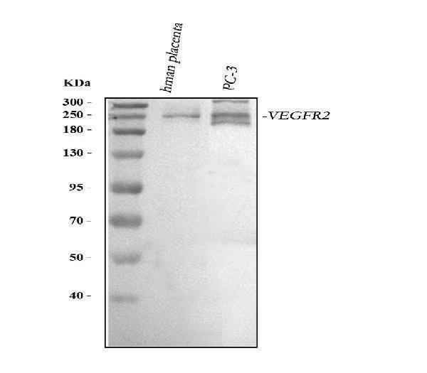
Western blot analysis of VEGF Receptor 2 using anti-VEGF Receptor 2 antibody (A00901-2).
Electrophoresis was performed on a 5-20% SDS-PAGE gel at 70V (Stacking gel) / 90V (Resolving gel) for 2-3 hours. The sample well of each lane was loaded with 30 ug of sample under reducing conditions.
Lane 1: human placenta tissue lysates,
Lane 2: human PC-3 whole cell lysates.
After electrophoresis, proteins were transferred to a nitrocellulose membrane at 150 mA for 50-90 minutes. Blocked the membrane with 5% non-fat milk/TBS for 1.5 hour at RT. The membrane was incubated with rabbit anti-VEGF Receptor 2 antigen affinity purified polyclonal antibody (Catalog # A00901-2) at 0.5 μg/mL overnight at 4°C, then washed with TBS-0.1%Tween 3 times with 5 minutes each and probed with a goat anti-rabbit IgG-HRP secondary antibody at a dilution of 1:5000 for 1.5 hour at RT. The signal is developed using an Enhanced Chemiluminescent detection (ECL) kit (Catalog # EK1002) with Tanon 5200 system. A specific band was detected for VEGF Receptor 2 at approximately 180-250 kDa. The expected band size for VEGF Receptor 2 is at 156 kDa.

Click image to see more details
Tumor necrosis factor-α induced protein 8 (TIPE) and vascular endothelial growth factor receptor 2 (VEGFR2) are highly expressed in CRC and act as oncogenes. (a) TIPE expression in five pairs of fresh human CRC tumor tissues and matched adjacent tissues analyzed by immunohistochemistry staining. (b) Representative results of TIPE expression in five pairs of fresh CRC tumor tissues and adjacent tissues as detected by Western blot; T indicates tumor tissue, and N indicates normal tissue. (c) Comparison of TIPE mRNA expression levels in human CRC tissues and matched adjacent tissues. TIPE mRNA expression was quantified by qRT-PCR and normalized to that in the matched adjacent normal tissues. (d) Through immunohistochemical staining, VEGFR2 expression was analyzed in five randomly selected pairs of CRC tumor tissues and matched adjacent tissues. (e) Comparison of VEGFR2 mRNA expression levels in human CRC tissues and matched adjacent tissues. VEGFR2 mRNA expression was quantified by qRT-PCR and normalized to that in the matched adjacent normal tissues.
Index in PubMed under a CC BY license. PMID: 31929755

Click image to see more details
Knockdown of TIPE downregulates VEGFR2 expression and inhibits cell proliferation, cell migration and angiogenesis. (a) qRT-PCR of TIPE expression in HCT116 cells infected with lentiviral shTIPE or control shRNA. TIPE mRNA expression was quantified by qRT-PCR and normalized to that in the shRNA control cells. (b) TIPE expression in HCT116 cells infected with lentiviral shTIPE or control shRNA according to Western blot analysis. (c) Expression of VEGFR2 in shTIPE and shRNA control HCT116 cells based on qRT-PCR assays. (d) Tumor cell culture conditioned medium (TCM) from HCT116 cells stably transduced with shTIPE or control shRNA inhibited the proliferation of HUVECs as determined by CCK-8 assays. (e) Representative immunofluorescence (IF) images demonstrated that the level of TIPE has an effect on the expression of microfilaments and initial pseudopod extension in the stably transduced HCT116 cell lines. Scale bar, 50 µm. (f) Representative images (left panel) and quantification (right panel) of tube formation of HUVECs treated with TCM derived from control HCT116 cells or HCT116 cells with stable TIPE knockdown. The data were collected from four independent experiments using different batches of cells. Scale bar, 500 μm. **p <0.01, ***p<0.001.
Index in PubMed under a CC BY license. PMID: 31929755

Click image to see more details
TIPE promotes VEGFR2-mediated angiogenesis by upregulating PDK1 expression and phosphorylation. (a) TIPE knockdown in HCT116 cells inhibits PDK1 phosphorylation according to Western blot analysis. (b) After cells were treated with a PDK1 inhibitor (GSK2334470), the expression levels of TIPE and PDK1 and their corresponding phosphorylation levels were detected by Western blot analysis. (c) After cells were treated with GSK2334470 and DMSO, the expression levels of VEGFR2 were tested and verified by Western blot analysis. (d) The proliferation of HUVECs cultured with TCM in the presence of GSK2334470 was determined by CCK-8 assays. (e) Tube formation by HUVECs treated with GSK2334470 was measured, and the results are expressed as the tubule length. Representative statistical results are shown. (f) Representative morphological images are shown. Scale bar, 500 µm. ns: no significance, **p <0.01, ***p<0.001. (g) Schematic diagram representing the role of TIPE and VEGFR2 in tumor angiogenesis in CRC.
Index in PubMed under a CC BY license. PMID: 31929755

Click image to see more details
IHC analysis of VEGF Receptor 2 using anti-VEGF Receptor 2 antibody (A00901-2).
VEGF Receptor 2 was detected in paraffin-embedded section of human intestinal cancer tissue. Heat mediated antigen retrieval was performed in citrate buffer (pH6, epitope retrieval solution) for 20 mins. The tissue section was blocked with 10% goat serum. The tissue section was then incubated with 1ug/ml rabbit anti-VEGF Receptor 2 Antibody (A00901-2) overnight at 4 Biotinylated goat anti-rabbit IgG was used as secondary antibody and incubated for 30 minutes at 37 The tissue section was developed using Strepavidin-Biotin-Complex (SABC)(Catalog # SA1022) with DAB as the chromogen.

Click image to see more details
IHC analysis of VEGF Receptor 2 using anti-VEGF Receptor 2 antibody (A00901-2).
VEGF Receptor 2 was detected in paraffin-embedded section of human lung cancer tissue. Heat mediated antigen retrieval was performed in citrate buffer (pH6, epitope retrieval solution) for 20 mins. The tissue section was blocked with 10% goat serum. The tissue section was then incubated with 1ug/ml rabbit anti-VEGF Receptor 2 Antibody (A00901-2) overnight at 4 Biotinylated goat anti-rabbit IgG was used as secondary antibody and incubated for 30 minutes at 37 The tissue section was developed using Strepavidin-Biotin-Complex (SABC)(Catalog # SA1022) with DAB as the chromogen.

Click image to see more details
IHC analysis of VEGF Receptor 2 using anti-VEGF Receptor 2 antibody (A00901-2).
VEGF Receptor 2 was detected in paraffin-embedded section of human placenta tissue. Heat mediated antigen retrieval was performed in citrate buffer (pH6, epitope retrieval solution) for 20 mins. The tissue section was blocked with 10% goat serum. The tissue section was then incubated with 1ug/ml rabbit anti-VEGF Receptor 2 Antibody (A00901-2) overnight at 4 Biotinylated goat anti-rabbit IgG was used as secondary antibody and incubated for 30 minutes at 37 The tissue section was developed using Strepavidin-Biotin-Complex (SABC)(Catalog # SA1022) with DAB as the chromogen.

Click image to see more details
IHC analysis of VEGF Receptor 2 using anti-VEGF Receptor 2 antibody (A00901-2).
VEGF Receptor 2 was detected in paraffin-embedded section of human mammary cancer tissue. Heat mediated antigen retrieval was performed in citrate buffer (pH6, epitope retrieval solution) for 20 mins. The tissue section was blocked with 10% goat serum. The tissue section was then incubated with 1ug/ml rabbit anti-VEGF Receptor 2 Antibody (A00901-2) overnight at 4 Biotinylated goat anti-rabbit IgG was used as secondary antibody and incubated for 30 minutes at 37 The tissue section was developed using Strepavidin-Biotin-Complex (SABC)(Catalog # SA1022) with DAB as the chromogen.
Specific Publications For Anti-VEGF Receptor 2/KDR Antibody Picoband® (A00901-2)
Loading publications
Recommended Resources
Here are featured tools and databases that you might find useful.
- Boster's Pathways Library
- Protein Databases
- Bioscience Research Protocol Resources
- Data Processing & Analysis Software
- Photo Editing Software
- Scientific Literature Resources
- Research Paper Management Tools
- Molecular Biology Software
- Primer Design Tools
- Bioinformatics Tools
- Phylogenetic Tree Analysis
Customer Reviews
Have you used Anti-VEGF Receptor 2/KDR Antibody Picoband®?
Share your experimental results or join a short interview to earn up to $1,000 in product credits or other rewards.
0 Reviews For Anti-VEGF Receptor 2/KDR Antibody Picoband®
Customer Q&As
Have a question?
Find answers in Q&As, reviews.
Can't find your answer?
Submit your question
4 Customer Q&As for Anti-VEGF Receptor 2/KDR Antibody Picoband®
Question
We ordered your anti-VEGF Receptor 2/KDR antibody for Flow Cytometry on lung in a previous project. I am using human, and We are going to use the antibody for ELISA next. We are interested in examining lung as well as umbilical vein in our next experiment. Could you please give me some suggestion on which antibody would work the best for ELISA?
Verified Customer
Verified customer
Asked: 2020-05-05
Answer
I took a look at the website and datasheets of our anti-VEGF Receptor 2/KDR antibody and it seems that A00901-2 has been validated on human in both Flow Cytometry and ELISA. Thus A00901-2 should work for your application. Our Boster satisfaction guarantee will cover this product for ELISA in human even if the specific tissue type has not been validated. We do have a comprehensive range of products for ELISA detection and you can check out our website bosterbio.com to find out more information about them.
Boster Scientific Support
Answered: 2020-05-05
Question
My boss were satisfied with the WB result of your anti-VEGF Receptor 2/KDR antibody. However we have been able to see positive staining in lung cell junction. endoplasmic using this antibody. Is that expected? Could you tell me where is KDR supposed to be expressed?
Verified Customer
Verified customer
Asked: 2018-06-20
Answer
Based on literature, lung does express KDR. Generally KDR expresses in cell junction. endoplasmic, isoform 1: cell membrane. Regarding which tissues have KDR expression, here are a few articles citing expression in various tissues:
Umbilical vein, Pubmed ID: 15815621, 1656371, 7559454
Boster Scientific Support
Answered: 2018-06-20
Question
We are currently using anti-VEGF Receptor 2/KDR antibody A00901-2 for human tissue, and we are well pleased with the ELISA results. The species of reactivity given in the datasheet says human. Is it true that the antibody can work on goat tissues as well?
Verified Customer
Verified customer
Asked: 2018-06-06
Answer
The anti-VEGF Receptor 2/KDR antibody (A00901-2) has not been tested for cross reactivity specifically with goat tissues, but there is a good chance of cross reactivity. We have an innovator award program that if you test this antibody and show it works in goat you can get your next antibody for free. Please contact me if I can help you with anything.
Boster Scientific Support
Answered: 2018-06-06
Question
We have been able to see staining in human lung. Any tips? Is anti-VEGF Receptor 2/KDR antibody supposed to stain lung positively?
L. Zhao
Verified customer
Asked: 2013-10-15
Answer
According to literature lung does express KDR. According to Uniprot.org, KDR is expressed in lung, umbilical vein, among other tissues. Regarding which tissues have KDR expression, here are a few articles citing expression in various tissues:
Umbilical vein, Pubmed ID: 15815621, 1656371, 7559454
Boster Scientific Support
Answered: 2013-10-15
