Click image to see more details
-

-
-
-
-
+9
Product Info Summary
| SKU: | M03036 |
|---|---|
| Size: | 100 μl |
| Reactive Species: | Human, Mouse, Rat |
| Host: | Rabbit |
| Application: | IP, IF, ICC, WB |
Customers Who Bought This Also Bought

Product info
Product Name
Anti-xCT Rabbit Monoclonal Antibody
SKU/Catalog Number
M03036
BM5318 is an alternative SKU for this antibody, used in previous lots.
Size
100 μl
Form
Liquid
Description
Boster Bio Anti-xCT Rabbit Monoclonal Antibody catalog # M03036. Tested in WB, ICC/IF, IP applications. This antibody reacts with Human, Mouse, Rat.
Storage & Handling
Store at -20°C for one year. For short term storage and frequent use, store at 4°C for up to one month. Avoid repeated freeze-thaw cycles.
Cite This Product
Anti-xCT Rabbit Monoclonal Antibody (Boster Biological Technology, Pleasanton CA, USA, Catalog # M03036)
Host
Rabbit
Contents
Rabbit IgG in stabilizing components, phosphate buffered saline, pH 7.4, 150mM NaCl, 0.02% sodium azide and 50% glycerol.
*This antibody is supplied in a stabilized formulation.
Compatibility with conjugation reactions depends on the chemistry of the conjugation method used.
For conjugation methods that are not compatible with the stabilizing components present in this formulation, a carrier-free antibody format is required.
Clonality
Monoclonal
Clone Number
ACGI-19
Isotype
Rabbit IgG
Immunogen
A synthesized peptide derived from human xCT
Reactive Species
M03036 is reactive to SLC7A11 in Human, Mouse, Rat
Observed Molecular Weight
55 kDa
Calculated molecular weight
55.4 kDa
Antibody Validation
Boster validates all antibodies on WB, IHC, ICC, Immunofluorescence, and ELISA with known positive control and negative samples to ensure specificity and high affinity, including thorough antibody incubations.
Application & Images
Applications
M03036 is guaranteed for IP, IF, ICC, WB Boster Guarantee
Recommend Dilution
WB 1:500-2000
ICC/IF 1:50-200
IP 1:50
Tested application
Suggested blocking solution with 5% non-fat milk or BSA; (*)Recommended protein loading: 20-40 µg per lane
Validation Images & Assay Conditions
Click image to see more details
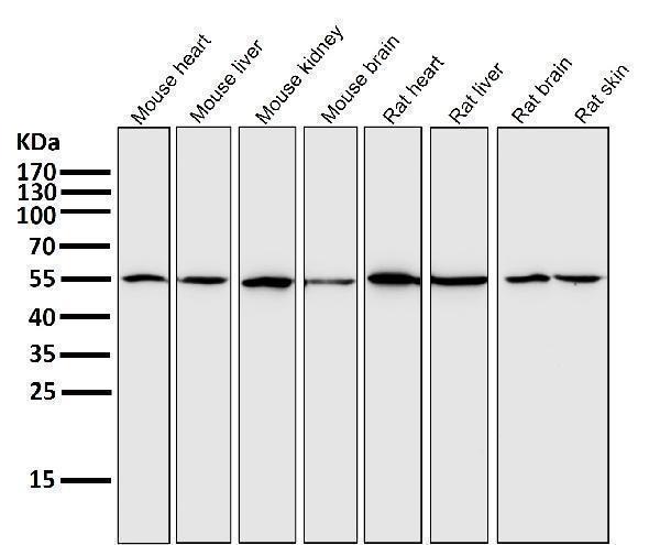
All lanes use the Antibody at 1:1000 dilution for 1 hour at room temperature.

Click image to see more details
All lanes use the Antibody at 1:1000 dilution for 1 hour at room temperature.

Click image to see more details
Immunofluorescent analysis using the Antibody at 1:50 dilution.

Click image to see more details
Western blot analysis of xCT expression in (1) HepG2 cell lysate; (2) Mouse brain lysate.

Click image to see more details
Immunofluorescent analysis using the Antibody at 1:150 dilution.

Click image to see more details
Western blot analysis of SLC7A11 using anti-SLC7A11 antibody (M03036).
Electrophoresis was performed on a 10% SDS-PAGE gel at 80V (Stacking gel) / 120V (Resolving gel) for 2 hours. The sample well of each lane was loaded with 30 μg of sample under reducing conditions.
Lane 1: Human U-87MG whole cell lysates,
Lane 2: Human LN229 whole cell lysates,
Lane 3: Rat C6 whole cell lysates,
Lane 4: Rat RG2 whole cell lysates.
After electrophoresis, proteins were transferred to a nitrocellulose membrane at 150 mA for 50-90 minutes. Blocked the membrane with 5% non-fat milk/TBS for 1.5 hour at RT. The membrane was incubated with rabbit anti-SLC7A11 antigen affinity purified monoclonal antibody (M03036) at a dilution of 1:1000 overnight at 4°C, then washed with TBS-0.1%Tween-20 3 times with 5 minutes each and probed with a goat anti-rabbit IgG-HRP secondary antibody (Catalog # BA1054) at a dilution of 1:5000 for 1.5 hour at RT. The signal is developed using an ECL Plus Western Blotting Substrate (Catalog # AR1196-200) with Tanon 5200 system. A specific band was detected for SLC7A11 at approximately 60 kDa. The expected band size for SLC7A11 is at 55 kDa.

Click image to see more details
Immunofluorescent analysis using the Antibody at 1:150 dilution.

Click image to see more details
Immunofluorescent analysis using the Antibody at 1:50 dilution.

Click image to see more details
ICC/IF analysis of SLC7A11 using anti-SLC7A11 antibody (M03036).
SLC7A11 was detected in an immunocytochemical section of rat C6 cells. The cells were fixed with 4% paraformaldehyde for 10 minutes and then treated with a membrane permeabilization agent (AR0205) for 5 minutes.The cells were blocked with 10% goat serum. And then incubated with rabbit anti-SLC7A11 Antibody (M03036) at a dilution of 1:50 overnight at 4°C. DyLight®488 Conjugated Goat Anti-Rabbit IgG (BA1127) was used as secondary antibody at 1:500 dilution and incubated for 30 minutes at 37°C. The section was counterstained with DAPI. Visualize using a fluorescence microscope and filter sets appropriate for the label used.

Click image to see more details
AKT inhibition induced MDA-MB-231 pyroptosis and decrease SLC7A11 expression. (A) Western blot analysis of NLRP3, GSDMD, caspase-1, AKT, p-AKT, PI3K, p-PI3K, mTOR and p-mTOR expression in MDA-MB-231 cells treated with AKT inhibitor or control buffer. * p <0.05, ** p < 0.01. Statistical analysis of the two groups were performed using two-tailed Student’s t test. The concentration of IL1β (B) and IL18 (C) in cell culture supernatant were all significantly elevated in the MK-2206-treated group, compared to the control group ( p = 0.0134 and p = 0.0015, respectively, t -test). (D) Analysis of pathways and co-expression data of SLC7A11, EHPA2, pyroptosis related genes and AKT/PI3K/mTOR related genes. Relative mRNA expression levels ( p = 0.0002, t -test) (E) and protein expression levels (F) of ferroptosis-associated gene SLC7A11 were all decreased in shEPHA2–1 group. Bars represent the mean ± SD.
Index in PubMed under a CC BY license. PMID: 40919148

Click image to see more details
SS attenuated the oxidation resistance of LUAD cells. (A, B) GSH or Cys was detected in Calu-1 or A549 cells, which were respectively treated with 10, 15, 20 μM or 20, 25, 30 μM SS for 6 h, and pretreated with or without Fer-1 (1 μM), the data statistic was shown in a histogram (*P < 0.05, **P < 0.01, ***P < 0.001). (C) Western blot analysis was used to detect the expressions of SLC7A11 and GPX4 in Calu-1 cells, which were treated with 10, 15, 20 μM SS for 6 h. (D) Quantitative analysis of gray value of the SLC7A11 and GPX4 blots. (E) Western blotting analysis was used to detect the expressions of SLC7A11 and GPX4 in Calu-1 cells, which were treated with 20 μM SS or 4 μM erastin, with or without Fer-1 (1 μM) for 6 h. (F) Quantitative analysis of gray value of the SLC7A11 and GPX4 blots.
Index in PubMed under a CC BY license. PMID: 35664792

Click image to see more details
AA and PC exerted synergistic effects on regulating GSH metabolism. (A) IHC analysis of SLC7A11 and GPX4 in the femur. (B) GSH/GSSG ratio in tibias. Data are expressed as the mean ± SD (n = 5). ** p < 0.01 compared with the OVX group. ## p < 0.01 compared with the AA/PC group.
Index in PubMed under a CC BY license. PMID: 39512823

Click image to see more details
M2c macrophages increase ferroptosis resistance in gastric cancer cells. a The CCK-8 method was used to detect the survival of gastric cancer cells (Hgc27 and MKN45) intervened with RSL3 for 24 h. b The CCK-8 method was used to detect the survival of gastric cancer cells (Hgc27 and MKN45) intervened with Fer-1 for 24 h. c The expression of SOD in different intervention groups. d The expression of MDA in different intervention groups. e The expression of GSH in different intervention groups. f The expression of TGFβ1 protein WB in different cell lines. g The expression results of TGFβ1 protein. h The expression of key ferroptosis proteins WB in different cell lines. i The expression results of FSP1 protein. j Expression results of DHODH protein. k Expression results of GPX4 protein. l SLC7A11 protein expression results. m The intervention of RSL3 on the expression of key ferroptosis protein WB in different co culture groups. n The expression results of GPX4 protein. o SLC7A11 protein expression results. p The WB expression of key proteins involved in ferroptosis in different intervention groups. q The expression results of GPX4 protein. r The expression results of SLC7A11 protein. s Fluorescence results of mitochondrial membrane potential in different intervention groups. Scale bar=50 μm. *p<0.05, **p<0.01, ***p<0.001.
Index in PubMed under a CC BY license. PMID: 39991579
Specific Publications For Anti-xCT Rabbit Monoclonal Antibody (M03036)
Loading publications
Recommended Resources
Here are featured tools and databases that you might find useful.
- Boster's Pathways Library
- Protein Databases
- Bioscience Research Protocol Resources
- Data Processing & Analysis Software
- Photo Editing Software
- Scientific Literature Resources
- Research Paper Management Tools
- Molecular Biology Software
- Primer Design Tools
- Bioinformatics Tools
- Phylogenetic Tree Analysis
Customer Reviews
Have you used Anti-xCT Rabbit Monoclonal Antibody?
Share your experimental results or join a short interview to earn up to $1,000 in product credits or other rewards.
0 Reviews For Anti-xCT Rabbit Monoclonal Antibody
Customer Q&As
Have a question?
Find answers in Q&As, reviews.
Can't find your answer?
Submit your question
8 Customer Q&As for Anti-xCT Rabbit Monoclonal Antibody
Question
We are currently using anti-xCT Monoclonal antibody M03036 for human tissue, and we are content with the WB results. The species of reactivity given in the datasheet says human, mouse, rat. Is it true that the antibody can work on goat tissues as well?
Verified Customer
Verified customer
Asked: 2020-01-31
Answer
The anti-xCT Monoclonal antibody (M03036) has not been validated for cross reactivity specifically with goat tissues, but there is a good chance of cross reactivity. We have an innovator award program that if you test this antibody and show it works in goat you can get your next antibody for free. Please contact me if I can help you with anything.
Boster Scientific Support
Answered: 2020-01-31
Question
What sample target on Western Blot application was tested with M03036?
Verified customer
Asked: 2020-01-03
Answer
Western blot analysis of xCT expression in (1) HepG2 cell lysate; (2) Mouse brain lysate.
Boster Scientific Support
Answered: 2020-01-16
Question
Is there a BSA free version of anti-xCT Monoclonal antibody M03036 available?
Verified Customer
Verified customer
Asked: 2019-12-19
Answer
I appreciate your recent telephone inquiry. I can confirm that some lots of this anti-xCT Monoclonal antibody M03036 are BSA free. For now, these lots are available and we can make a BSA free formula for you free of charge. It will take 3 extra days to prepare. If you require this antibody BSA free again in future, please do not hesitate to contact me and I will be pleased to check which lots we have in stock that are BSA free.
Boster Scientific Support
Answered: 2019-12-19
Question
Would M03036 anti-xCT Monoclonal antibody work on parafin embedded sections? If so, which fixation method do you recommend we use (PFA, paraformaldehyde, other)?
Verified Customer
Verified customer
Asked: 2019-07-08
Answer
It shows on the product datasheet, M03036 anti-xCT Monoclonal antibody as been tested on IP. It is best to use PFA for fixation because it has better tissue penetration ability. PFA needs to be prepared fresh before use. Long term stored PFA turns into formalin, as the PFA molecules congregate and become formalin.
Boster Scientific Support
Answered: 2019-07-08
Question
I was wanting to use your anti-xCT Monoclonal antibody for IP for human tongue on frozen tissues, but I want to know if it has been validated for this particular application. Has this antibody been validated and is this antibody a good choice for human tongue identification?
Verified Customer
Verified customer
Asked: 2019-02-25
Answer
It shows on the product datasheet, M03036 anti-xCT Monoclonal antibody has been validated for IP, WB on human, mouse, rat tissues. We have an innovator award program that if you test this antibody and show it works in human tongue in IHC-frozen, you can get your next antibody for free.
Boster Scientific Support
Answered: 2019-02-25
Question
I have attached the WB image, lot number and protocol we used for tongue using anti-xCT Monoclonal antibody M03036. Please let me know if you require anything else.
Verified Customer
Verified customer
Asked: 2017-12-01
Answer
Thank you very much for the data. Our lab team are working to resolve this as quickly as possible, and we appreciate your patience and understanding! You have provided everything we needed. Please let me know if there is anything you need in the meantime.
Boster Scientific Support
Answered: 2017-12-01
Question
Is this M03036 anti-xCT Monoclonal antibody reactive to the isotypes of SLC7A11?
M. Gonzalez
Verified customer
Asked: 2017-05-12
Answer
The immunogen of M03036 anti-xCT Monoclonal antibody is A synthesized peptide derived from human xCT Sodium-independent, high-affinity exchange of anionic amino acids with high specificity for anionic form of cystine and glutamate. Could you tell me which isotype you are interested in so I can help see if the immunogen is part of this isotype?
Boster Scientific Support
Answered: 2017-05-12
Question
Will anti-xCT Monoclonal antibody M03036 work on feline WB with placenta?
L. Zhao
Verified customer
Asked: 2016-04-01
Answer
Our lab technicians have not validated anti-xCT Monoclonal antibody M03036 on feline. You can run a BLAST between feline and the immunogen sequence of anti-xCT Monoclonal antibody M03036 to see if they may cross-react. If the sequence homology is close, then you can perform a pilot test. Keep in mind that since we have not validated feline samples, this use of the antibody is not covered by our guarantee. However we have an innovator award program that if you test this antibody and show it works in feline placenta in WB, you can get your next antibody for free.
Boster Scientific Support
Answered: 2016-04-01
