Click image to see more details
-

-
-
-
-
+7
Product Info Summary
| SKU: | M00116 |
|---|---|
| Size: | 100 μl |
| Reactive Species: | Human |
| Host: | Rabbit |
| Application: | Flow Cytometry, IP, IF, IHC, ICC, WB |
Customers Who Bought This Also Bought

Product info
Product Name
Anti-YAP1/Yap Rabbit Monoclonal Antibody
SKU/Catalog Number
M00116
BM4253 is an alternative SKU for this antibody, used in previous lots.
Size
100 μl
Form
Liquid
Description
Boster Bio Anti-YAP1/Yap Rabbit Monoclonal Antibody catalog # M00116. Tested in WB, IHC, ICC/IF, IP, Flow Cytometry applications. This antibody reacts with Human.
Storage & Handling
Store at -20°C for one year. For short term storage and frequent use, store at 4°C for up to one month. Avoid repeated freeze-thaw cycles.
Cite This Product
Anti-YAP1/Yap Rabbit Monoclonal Antibody (Boster Biological Technology, Pleasanton CA, USA, Catalog # M00116)
Host
Rabbit
Contents
Rabbit IgG in stabilizing components, phosphate buffered saline, pH 7.4, 150mM NaCl, 0.02% sodium azide and 50% glycerol.
*This antibody is supplied in a stabilized formulation.
Compatibility with conjugation reactions depends on the chemistry of the conjugation method used.
For conjugation methods that are not compatible with the stabilizing components present in this formulation, a carrier-free antibody format is required.
Clonality
Monoclonal
Clone Number
CIE-25
Isotype
Rabbit IgG
Immunogen
A synthesized peptide derived from human YAP1
Reactive Species
M00116 is reactive to YAP1 in Human
Observed Molecular Weight
70 kDa
Calculated molecular weight
54.5 kDa
Antibody Validation
Boster validates all antibodies on WB, IHC, ICC, Immunofluorescence, and ELISA with known positive control and negative samples to ensure specificity and high affinity, including thorough antibody incubations.
Application & Images
Applications
M00116 is guaranteed for Flow Cytometry, IP, IF, IHC, ICC, WB Boster Guarantee
Recommend Dilution
WB 1:1000-5000
IHC 1:50-200
ICC/IF 1:50-200
IP 1:50
FC 1:50
Tested application
Suggested blocking solution with 5% non-fat milk or BSA; (*)Recommended protein loading: 20-40 µg per lane
Use TE buffer pH 9.0 for antigen retrieval; (*) citrate buffer pH 6.0 is an alternative.
Validation Images & Assay Conditions
Click image to see more details
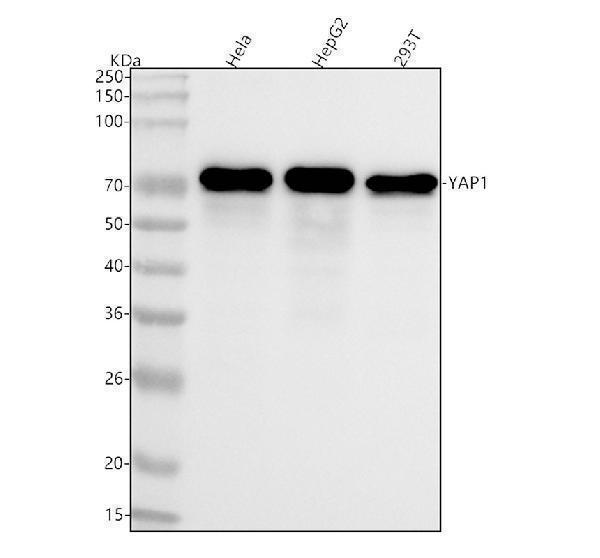
Western blot analysis of YAP1 using anti-YAP1 antibody (M00116).
Electrophoresis was performed on a 5-20% SDS-PAGE gel at 70V (Stacking gel) / 90V (Resolving gel) for 2-3 hours. The sample well of each lane was loaded with 30 ug of sample under reducing conditions.
Lane 1: human Hela whole cell lysates,
Lane 2: human HepG2 whole cell lysates,
Lane 3: human 293T whole cell lysates.
After electrophoresis, proteins were transferred to a nitrocellulose membrane at 150 mA for 50-90 minutes. Blocked the membrane with 5% non-fat milk/TBS for 1.5 hour at RT. The membrane was incubated with rabbit anti-YAP1 antigen affinity purified monoclonal antibody (Catalog # M00116) at 1:5000 overnight at 4°C, then washed with TBS-0.1%Tween 3 times with 5 minutes each and probed with a goat anti-rabbit IgG-HRP secondary antibody at a dilution of 1:500 for 1.5 hour at RT. The signal is developed using an Enhanced Chemiluminescent detection (ECL) kit (Catalog # EK1002) with Tanon 5200 system. A specific band was detected for YAP1 at approximately 70 kDa. The expected band size for YAP1 is at 54 kDa.

Click image to see more details
Immunohistochemical analysis of paraffin-embedded human uterus, using YAP1 Antibody.

Click image to see more details
YAP knockdowned significantly decreased YAP expression at mRNA ( a ) and protein ( b ) levels in Hep-2 cells. c Immunofluorescence (bottom panel) was used to detect the expression of YAP (red). DAPI (blue) was used to stain the nuclei. The fluorescence intensity of YAP was stronger in YAP-NC groups, and was weaker in cells transfected with shRNA. One representative experiment out of the three performed is shown (80X). Experiments were repeated three times, and data are shown as the mean ± SD, ** P < 0.01 ( a ), ** P < 0.01 ( b )
Index in PubMed under a CC BY license. PMID: 31269911

Click image to see more details
YAP knockdowned suppresses cell proliferation, invasion and migration. Silenced YAP inhibited cell proliferation in Hep-2 cells by CCK-8 (top panel). a The OD value at 450 nm for 5 days. Hep-2 with no treatment (Control), Non-nonspecific shRNA treatment (YAP-NC), YAP knockdown (YAP-shRNA) groups, and cDNA overexpression after YAP knockdown (YAP-shRNA+YAP-cDNA), * P < 0.05. b Colony formation assays (second panel) were performed to evaluate the proliferative capability of control, YAP-NC, YAP-shRNA and YAP-shRNA+YAP-cDNA cell groups. A representative image is shown, and a statistical comparison of the indicated groups was performed across four independent experiments, ** P < 0.01. c Wound healing assays (third panel) were performed to explore the migration capability, and solid lines represented the wound edges. Images were captured by using light microscopy (40X). The migration index was calculated as described in the Materials and Methods (control, YAP-NC, YAP-shRNA and YAP-shRNA+YAP-cDNA). Statistical analysis is shown, ** P < 0.01
Index in PubMed under a CC BY license. PMID: 31269911

Click image to see more details
Silencing of YAP promotes cell apoptosis and induces cell cycle arrest. a Guava easyCyte12 was used to investigate differences in cell cycle (top panel) distribution following YAP silencing or overexpression in Hep-2. Silenced YAP drove G2/M arrest in control, YAP-NC, YAP-shRNA and YAP-shRNA+YAP-cDNA cell groups. In addition, when YAP knockdown Hep-2 cells overexpressed YAP, the G2/M phase distribution was decreased, ** P < 0.01; b Apoptotic cells (medium panel) were analysed by Guava easyCyte12 via staining of annexin PE/7-AAD. The percentage of apoptotic cells is shown. ** P < 0.01
Index in PubMed under a CC BY license. PMID: 31269911

Click image to see more details
a Targeted silencing of YAP inhibits the expression of mRNA and protein levels. a qRT-PCR was used to analyse the mRNA expression of YAP, GSK-3β, DDK1, E-cadherin, β-catenin and vimentin in Hep-2 cell lines after transfection or not, and GAPDH was used as the internal control. Experiments were repeated ten times, and data are shown as the mean ± SD, ** P < 0.01. b Western blot assay was employed to investigate the expression of YAP, β-catenin, E-cadherin, vimentin, DDK1 and GSK-3β. WB results indicated that the expression of vimentin and β-catenin were downregulated with YAP knockdown and were upregulated when cells were administered YAP-cDNA. Representative images are shown. Statistical analysis of the relative optical density of each band is shown. GAPDH was used as an internal control, ** P < 0.01. c Model of the role of YAP in laryngeal cancer
Index in PubMed under a CC BY license. PMID: 31269911

Click image to see more details
a Targeted silencing of YAP suppresses laryngeal cancer tumourigenicity and metastasis in vivo. The effects of silenced YAP on tumour suppression in vivo. Images of tumours (left panel) formed in nude mice injected subcutaneously with Hep-2 cells transfected with control, negative vector, and YAP-shRNA. Fluorescence images (right panel) of tumours captured on the IVIS system 21 days after subcutaneous injection. Cells of each group were stably transfected with luciferase. Tumours with YAP knockdown were smaller compared to the other groups. Tumour growth curves are plotted. ** P < 0.01. b A pulmonary metastasis model (left panel) was established after 3 weeks of the indicated treatment. Images from the pulmonary metastasis model and the corresponding statistical analysis are shown. ** P < 0.01
Index in PubMed under a CC BY license. PMID: 31269911

Click image to see more details
Immunohistochemistry staining for YAP, KI-67 and Bcl-2 protein in xenograft tumour model and pulmonary metastasis model. Cytoplasmic staining was considered to be positive for YAP, KI-67 and Bcl-2. b & f Higher YAP expression in both xenograft tumour and pulmonary models. c & d High YAP expression mainly in the nucleus of LSCC. e & f Low YAP expression in LSCC in both the cytoplasm and nucleus of tumour cells. g & h Low expression of YAP protein mainly in the nucleus of LSCC. [A, a, E, e] × 40; [B, b, C, c, D, d, F, f, G, g, H, h] × 100
Index in PubMed under a CC BY license. PMID: 31269911

Click image to see more details
Immunofluorescent analysis of MCF7 cells, using YAP1 Antibody .

Click image to see more details
Immunofluorescent analysis using the Antibody at 1:50 dilution.

Click image to see more details
Immunofluorescent analysis using the Antibody at 1:150 dilution.
Specific Publications For Anti-YAP1/Yap Rabbit Monoclonal Antibody (M00116)
Loading publications
Recommended Resources
Here are featured tools and databases that you might find useful.
- Boster's Pathways Library
- Protein Databases
- Bioscience Research Protocol Resources
- Data Processing & Analysis Software
- Photo Editing Software
- Scientific Literature Resources
- Research Paper Management Tools
- Molecular Biology Software
- Primer Design Tools
- Bioinformatics Tools
- Phylogenetic Tree Analysis
Customer Reviews
Have you used Anti-YAP1/Yap Rabbit Monoclonal Antibody?
Share your experimental results or join a short interview to earn up to $1,000 in product credits or other rewards.
0 Reviews For Anti-YAP1/Yap Rabbit Monoclonal Antibody
Customer Q&As
Have a question?
Find answers in Q&As, reviews.
Can't find your answer?
Submit your question
5 Customer Q&As for Anti-YAP1/Yap Rabbit Monoclonal Antibody
Question
I see that the anti-YAP1/Yap Rabbit Monoclonal antibody M00116 works with WB, what is the protocol used to produce the result images on the product page?
Verified Customer
Verified customer
Asked: 2019-09-05
Answer
You can find protocols for WB on the "support/technical resources" section of our navigation menu. If you have any further questions, please send an email to support@bosterbio.com
Boster Scientific Support
Answered: 2019-09-05
Question
I was wanting to use your anti-YAP1/Yap Rabbit Monoclonal antibody for WB for human esophagus on frozen tissues, but I want to know if it has been validated for this particular application. Has this antibody been validated and is this antibody a good choice for human esophagus identification?
Verified Customer
Verified customer
Asked: 2018-09-12
Answer
As indicated on the product datasheet, M00116 anti-YAP1/Yap Rabbit Monoclonal antibody has been validated for IP, IF, IHC, WB on human tissues. We have an innovator award program that if you test this antibody and show it works in human esophagus in IHC-frozen, you can get your next antibody for free.
Boster Scientific Support
Answered: 2018-09-12
Question
Can you help my question with product M00116, anti-YAP1/Yap Rabbit Monoclonal antibody. I was wondering if it would be possible to conjugate this antibody with biotin. I would need it to be without BSA or sodium azide. I am planning on using a buffer exchange of sodium azide with PBS only. Would there be problems for me to conjugate the antibody and store it in -20 degrees in small aliquots?
E. Mitchell
Verified customer
Asked: 2018-05-10
Answer
It is not recommended storing this antibody with PBS buffer only in -20 degrees. If you want to store it in -20 degrees it is best to add some cryoprotectant like glycerol. If you want carrier free M00116 anti-YAP1/Yap Rabbit Monoclonal antibody, we can provide it to you in a special formula with trehalose and/or glycerol. These molecules will not interfere with conjugation chemistry and provide a good level of protection for the antibody from degradation. Please be sure to specify this in your purchase order.
Boster Scientific Support
Answered: 2018-05-10
Question
Is this M00116 anti-YAP1/Yap Rabbit Monoclonal antibody reactive to the isotypes of YAP1?
T. Brown
Verified customer
Asked: 2015-08-12
Answer
The immunogen of M00116 anti-YAP1/Yap Rabbit Monoclonal antibody is A synthesized peptide derived from human YAP1. Could you tell me which isotype you are interested in so I can help see if the immunogen is part of this isotype?
Boster Scientific Support
Answered: 2015-08-12
Question
Would anti-YAP1/Yap Rabbit Monoclonal antibody M00116 work for WB with esophagus?
R. Moore
Verified customer
Asked: 2013-08-16
Answer
According to the expression profile of esophagus, YAP1 is highly expressed in esophagus. So, it is likely that anti-YAP1/Yap Rabbit Monoclonal antibody M00116 will work for WB with esophagus.
Boster Scientific Support
Answered: 2013-08-16
