This website uses cookies to ensure you get the best experience on our website.
- Table of Contents
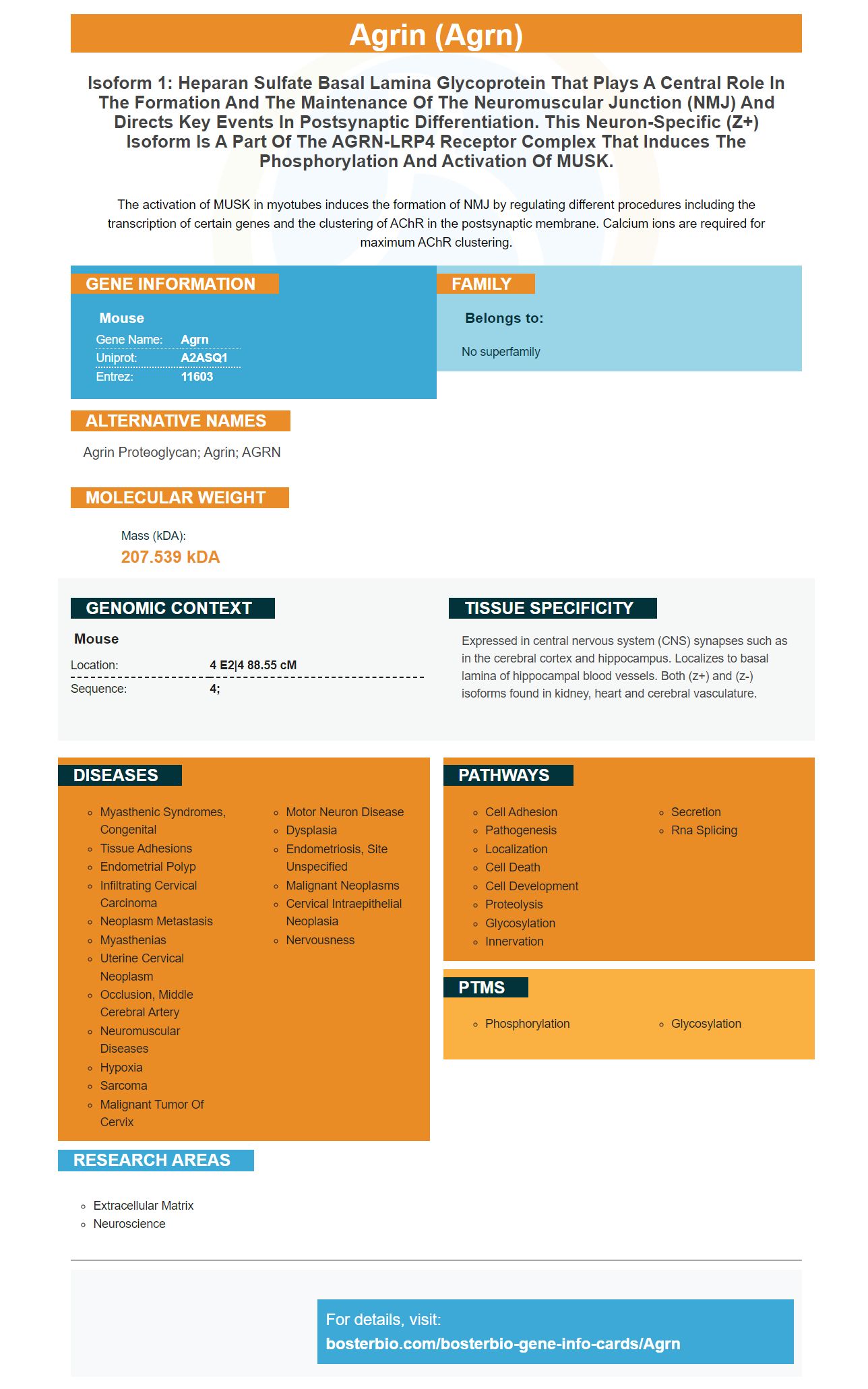
Facts about Agrin.
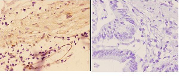
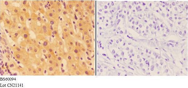
The activation of MUSK in myotubes induces the formation of NMJ by regulating different procedures including the transcription of certain genes and the clustering of AChR in the postsynaptic membrane. Calcium ions are required for maximum AChR clustering.
| Mouse | |
|---|---|
| Gene Name: | Agrn |
| Uniprot: | A2ASQ1 |
| Entrez: | 11603 |

| Belongs to: |
|---|
| No superfamily |

agrin proteoglycan; Agrin; AGRN
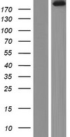
Mass (kDA):
207.539 kDA

| Mouse | |
|---|---|
| Location: | 4 E2|4 88.55 cM |
| Sequence: | 4; |
Expressed in central nervous system (CNS) synapses such as in the cerebral cortex and hippocampus. Localizes to basal lamina of hippocampal blood vessels. Both (z+) and (z-) isoforms found in kidney, heart and cerebral vasculature.


PMID: 8653787 by Glass D.J., et al. Agrin acts via a MuSK receptor complex.
PMID: 9188458 by Smith M.A., et al. Selective regulation of agrin mRNA induction and alternative splicing in PC12 cells by Ras-dependent actions of nerve growth factor.