This website uses cookies to ensure you get the best experience on our website.
- Table of Contents
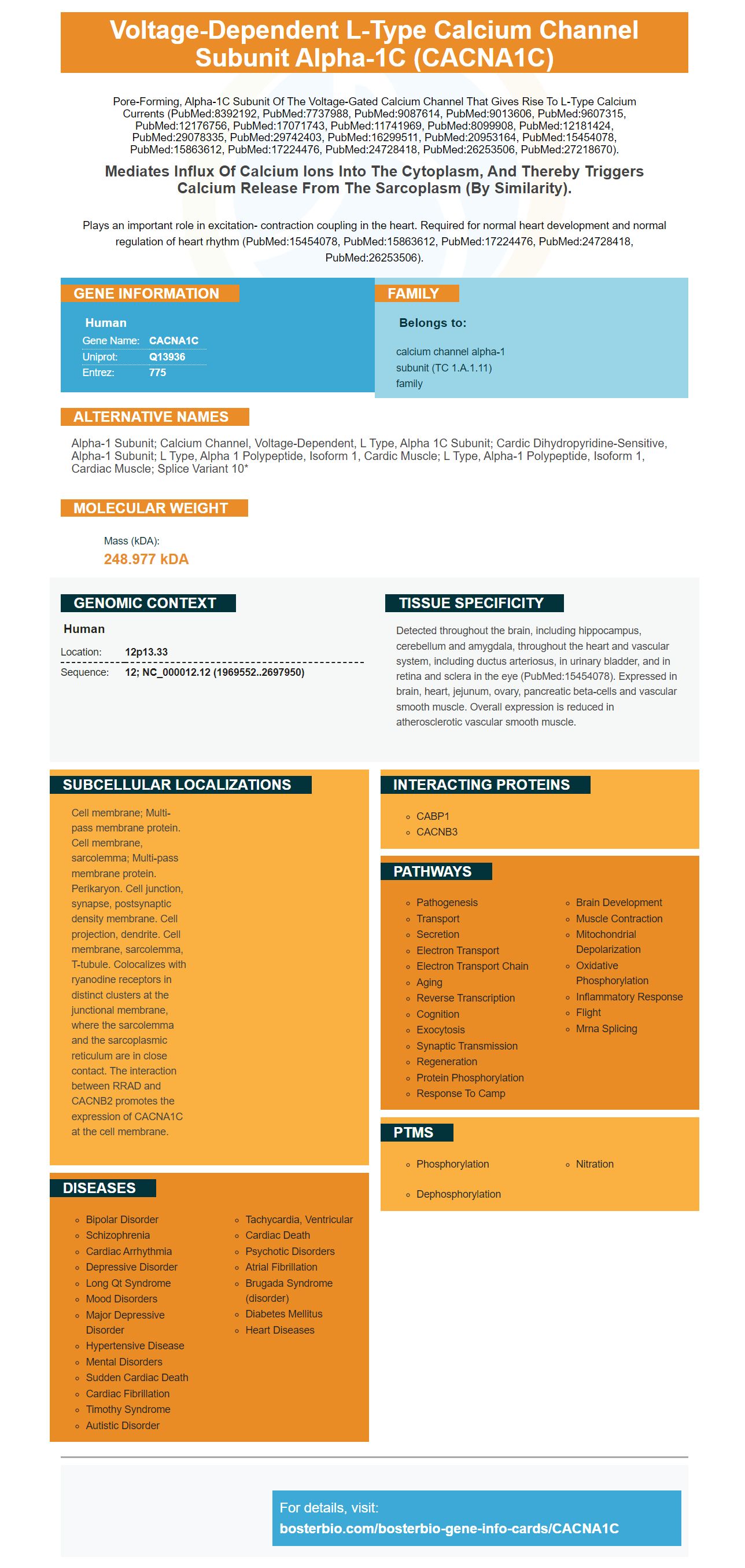
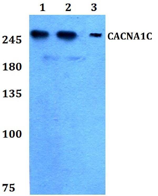
Facts about Voltage-dependent L-type calcium channel subunit alpha-1C.
Pore-forming, alpha-1C subunit of the voltage-gated calcium channel that gives rise to L-type calcium currents (PubMed:8392192, PubMed:7737988, PubMed:9087614, PubMed:9013606, PubMed:9607315, PubMed:12176756, PubMed:17071743, PubMed:11741969, PubMed:8099908, PubMed:12181424, PubMed:29078335, PubMed:29742403, PubMed:16299511, PubMed:20953164, PubMed:15454078, PubMed:15863612, PubMed:17224476, PubMed:24728418, PubMed:26253506, PubMed:27218670).
Mediates influx of calcium ions into the cytoplasm, and thereby triggers calcium release from the sarcoplasm (By similarity).Plays an important role in excitation- contraction coupling in the heart. Required for normal heart development and normal regulation of heart rhythm (PubMed:15454078, PubMed:15863612, PubMed:17224476, PubMed:24728418, PubMed:26253506).
Human | |
---|---|
Gene Name: | CACNA1C |
Uniprot: | Q13936 |
Entrez: | 775 |
Belongs to: |
---|
calcium channel alpha-1 subunit (TC 1.A.1.11) family |
alpha-1 subunit; calcium channel, voltage-dependent, L type, alpha 1C subunit; cardic dihydropyridine-sensitive, alpha-1 subunit; L type, alpha 1 polypeptide, isoform 1, cardic muscle; L type, alpha-1 polypeptide, isoform 1, cardiac muscle; splice variant 10*
Mass (kDA):
248.977 kDA
Human | |
---|---|
Location: | 12p13.33 |
Sequence: | 12; NC_000012.12 (1969552..2697950) |
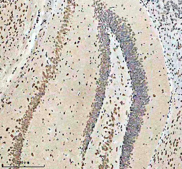
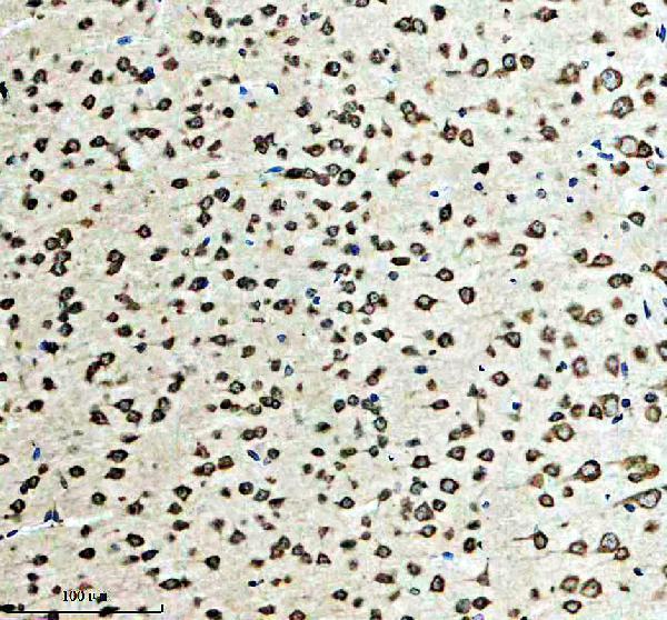
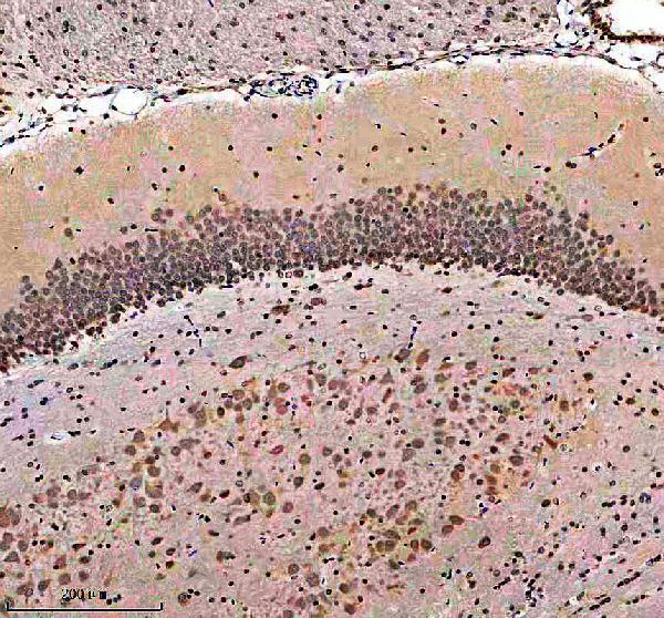
Detected throughout the brain, including hippocampus, cerebellum and amygdala, throughout the heart and vascular system, including ductus arteriosus, in urinary bladder, and in retina and sclera in the eye (PubMed:15454078). Expressed in brain, heart, jejunum, ovary, pancreatic beta-cells and vascular smooth muscle. Overall expression is reduced in atherosclerotic vascular smooth muscle.
Cell membrane; Multi-pass membrane protein. Cell membrane, sarcolemma; Multi-pass membrane protein. Perikaryon. Cell junction, synapse, postsynaptic density membrane. Cell projection, dendrite. Cell membrane, sarcolemma, T-tubule. Colocalizes with ryanodine receptors in distinct clusters at the junctional membrane, where the sarcolemma and the sarcoplasmic reticulum are in close contact. The interaction between RRAD and CACNB2 promotes the expression of CACNA1C at the cell membrane.
If you are interested in CACNA1C detection then you should take a look at the Boster Bio Anticaveolin-1/CAV1 Marker. This marker can detect the protein in all kinds of biological samples. The advantages of using this marker are discussed in this article. All scientists will benefit from the information contained in this article. These benefits are accessible to all scientists in the world.
A new product, Boster Bio Anti-Caveolin-1/CAV1 Marker can be used to detect Caveolin-1. The molecule binds directly with G-protein alpha subunits , and is able to regulate their activity. Although the molecular reasons for its involvement in liver injury and repair are not fully understood, it is important. Caveolin-1, among its many functions, is involved in T-cell growth, NFKappaB signaling, and CTNNB1 signaling via the Wnt pathway. It interacts with syntaxin GLIPR2, syntaxin, and the rotaviruses DPP4 A NSP4, BMX, and BMX.
Cav1 activation is necessary for the inflammatory response of many pathogens. It has been proven to reduce the inflammatory response to LPS by inhibiting the activation of nuclear factor kB. The stimuli Cav1 is exposed to may alter its role. This antibody, known as CCl4-CAV1, is available in a variety of concentrations to detect Cav1 expression.
The immunohistochemical detection for Cav-1 is a sensitive and precise method to determine the presence of this protein in your samples. In the dark tissues, the tissues were stained with CD68 and CD34 to visualize the presence of Cav-1 and to determine the amount of fluorescence. Once these conditions have been fulfilled, the Boster Bio Anti-Caveolin-1 marker can be utilized.
Boster Bio Anti Caveolin-1 antibody PA1514 reacts with mouse, human, and the rat Caveolin-1. The Boster Bio Anti-Caveolin-1 Marker reacts with Human, Mouse, and the Caveolin-1 protein of the Rat. These results are reliable and precise. Its cost is also competitive. It is also available online.
This high-throughput ELISA Kit provides a convenient and accurate method to test Caveolin-1 protein expression in cells. It can be used to screen tests or studies to investigate the effects of different treatments. This includes activators, inhibitors, and the ligands. The results of the kit will be presented in terms of the levels of proteins in the sample. It will also show whether the treatment was efficient.
The CACNA1C gene instructs the body on how to create calcium channels which transport positively charged calcium atoms into cells. Calcium ions play an important role in various cell functions, including cell-to-cell communication as well as the tensing process of muscle fibers and the regulation of certain genes. Calcium is also crucial in the development of the brain and bones prior to the time of birth. Therefore, individuals with this gene are at a greater likelihood of developing schizophrenia and it has been proven to be genetically linked with certain forms of depression.
Biological tests that detect CACN1C have been developed to identify this gene variant. It has been shown to be involved in the development of TS, a rare multisystem disorder characterized by severe QT extension, syndactyly and a variety of organ system dysfunctions, and it often causes early death in children. People with CACNA1C mutations are unlikely to have any of these traits like syndactyly or cognitive impairments.
The presence of the risk allele has been shown to increase the risk of this mutation. Researchers discovered that carriers of the risk allele had higher levels the gene's mRNA, indicating that this specific transcript could affect the function of certain brain regions. Calcium channel protein plays an important role in neuronal function and development. It is responsible for maintaining connectivity throughout adulthood, and establishing connectivity during developmental stages. The development of new therapies could be made simpler by changes in gene expression.
Recent genetic studies have identified CACNA1C as a risk gene for affective disorders. The principal SNP rs1006737 is linked to an increase in mRNA expression of CACNA1C as well as increased calcium signaling. This gene encodes the pore-forming component of CaV1.2 which is the main voltage-gated calcium channel. The channel connects Ca2+ influx with neuronal gene transcription, and is thought to play an important part in synaptic reorganization, dendritic growth, and cell survival.
Molecular analysis of CACNA1C has revealed the existence of numerous pathogenic variants within the gene. Certain variants contribute to the development of various organs including the CNS bones, CNS, and glucose metabolism. However the genetic analysis of CACNA1C requires the complete sequencing of the entire genome region to identify the exact diagnosis. In the absence of germ cell diagnosis, results from genetic tests could not be consistent.
GWAS-confirmed the risk of psychiatric illness. The gene is involved in multiple pathways of oxidative stress, and specifically, it affects mitochondrial function and integrity. Many studies have demonstrated CACNA1C to be associated with schizophrenia. These findings highlight the potential benefits of CACNA1C genetic tests for diagnosing mood disorders and other disorders. CACNA1C is an accurate genetic test that should be considered when looking for one.
If CACNA1C is detected in an individual in the family A genetic test is able to detect it. A EKG test can help identify relatives at risk. Additionally, genetic testing can also be conducted for women who are pregnant. To determine whether CACNA1C is affecting the pregnancy process, the results of genetic tests should be confirmed. This can help provide a more accurate prediction of the risk of having a baby.
PMID: 1316612 by Soldatov N.M.; Molecular diversity of L-type Ca2+ channel transcripts in human fibroblasts.
PMID: 8392192 by Schultz D., et al. Cloning, chromosomal localization, and functional expression of the alpha-1 subunit of the L-type voltage-dependent calcium channel from normal human heart.