This website uses cookies to ensure you get the best experience on our website.
- Table of Contents
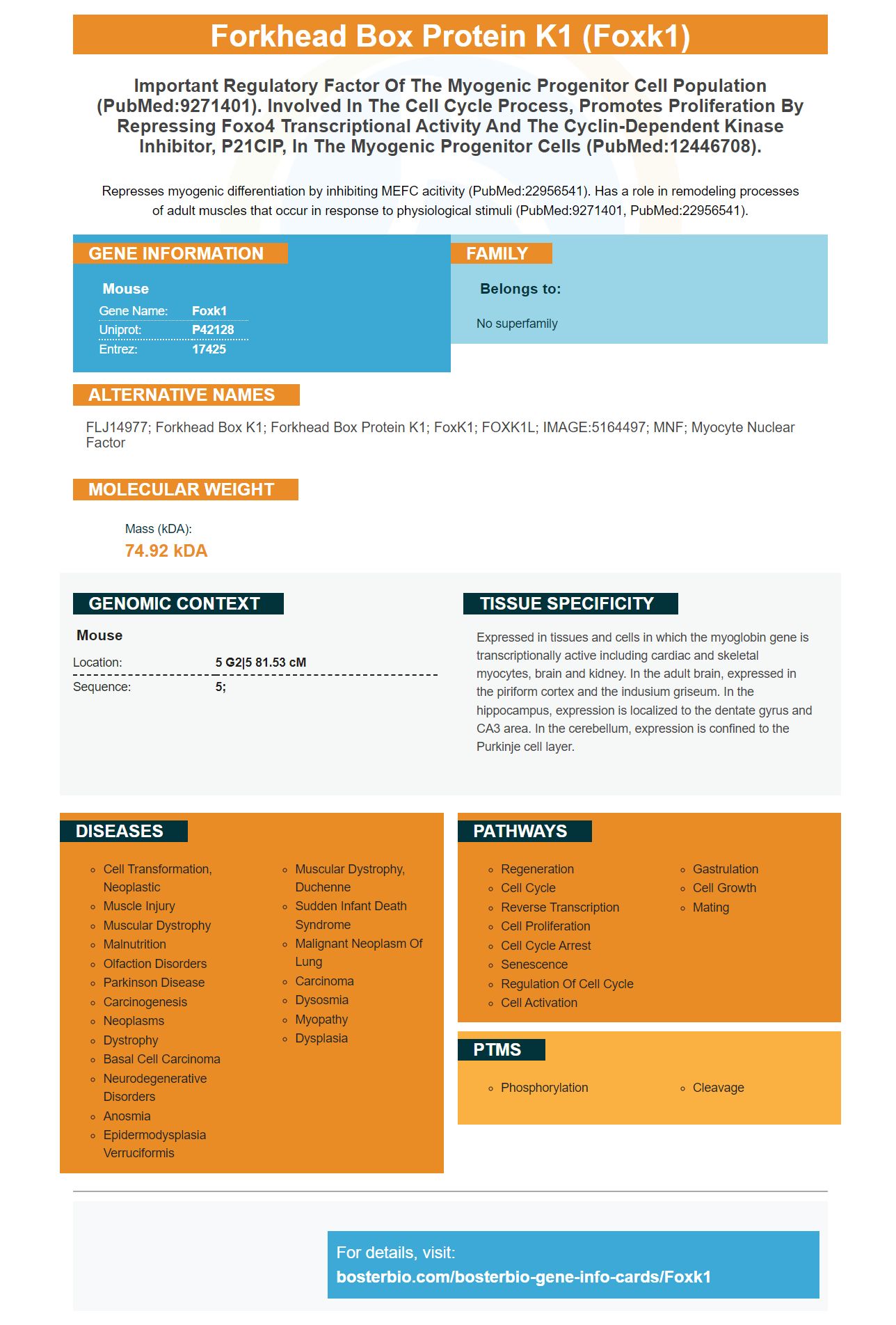
Facts about Forkhead box protein K1.
Represses myogenic differentiation by inhibiting MEFC acitivity (PubMed:22956541). Has a role in remodeling processes of adult muscles that occur in response to physiological stimuli (PubMed:9271401, PubMed:22956541).
Mouse | |
---|---|
Gene Name: | Foxk1 |
Uniprot: | P42128 |
Entrez: | 17425 |
Belongs to: |
---|
No superfamily |
FLJ14977; forkhead box K1; forkhead box protein K1; FoxK1; FOXK1L; IMAGE:5164497; MNF; Myocyte nuclear factor
Mass (kDA):
74.92 kDA
Mouse | |
---|---|
Location: | 5 G2|5 81.53 cM |
Sequence: | 5; |
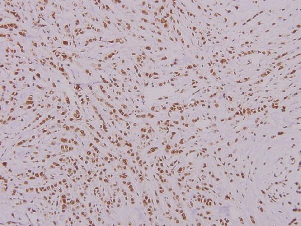
Expressed in tissues and cells in which the myoglobin gene is transcriptionally active including cardiac and skeletal myocytes, brain and kidney. In the adult brain, expressed in the piriform cortex and the indusium griseum. In the hippocampus, expression is localized to the dentate gyrus and CA3 area. In the cerebellum, expression is confined to the Purkinje cell layer.
If you're looking to use a FOXK1 marker, then read this article. In this article, we'll review SNHG1, FOXK1, LINC01503, ELIS, and a LINC01503 probe. This article will aid you in making the right choice for your research. In addition, we'll explain how to get the most precise results from ELISA.
Research studies can benefit by using research studies can benefit from the use of FOXK1 marker. It is helpful in the identification of biomarkers in a variety of fields such as development biology, cancer neurosciences, inflammation, and cancer. This marker is highly sensitive to picograms, and even at picogram-levels. Researchers from a variety of industries use this product for their research. The company offers a variety of kits for antibodies and ELISA kits for various uses. The kits are available through tebubio.
The SNHG1 gene blocks miR-376a and enhances FOXK1/Snail's expression. The results suggest that FOXK1 and Snail could be therapeutic targets for HCC. The results need to be confirmed using different animal models and cell types. FOXK1 (a potential therapeutic target) and Snail (a multiple role in HCC) are not yet known.
FOXK1's downstream target is gene Snail. JASPAR has identified three binding sites for Snail. Then the Snail expression was high in both HCC tissue and cell lines. In addition, immunohistochemistry revealed that FOXK1 was overexpressed in HOSEpiC cells. After determining the expression level cells were transfected with the sh-FOXK1–neo plasmid.
Sh-SNHG1-1 significantly reduced the expression of SNHG1-1 and resulted in a decrease in proliferative, migration, and invasion. The suppression of SNHG1 led to an increase in the expression of other EMTand migration-related proteins, including Bax or Bcl-2. The cells went into apoptosis and then ended up dying. This is why they were able to express the FOXK1 gene using a variety.
Many tissues of our body contain the FOXK1 marker. Most significantly it is found in the mGC tissues. Higher levels of FOXK1 were associated with decreased OS, according to a study by Kaplan-Meier. FOXK1 overexpression was strongly linked to a poor prognosis in GC patients. Here are the best uses of the FOXK1 marker.
It is believed that FOXK1 can promote the invasion and migration of GC cells. Using a substantive anti-FOXK1 antibody, researchers analyzed the DNA-binding region of FOXK1. MAZ was the most compatible with a consensus GGGGGG motif. Bioinformatics analysis revealed that MAZ had two binding sites in the FOXK 5'-UTR. Both genes are expressed in the TCGA data set for GC.
The FOXK1 marker is used to test the effects of FOXK1 inhibition upon the growth and development of tumor cells. The marker can be used in a variety of cancer types such as melanoma and Alzheimer's disease. In addition, FOXK1 is expressed in many tissues, including the cholangitis and glioma. This allows researchers to examine the role of FOXK1 in the development of the disease.
In this study, two pathologists assessed the expression of FOXK1 in human tumor cells. They scored tumor tissues using two parameters: intensity of staining and the percentage of immunoreactive cells. The staining intensity was graded as zero (negative) or one (weak), or two ("moderate"). The percentage of stained cells ranged between 51 and 100%, which equates to between 0% and 100 percent. These two parameters were then combined to calculate the final score.
The ELISA for the FOXK1 (Forkhead box K1) marker is a powerful instrument to assess breast cancer cell growth and invasion. Certain cancers trigger the activity of FOXK1 which includes lung, glioma, as well as gastric. However, its exact role isn't clear. Understanding how FOXK1 regulates cell growth is vital to understand its function.
ELISA for FOXK1 allows quantitative measurement of the FOXK1 protein in serum, plasma, cell culture supernatants and tissue homogenates. Human FOXK1 regulates EMT and is a key gene in breast cancer. FOXK1 increase the expression of certain cell proliferation markers , such as snail or slug. The ELISA kit that detects the FOXK1 marker has high sensitivity and precision and is highly recommended for monitoring the progression of disease.
An ELISA for the FOXK1 markers utilizes monoclonal antibodies that target specific proteins. The primary antibody removes the protein from solution, while the secondary mAb provides a signal indicating that the protein is present. The ELISA kit comes with a standard concentration for the protein recombinant, which allows you to create an ELISA standard curve. Compare the signals from one sample with the signal from another sample when interpreting the results.
The ELISA test for FOXK1 is highly sensitive and specific and is suitable for research and clinical purposes. FOXK1 is one of the FOX transcription factors family and regulates cell cycle progression via interaction with the inhibitor of cyclin dependent kinase, P21. It regulates myogenic stem cells growth and differentiation.
A new reagent for DNA microarrays based on the FOXK1 gene, the Boster Bio LINC01503 Probe is approved by the U.S. Food and Drug Administration (FDA). The probe is specifically designed to target the FOXK1 gene, which regulates the expression of many other proteins. It also recognizes expression of other FOXK1 markers, such as the kinase SFPQ. In the present study, LINC01503 inhibits the growth of NPC tumors in vivo when compared to scramble controls. In addition, tumors containing shLINC01503 had a significantly lower size than those in the control group. This conclusion was further supported by data on the weight of tumors. A FISH-IHC test also conducted to confirm this finding.
The RNA was transscribed in vitro and biotin-labeled to detect FOXK1 protein. Thermo Fisher Scientific used the Pierce(tm) 3' RNA End Desthiobiotinylation Kit to label the RNA. The bound proteins were determined by western blotting and mass spectrometry.
Expression of LINC01503 was assessed in paraffin-embedded tumor tissues from NPC patients. Based on the FOXK1 marker, patients were divided into two groups. Patients with high FOXK1 expression had significantly lower overall survival rates and shorter time to live without illness than patients who had low expression. However there was no evidence of an association between LINC01503 and clinical characteristics.
Boster Bio's FOXK1 ELISA Kits are high-throughput and easy to use for monitoring the expression profile FOXK1 protein in cells and in tissues that have been cultured. They can be utilized by researchers to screen for activators, drugs and treatments. This kit cannot be purchased separately, however Boster researchers are able to share the results of their samples that are not validated and receive product credits for their research.
These ELISA kits are based on Picokine(tm) which is the highest-sensitivity ELISA platform. These ELISA kits have been validated for use with various samples and attain sensitivities down to the picogram level. Images and validation procedures are available upon request. Picoband, a polymer-based secondary antibody that is revolutionary can help save 30 minutes of IHC. Technical assistance is provided by Sanbio the company that is one of the BeNeLux distributor.
Recent research published in Cell Reports indicates that IHC-optimized monoclonals against FOXK1 protein are particularly effective in detecting a specific epitope within cells. The antibodies are highly specific and can recognize multiple epitopes of a single antigen. Monoclonal antibody have similar properties like polyclonal, however they are less expensiveand more robust over a variety of salt and pH levels, better able to stand up to the fluctuations from batch-to-bath, and more prone to fluctuations that occur sporadically.
Besides detecting specific epitopes, IHC-optimized monoclonals can be utilized for various applications, including classifying cancerous tumors, determining the place of metastasis and identifying malignant cells. These antibodies can detect molecular alterations such as ATRX and IDh2 mutations. This makes them a great choice for the diagnostics of tumors.
The affinity purified immunogen polyclonal antibodies diminish background staining and improve their specificity. They are therefore more effective in finding specific antibodies. Because they bind to more specific antigens to target, and less non-specific antibodies pass through the column, which means that the process is more efficient. This enriches the antigen-specific, polyclonal antibody group, which makes them more sensitive in finding a specific type antigen.
A common issue that is encountered with mouse monoclonal antibodies is the intense background staining which occurs. This is due to secondary antimouse antibody binding to the endogenous mouse tissues. This issue has been solved by a variety of methods. To lessen background staining one option is to use monovalent antimouse antibodies. Another approach is to use a goat antimouse that is conjugated with biotin to minimize background staining.
PMID: 8007964 by Bassel-Duby R., et al. Myocyte nuclear factor, a novel winged-helix transcription factor under both developmental and neural regulation in striated myocytes.
PMID: 9271401 by Yang Q., et al. Transient expression of a winged-helix protein, MNF-beta, during myogenesis.