This website uses cookies to ensure you get the best experience on our website.
- Table of Contents
7 Citations 16 Q&As
3 Citations 16 Q&As
6 Citations 15 Q&As
7 Citations 12 Q&As
6 Citations
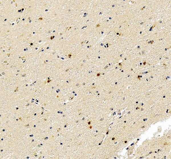
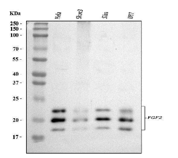
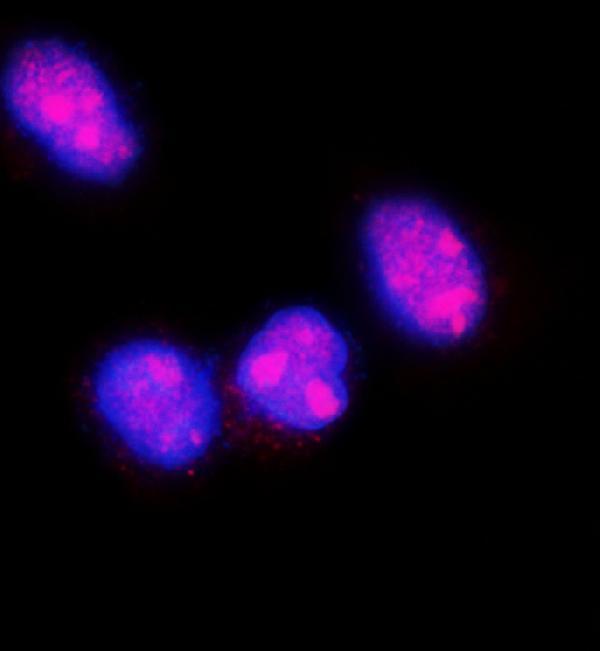
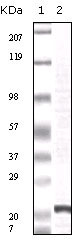
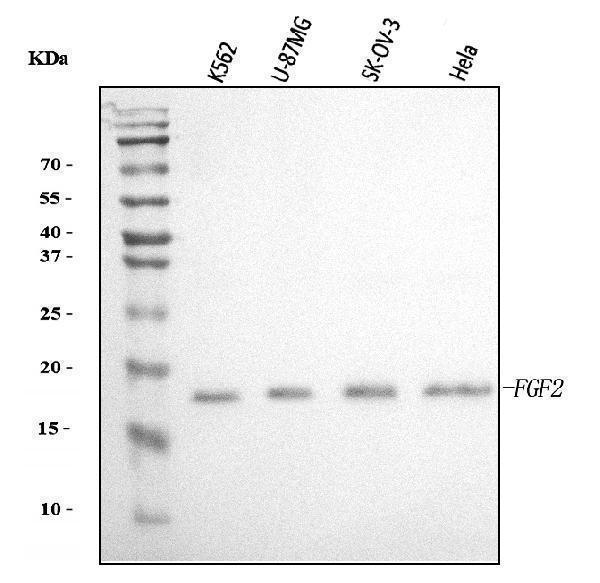
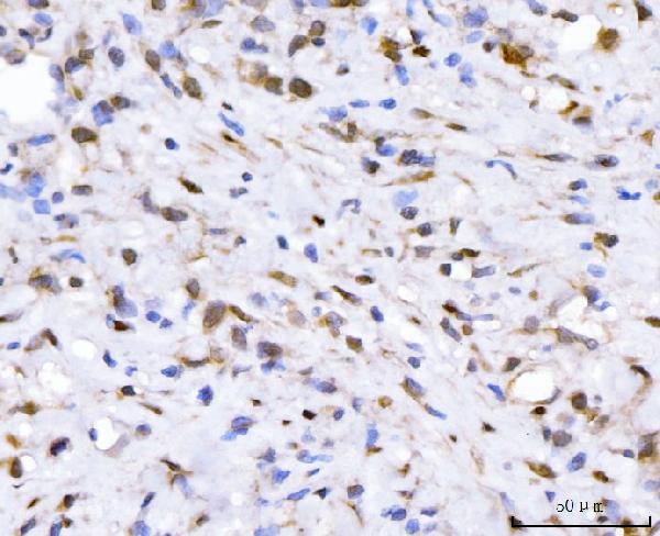
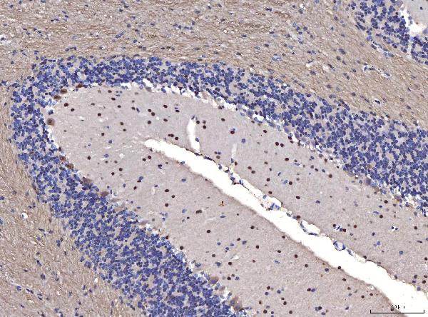
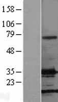
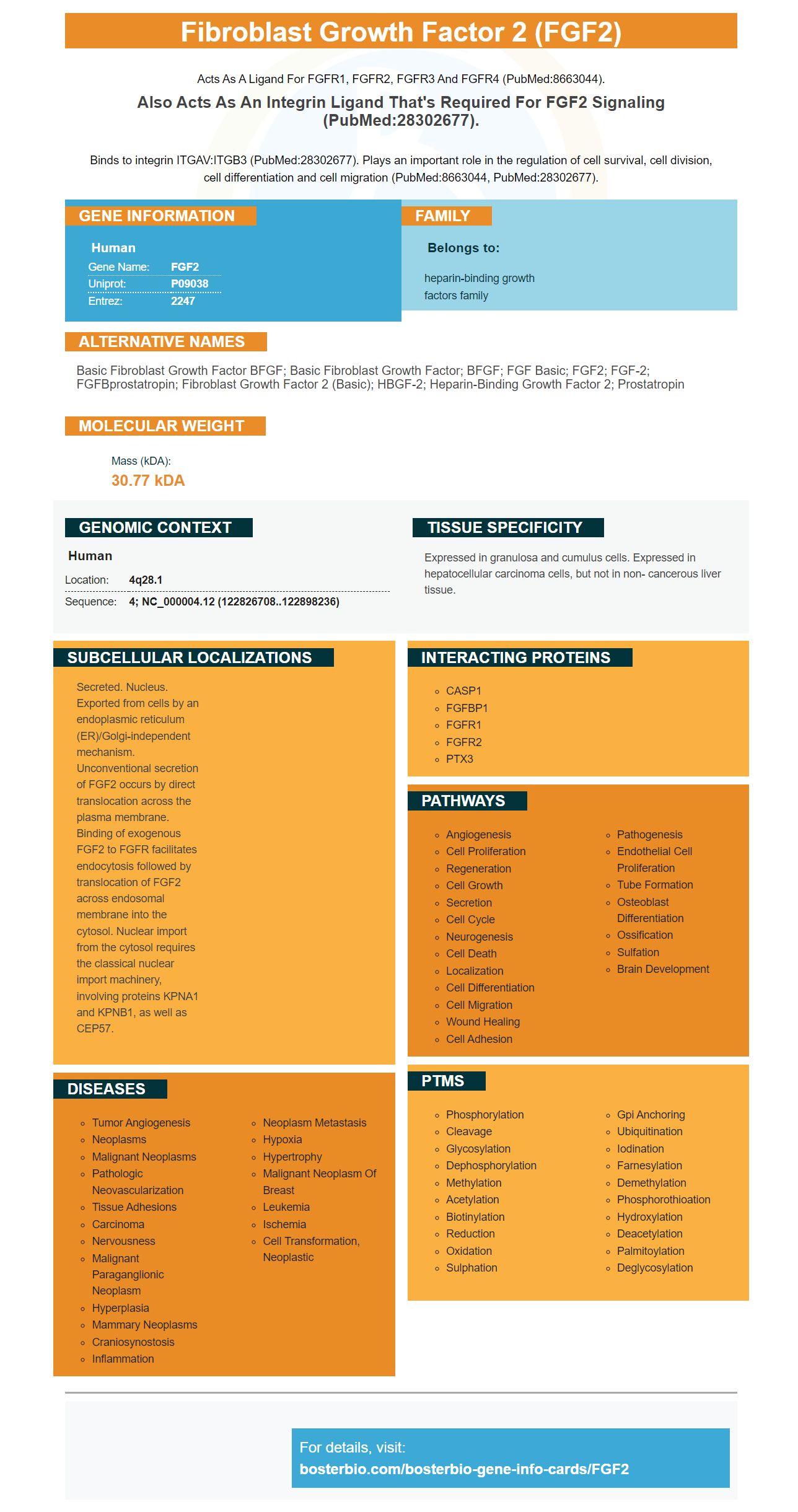
Facts about Fibroblast growth factor 2.
Acts as a ligand for FGFR1, FGFR2, FGFR3 and FGFR4 (PubMed:8663044).
Also acts as an integrin ligand that's required for FGF2 signaling (PubMed:28302677).Binds to integrin ITGAV:ITGB3 (PubMed:28302677). Plays an important role in the regulation of cell survival, cell division, cell differentiation and cell migration (PubMed:8663044, PubMed:28302677).
Human | |
---|---|
Gene Name: | FGF2 |
Uniprot: | P09038 |
Entrez: | 2247 |
Belongs to: |
---|
heparin-binding growth factors family |
basic fibroblast growth factor bFGF; Basic fibroblast growth factor; bFGF; FGF basic; FGF2; FGF-2; FGFBprostatropin; fibroblast growth factor 2 (basic); HBGF-2; heparin-binding growth factor 2; Prostatropin
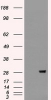
Mass (kDA):
30.77 kDA
Human | |
---|---|
Location: | 4q28.1 |
Sequence: | 4; NC_000004.12 (122826708..122898236) |
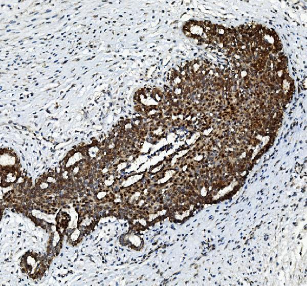
Expressed in granulosa and cumulus cells. Expressed in hepatocellular carcinoma cells, but not in non- cancerous liver tissue.
Secreted. Nucleus. Exported from cells by an endoplasmic reticulum (ER)/Golgi-independent mechanism. Unconventional secretion of FGF2 occurs by direct translocation across the plasma membrane. Binding of exogenous FGF2 to FGFR facilitates endocytosis followed by translocation of FGF2 across endosomal membrane into the cytosol. Nuclear import from the cytosol requires the classical nuclear import machinery, involving proteins KPNA1 and KPNB1, as well as CEP57.
FGF2 is a growth factor. It is also called fibroblast growth factors, or bFGF. The body produces this hormone to stimulate various processes, including healing and cell regeneration. Despite its significance in human biology, research is restricted. A new antibody that targets FGF2 was recently launched. It is anticipated to revolutionize research on human cell regeneration.
Fibroblast growth factors 2 (bFGP) is a protein that is utilized in cell culture to study purposes. Its role in the development of neural cells and function is not clear but it could contribute to the development of pathological conditions such as osteoporosis. The biological functions of bFGP include stimulation of cell proliferation and angiogenesis. FGF basic may play a crucial role in the prevention and treatment of a wide range of diseases and disorders.
Two major effects of bFGF for skeletal health are listed below. First, it hinders RUNX2 mRNA expression. It also blocks BGLAP's mRNA expression. RUNX2 regulates osteo/odontogenic development and function. BGLAP encodes OCN, an extracellular matrix protein found in bone that plays a significant role in mineralization. Additionally, it could also play a role in endocrine functions that are more extensive. Additionally, previous studies have implicated bFGF in the inhibition of osteo/odontogenic differentiation in SHEDs. Further, bFGF reduced the expression of BGLAP in stem cells derived from the apicalpapilla.
bFGF is a hormone which has a variety of physiological functions. It has been utilized to repair bone, periodontal tissues, and preserve the alveolar ridge. In vivo, bFGF is thought to have an anabolic impact. The exposure to bFGF proteins encourages osteogenic differentiation of local stem cells. Furthermore it regulates angiogenesis, cell migration, and proliferation and may facilitate healing in the vivo.
BMC boosts the production of FGF-2 within regenerating nerve tissue. The treatment increases glial cell growth, which in turn improves neuronal survival. Increased levels of FGF-2 were seen in lesions of the optic nerve's crush. This is a sign of tissues that are regenerating. However, bFGF cannot be used to detect nerve tissue.
It is interesting to note that the FGF-2 gene is also present in bone marrow-derived cells. SCs and lumbar spinal astrocytes release FGF-2. When bone marrow-derived cells express the hormone, SCs and glial cells multiply, which may contribute to regeneration. FGF-2 has been demonstrated to be a reliable indicator for regeneration of nerves.
The FGF8 gene is involved in a range of biological processes, including morphogenesis as well as embryogenesis. The protein is involved in gastrulation as well as limb development in mice. It is crucial for limb morphogenesis in chicks. In humans, FGF8 has been found to regulate the formation of craniofacial structures, as well as the salivary glands. A decrease in FGF8 expression could lead to the degeneration of molars, inability close the mouth and a smaller mouth.
In the menisctomized knee joint FGF8 stimulates the growth of osteoblasts and synovial cells and aids in the degrading of ECM and chondrocytes. In the laboratory, FGF8 injection reduced the weight of a rat patella. FGF8 injection led to reduction in bone mass of the patella in rats. In the MIA OA model, treatment with anti-FGF8 antibody slowed bone loss. These findings suggest a possible role for FGF8 in pathophysiology of OA.
In a rabbit study FGF8 increased the release of prostaglandin E2 and matrix metalloproteinase-3. This protein also promotes the growth of rabbit synovial cells. The antibody against FGF8 was able to stop the bone loss that FGF8 causes however, the results were not statistically significant. It is important to note that FGF8 is not an effective therapy for treating inflammatory arthritis.
FGF8 immunohistochemistry can detect the protein. Boster Bio has prepared anti-FGF8 antibody PA1216 for IHC-P and Western Blotting. Anti-FGF8 antibody reacts to Human, Mouse, and Rat. However, in some instances it is used to detect the protein within cells. Boster Bio's Fibroblast Growth Factor-8 (FGF8) marker is an accurate FGF8 immunoassay.
The FGF8 gene gives instructions for the fibroblast growth factor. The FGF8 gene is involved in a variety of biological processes that involve cell division and the regulation of cell growth, and development before birth. FGF8 connects to its receptor on the cell's surface. It initiates a series chemical reactions inside the cell. These results provide valuable information about the development and maintenance of the various body structures.
Fibroblast growth factor 10 (FGF10) is part of the heparin-binding glycoproteins group that has diverse biological functions towards cells. It is a heparin-binding protein with preference for FGF receptors 2 and 3, and also Chemotactic properties. It is important in the process of tissue repair as well as in embryonic growth and tumor growth. It also exhibits chemotactic capabilities and is a significant influencer of adipogenesis, as well as the development of different tissues.
While FGFs have been shown to be functional in a variety of cell types and in the targeted tissues in vitro, few studies have focused on their delivery mechanism. Apart from being used in medical implants, FGFs have been tested as biomaterials in collaboration with various medical tissues for regeneration, including bone, skin, and cartilage. These agents are specifically bound with FGFs and designed to be encapsulated within hydrogels and porous scaffolds.
FGFs biochemical composition differs from one species to the next. FGF10 is a neurotrophic factor with a variety of functions, including neurite extension and survival of neurons. Additionally, it promotes the regeneration of the skeletal muscles. It may be useful for repair of damaged muscles. This supplement could also be used to treat inflammation, injuries, scarring, and other conditions.
The gene that is responsible for FGF9 expression is known to be involved in the formation of the lips and palate clefts. Mice with Fgf9 knockouts have changes in the growth of the palatal and fusion, and their heads are narrower. The staining of the skull also shows early partial ossification. This means that Fgf9-deficient mice have lower bone density than normal mice.
The FGF signaling pathway is triggered by activating FGFRs. These receptors function as docking places for signaling molecules which trigger specific cells' responses. Activated FGFRs activate signaling complexes that then trigger a sequence of phosphorylation processes. The RAS-MAP kinase pathway is most well-known, followed by the PI3 kinase/AKT pathways.
The pathophysiology behind PSD is complex, it has been linked to the high levels of FGF9. In the hippocampus, FGF9 is upregulated, and the time of onset of PSD coincides with the optimal period for recovery of neurological function. This could suggest that FGF9 may be a unique modulator for the negative effect. Fibroblast growth factor 9 is a treatment option for poststroke depression, could be beneficial.
The ERK1/2 phosphorylation response to FGF2 was examined in primary fibroblasts treated with or without heparin. Activation of ERK1/2 was observed in a sigmoidal manner within 5 min and decreased after 60 minutes. These findings suggest that heparin plays an key roles in modulating FGFR responsiveness.
The melting temperature of FGF2 variants is determined by measuring their reactivity to Heparin, a well-known binding partner. Data were fitted to sigmoidal curves and color maps were generated. Two-way ANOVA was used to examine the data. The FGF2-STAB variants showed a lower affinity for heparin, compared to wild-type FGF2.
The basic fibroblast growth factor is a cytokine, which has wide mitogenic and cell survival capabilities. It is found in the basement membranes of normal tissues, as well as in the subendothelial layer of blood vessels. In the absence of a signal protein, this protein remains membrane bound. The FGF2 recombinant FGF2 marker has high specificity.
The Bradford reagent was used for molecular analyses. SDS-PAGE was used. The protein solution was lyophilized at 1 mg/ml before being stored at 4degC. After reconstitution, the solution was sterilely processed through a 0.22 mm filter. The solution was then dilute to 100 mg/ml FGF2 with PBS. The protein solution was mixed with bovine serum albumin and used in experiments.
The FGF2 marker was associated with the expression of NRG1 in PHNs. This protein is also associated with cognitive function and has been hypothesized to prevent cognitive decline in the process of aging. A decrease in FGF2 levels was observed in PHNs and was caused by inhibition of ErbB4 and NRG1 signals. The anti-NRG1 antibody slowed synaptic activity.
After 24 hours of FGF2 treatment the RNA was isolated from myelinating cells. The cDNA was made using a reverse transcription kit using a cDNA template. The cDNA was used to conduct real-time PCR. The reaction involved one ml of DNA template 10ng of cDNA, as well as 50 pmol/ml of primers.
PMID: 3472745 by Abraham J.A., et al. Human basic fibroblast growth factor: nucleotide sequence, genomic organization, and expression in mammalian cells.
PMID: 3780670 by Abraham J.A., et al. Human basic fibroblast growth factor: nucleotide sequence and genomic organization.
*Showing only the more recent 20. More publications can be found for each product on its corresponding product page