This website uses cookies to ensure you get the best experience on our website.
- Table of Contents
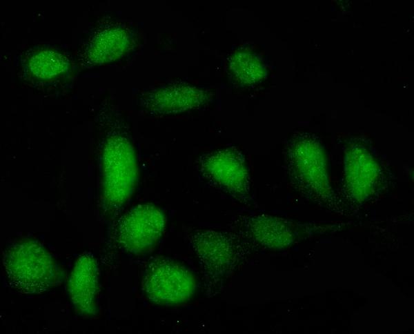
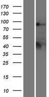
Facts about Tyrosine-protein phosphatase non-receptor type 22.
Dephosphorylates LCK at its triggering'Tyr-394' residue (By similarity). Dephosphorylates ZAP70 at its triggering'Tyr-492' residue (By similarity).
Mouse | |
---|---|
Gene Name: | Ptpn22 |
Uniprot: | P29352 |
Entrez: | 19260 |
Belongs to: |
---|
protein-tyrosine phosphatase family |
EC 3.1.3.48; Hematopoietic cell protein-tyrosine phosphatase 70Z-PEP; Lymphoid phosphatase; lymphoid-specific protein tyrosine phosphatase; Lyp; Lyp1; Lyp2; LyPTP; PEP; PEST-domain phosphatase; protein tyrosine phosphatase, non-receptor type 22 (lymphoid); protein tyrosine phosphatase, non-receptor type 8; PTPN22; PTPN8 (former); PTPN8LYP1; tyrosine-protein phosphatase non-receptor type 22
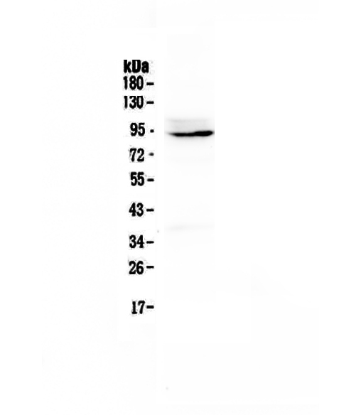
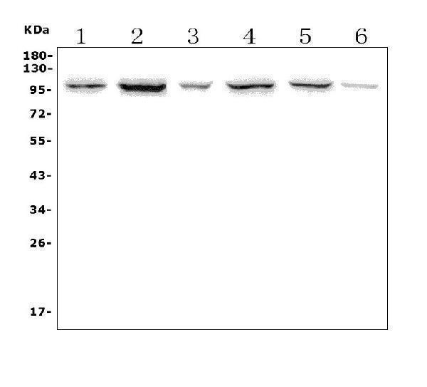
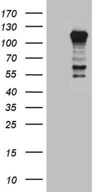
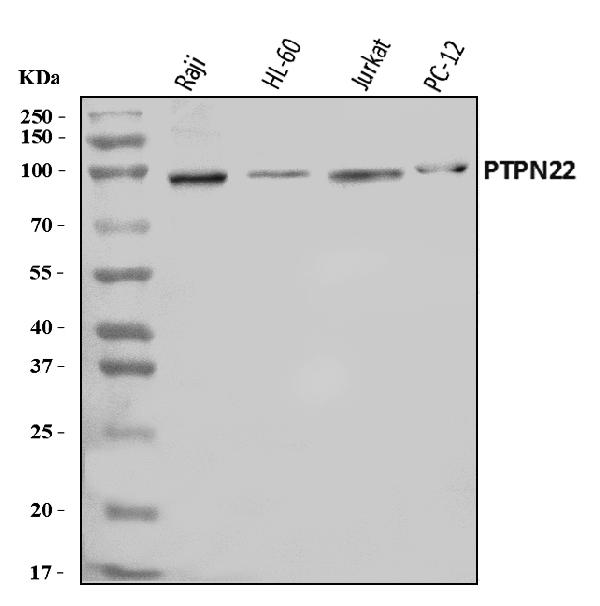
Mass (kDA):
89.714 kDA
Mouse | |
---|---|
Location: | 3|3 F2.2 |
Sequence: | 3; |
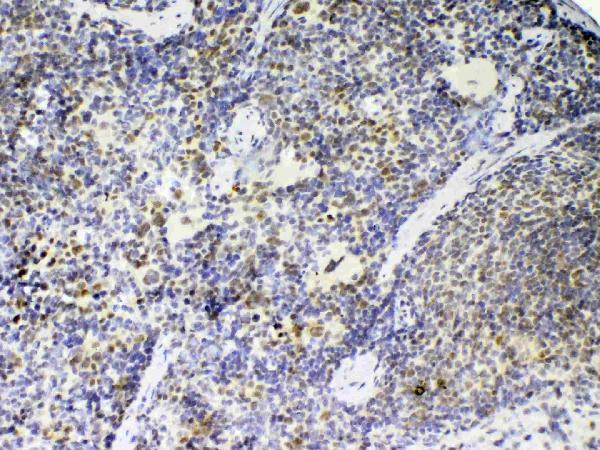
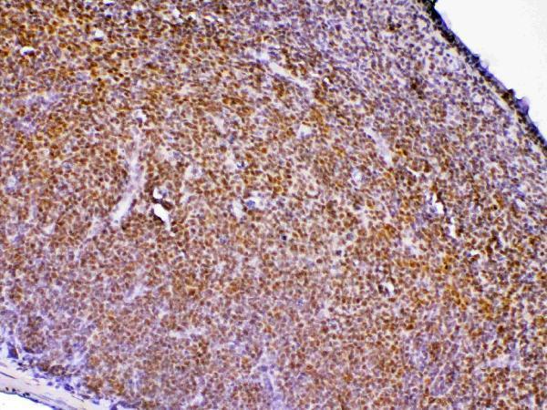
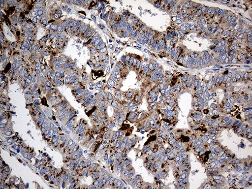
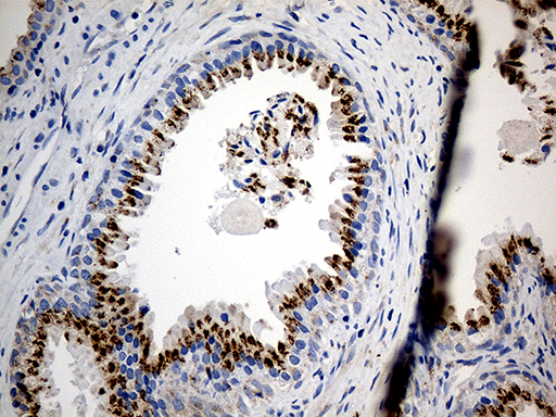
Spleen, thymus, lymph node and bone marrow.
This article will discuss Boster Bio's Anti-TSH Receptor/TSHR Antibody Picoband(tm) and its history and its applications. This article applies to scientists from all over the world and isn't specific to any particular area of study. It is applicable to scientists from all fields. There are exceptions like cases involving species or applications.
The Boster Bio Anti–TSH Recept (or TSHR Antibody picoband(t),) is designed for the detection of TSH and TSHR biomarkers across a variety of tissues and species. It has a high level sensitivities, reaching picogram levels. Boster Bio Immunological Reagents may be purchased via tebubio.
Boster Bio developed and tested this anti-TSH receptor/TSHR antibody using rabbit polyclonal antibodies. It is approved for Western Blot. It has been tested on Rats, Mouse and Human. It is a tool that can be used for a wide range of applications and is suitable for WB applications. The peptides are readily available and prices differ based on their length.
The PTPN22 gene encodes a cytoplasmic lymphoid-specific phosphatase. This protein is a blocker of activation of T cells. This enzyme has a resemblance N-terminus as the classic non-receptor phosphatases. The PTPN22 protein has a variety of applications in biology. It has recently been used to identify cancerous cells in the immune system.
PTPN22 is linked to a negative regulatory function in the immune response against tumors. However, the exact mechanism of action remains undetermined. Recent research suggests that a germline PTPN22 variation is associated with a lower risk of developing cancer. In addition, Ptpn22 expression has been associated with the regulation of immune function in a variety of cancer types. When it is expressed, tumors show increased numbers of macrophages, natural killer cells, and T cells.
PTPN22 has identified several single nucleotide polymorphisms. One non-synonymous SNP has been recorded in public databases. At nucleotide 1858 cystine is replaced by thymidine, leading to a tryptophan residue at codon 620. Codon 620 is located in the P1 proline-rich motif, which binds Lyp and Csk. This is the reason why R620W substitution could disrupt the binding process.
Although T2D patients share common genetic marker, PTPN22 CNVs are distinct for each person. For instance, in T2D patients, patients with PTPN22 CNV have a lower copy number than individuals without the disease. Healthy controls have an average CN number of nine. These individuals are different from T2D patients in terms copy number. Therefore, this gene may be useful in identifying patients with T2D.
The PTPN22R620W gene, in addition to the SSc connection is also linked to the development of autoimmune disorders like systemic sclerosis. Other rare polymorphisms within PTPN22 may be linked to autoimmune disorders. Additionally, SNPs in the 3'untranslated regions or in a haplotype block that is similar to the one may be related. Many groups have examined the relationship between PTPN22 gene polymorphisms and SLE in humans. The majority of studies conducted in case-control confirm this connection.
Other variables that are associated with the gluconic metabolic process, in addition to the PTPN22 genes, include plasma glucose levels and insulin levels. The PTPN22 gene could be a promising biomarker to diagnose T2D. This gene could eventually result in more precise and prompt diagnosis. Although the genetics of T2D can be complicated but the PTPN22 genes are used in a variety of ways in medical practice. The copy number of the gene is also associated with fasting glucose levels in the plasma.
The PTPN2 gene is involved in controlling the inflammatory response. The gene is lost and causes a faulty T-cell differentiation and decreased tolerance to self-antigens, as well as to commensal bacteria. It is also as a result of its loss that PTPN2 gene produces mediators of inflammation. PTPN22 is also a major component in regulating the differentiation CD4+ T cells. Loss of the gene impairs this process, leading to chronic intestinal inflammation.
Studies have previously demonstrated that the PTPN22 gene has been involved in autoimmune diseases. In addition, this gene functionally regulated by ROS through its noncatalytic C129 sequence. Although this particular residue is expected to affect the function of PTPN22'sgenes, other modifications could be possible. This makes it difficult to focus on PTPs in vivo, however recent advances in targeting the redox status of T lymphocytes could be a novel strategy.
Previous studies have demonstrated that PTPN22 gene mutations can affect the progression and development of LAAS. They found no correlation between the R620W gene and MS risk. HLA DR3 DQ2 haplotype was also associated with higher disease risks in males than in females. However there was no interaction between PTPN22 gene and CTLA4 gene in Finnish individuals.
However, the relationship between PTPN22 gene and type I diabetes cannot be explained by the -1123C promoter SNP, nor the 1858C-T variant. In mouse models of inflammation it was observed that the mutant PTPN22C129S had enhanced inhibitory effects. To determine the significance of this gene further research is required. This marker isn't sufficiently sensitive to determine if someone is with type I diabetes.
A recent study investigated the association of the PTPN22 gene with the development of beta-cell-specific autoimmunity. However it was not found to influence the development of GADA and IA-2A. This suggests that genetic influences on these autoantibodies may be complicated by the presence of other autoantibodies. Cox regression is able to adjust for other factors. HLA class II patients also showed a link between PTPN22 appearance and IAA.
Comparing the genotype frequencies of children born with the ICA genotype as well as those with the CT or CC genotypes was used to determine the impact of the PTPN22 polymorphism on IAA risk. The study did not reveal any significant differences in the age-dependent heterogeneity. The TT genotype is at a higher risk for ICA than the CT and CC genotypes.
The PTPs' redox regulation is controlled by the balance between catalytic Cys residues and inhibitory oxidation. This balance is maintained by thioredoxins, but glutathionylation in PTPs is also reported. PTPN22C129S has slower phosphorylation kinetics than the wild-type PTP22. This does not mean that the enzyme has been destroyed however, it could indicate that it is under redox control.
PTPN22 is present in all types of hematopoietic cells. It is also a negative regulator of T cell signaling. The R620W variation in PTPN22 has been associated with an increased risk of developing autoimmune disorders. This mutation prevents PTPN22 from binding to the C-terminal SRC Kinase. This mutation blocks downstream signaling and acts as an antagonistic regulator of T-cell responses.
PMID: 1373816 by Matthews R.J., et al. Characterization of hematopoietic intracellular protein tyrosine phosphatases: description of a phosphatase containing an SH2 domain and another enriched in proline-, glutamic acid-, serine-, and threonine-rich sequences.
PMID: 8890164 by Cloutier J.-F., et al. Association of inhibitory tyrosine protein kinase p50csk with protein tyrosine phosphatase PEP in T cells and other hemopoietic cells.