Click image to see more details
Product Info Summary
| SKU: | BA1101 |
|---|---|
| Size: | 0.5ml |
| Reactive Species: | Mouse |
| Host: | Goat |
| Application: | Flow Cytometry, IF, WB |
Customers Who Bought This Also Bought


Product info
Product Overview
| Product Name | Goat Anti-Mouse IgG (H+L) Secondary Antibody, FITC Conjugate |
|---|---|
| Synonyms | FITC-conjugated Goat Anti-mouse IgG; Goat Anti-Mouse IgG-FITC Secondary Antibody; Fluorescein-labeled Goat Anti-Mouse IgG Secondary Antibody;ee |
| Description | Goat Anti-Mouse IgG (H+L) Secondary Antibody, FITC Conjugate, for detection, localization and quantification of target proteins in a sample via indirect immunofluorescence in IHC-P, IHC-F, ICC, or FCM |
| Reagent Type | Fluorophore-conjugated secondary antibody |
| Label | FITC |
| Host | Goat |
| Target Species | Mouse |
| Antibody Class | IgG |
| Clonality | Polyclonal |
| Immunogen | Whole molecule mouse IgG |
| Purification | Immunoaffinity chromatography |
| Solid Phase Adsorbtion | Human serum proteins |
| Specificity | Mouse IgG specific |
| Form Supplied | No cross-reactivity with human/bovine/rabbit IgG |
| Formulation | 0.5 mg FITC-conjugated secondary antibody
0.01 M PBS (PH 7.4) 5 mg/mL BSA 50% glycerol |
| Pack Size | 0.5 ml |
| Concentration | 1 mg/ml |
| Application | IF, Flow Cytometry
*Our Boster Guarantee covers the use of this product in the above marked tested applications. |
| Storage | At -20˚C for one year from date of receipt. Avoid repeated freezing and thawing. Protect from light. |
| Shipping | Ships with gel ice. |
| Precautions | FOR RESEARCH USE ONLY. NOT FOR DIAGNOSTIC OR CLINICAL USE |
Assay Information
| Sample Type | Cell suspension, FFPE tissue sections, thawed frozen samples |
|---|---|
| Assay Type | Immunoanalytical |
| Assay Purpose | Protein detection/quantification |
| Technique | Immunodetection of target antibody with HRP reporter enzyme |
| Equipment Needed | Excitation light source; Filter set and detector: fluorescence microscope (can be combined with confocal microscope), fluorescence plate-reader, flow cytometer, or cell sorter; |
Main Advantages
| Specificity | High signal-to-noise ratio |
|---|---|
| Fast | Fewer number of processing steps - no need for adding a substrate; Less optimization required compared to enzymatic detection; Generates strong signals in a relatively short time span; Fluorescence can be observed directly |
| High Signal Amplification | Multiple secondary antibodies can bind to a single primary antibody; Multiple FITC molecules bind to a single secondary antibody; |
| High Image Quality | High-resolution compatibility with fluorescent microscopy; compatible with multi-planar microscopy |
| Precise target localization | Sharp, precise signal development |
| Multiplex Compatibility | Colocalization studies: use primary antibodies from different host species for simultaneous detection by fluorophore-conjugated secondary antibodies; use multiple differently colored fluorophores in the same experiment for target differentiation |
| Quantifieable | Rapid and precise quantitative analysis of fluorescent signal |
Background
Most commonly, secondary antibodies are generated by immunizing the host animal with a pooled population of immunoglobulins from the target species. The host antiserum is then purified through immunoaffinity chromatography to remove all host serum proteins, except the specific antibody of interest. Purified secondary antibodies are further solid phase adsorbed with other species serum proteins to minimize cross-reactivity in tissue or cell preparations, and are then modified with antibody fragmentation, label conjugation, etc., to generate highly specific reagents. Secondary antibodies can be conjugated to a large number of labels, including enzymes, biotin, and fluorescent dyes/proteins. Here, the antibody provides the specificity to locate the protein of interest, and the label generates a detectable signal. The label of choice depends upon the experimental application.
Immunofluorescence is a technique used for light microscopy with a fluorescence microscope which utilizes fluorescent dyes as reporters. It is being employed in a variety of applications such as cellular imaging and flow cytometry and is commonly used to visualize the distribution of target molecules through a sample, to detect protein location and activation, to identify protein complex formation and conformational changes, to monitor biological processes in vivo.
Fluorescent dyes (also known as fluorochromes, fluorophores, or simply fluors) are molecules that can absorb light of a specific energy and wavelength, thereby undergoing excitation, and then re-emit it at a lower energy and longer wavelength upon returning to the ground state.
Product Images
Validation Images & Assay Conditions
Click image to see more details
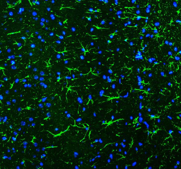
GFAP was detected in paraffin-embedded sections of rat brain tissues using mouse anti-GFAP Antigen Affinity purified monoclonal antibody (Catalog # M00213-8). FITC Conjugated Goat Anti-Mouse IgG (BA1101) was used to detect the primary antibody.
Specific Publications For BA1101
Loading publications
Customer Reviews
Have you used Goat Anti-Mouse IgG (H+L) Secondary Antibody, FITC Conjugated?
Submit a review and receive an Amazon gift card.
- $30 for a review with an image
0 Reviews For Goat Anti-Mouse IgG (H+L) Secondary Antibody, FITC Conjugated
Customer Q&As
Have a question?
Find answers in Q&As, reviews.
Can't find your answer?
Submit your question
