Click image to see more details
Product Info Summary
| SKU: | PA2068 |
|---|---|
| Size: | 100 μg/vial |
| Reactive Species: | Human, Mouse, Rat |
| Host: | Rabbit |
| Application: | Flow Cytometry, IF, ICC, WB |
Customers Who Bought This Also Bought

Product info
Product Name
Anti-Abl interactor 2 ABI2 Antibody Picoband®
SKU/Catalog Number
PA2068
BA3031-2 is an alternative SKU for this antibody, used in previous lots.
Size
100 μg/vial
Form
Lyophilized
Description
Boster Bio Anti-Abl interactor 2 ABI2 Antibody catalog # PA2068. Tested in Flow Cytometry, ICC/IF, WB applications. This antibody reacts with Human, Mouse, Rat. The brand Picoband indicates this is a premium antibody that guarantees superior quality, high affinity, and strong signals with minimal background in Western blot applications. Only our best-performing antibodies are designated as Picoband, ensuring unmatched performance.
Storage & Handling
Store at -20˚C for one year from date of receipt. After reconstitution, at 4˚C for one month. It can also be aliquotted and stored frozen at -20˚C for six months. Avoid repeated freeze-thaw cycles.
Cite This Product
Anti-Abl interactor 2 ABI2 Antibody Picoband® (Boster Biological Technology, Pleasanton CA, USA, Catalog # PA2068)
Host
Rabbit
Contents
Each vial contains 4 mg Trehalose, 0.9 mg NaCl and 0.2 mg Na2HPO4.
Clonality
Polyclonal
Isotype
Rabbit IgG
Immunogen
A synthetic peptide corresponding to a sequence at the N-terminus of human ABI2, identical to the related mouse and rat sequences.
Cross-reactivity
No cross-reactivity with other proteins
Reactive Species
PA2068 is reactive to ABI2 in Human, Mouse, Rat
Observed Molecular Weight
56-70 kDa
Calculated molecular weight
55.7 kDa
Background of ABI2
ABI2 (ABL Interactor 2), is a protein that in humans is encoded by the ABI2 gene. By analysis of a YAC and a BAC, Machado et al. (2000) mapped the ABI2 gene to 2q31-q33. ABI2 possesses a basic N terminus with homology to a homeodomain protein; a central serine-rich region; 3 PEST sequences, which are implicated in susceptibility to protein degradation; several proline-rich stretches; and an acidic C terminus with multiple phosphorylation sites and an SH3 domain. Dai and Pendergast (1995) suggested that the ABI proteins may function to coordinate the cytoplasmic and nuclear functions of the ABL1 tyrosine kinase.
Antibody Validation
Boster validates all antibodies on WB, IHC, ICC, Immunofluorescence, and ELISA with known positive control and negative samples to ensure specificity and high affinity, including thorough antibody incubations.
Application & Images
Applications
PA2068 is guaranteed for Flow Cytometry, IF, ICC, WB Boster Guarantee
Recommend Dilution
| Application | Dilution | Species |
|---|---|---|
| Western blot | 0.1-0.5μg/ml | Human, Mouse, Rat |
| Immunocytochemistry/Immunofluorescence | 5 μg/ml | Human |
| Flow Cytometry(Fixed) | 1-3 μg/1x106 cells | Human |
Tested application
Suggested blocking solution with 5% non-fat milk or BSA; (*)Recommended protein loading: 20-40 µg per lane
Validation Images & Assay Conditions
Click image to see more details
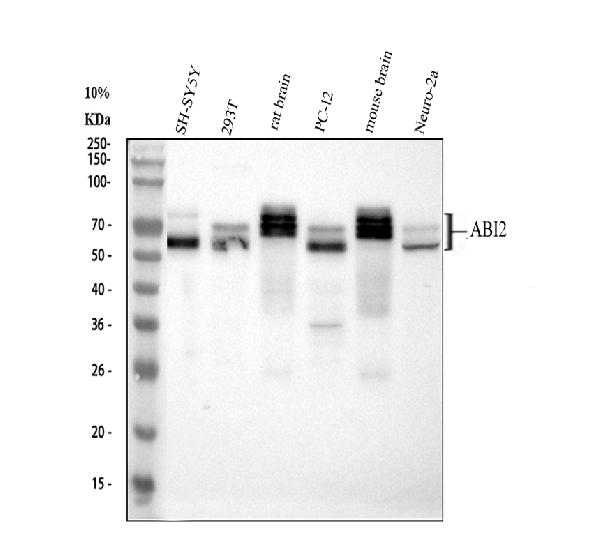
Western blot analysis of ABI2 using anti-ABI2 antibody (PA2068).
Electrophoresis was performed on a 10% SDS-PAGE gel at 80V (Stacking gel) / 120V (Resolving gel) for 2 hours. The sample well of each lane was loaded with 30 ug of sample under reducing conditions.
Lane 1: human SH-SY5Y whole cell lysates,
Lane 2: human 293T whole cell lysates,
Lane 3: rat brain tissue lysates,
Lane 4: rat PC-12 whole cell lysates,
Lane 5: mouse brain tissue lysates,
Lane 6: mouse Neuro-2a whole cell lysates.
After electrophoresis, proteins were transferred to a nitrocellulose membrane at 150 mA for 50-90 minutes. Blocked the membrane with 5% non-fat milk/TBS for 1.5 hour at RT. The membrane was incubated with rabbit anti-ABI2 antigen affinity purified polyclonal antibody (Catalog # PA2068) at 0.5 μg/mL overnight at 4°C, then washed with TBS-0.1%Tween 3 times with 5 minutes each and probed with a goat anti-rabbit IgG-HRP secondary antibody at a dilution of 1:5000 for 1.5 hour at RT. The signal is developed using an ECL Plus Western Blotting Substrate (Catalog # AR1196-200) with Tanon 5200 system. A specific band was detected for ABI2 at approximately 56-70 kDa. The expected band size for ABI2 is at 56 kDa.

Click image to see more details
IF analysis of ABI2 using anti-ABI2 antibody (PA2068) and anti-Tubulin Alpha antibody (M03989-3).
ABI2 was detected in immunocytochemical section of A549 cell. Enzyme antigen retrieval was performed using IHC enzyme antigen retrieval reagent (AR0022) for 15 mins. The cells were blocked with 10% goat serum. And then incubated with 5 μg/mL rabbit anti-ABI2 Antibody (PA2068) and mouse anti-Tubulin Alpha antibody (M03989-3) overnight at 4°C. DyLight®488 Conjugated Goat Anti-Rabbit IgG (BA1127) and Cy3 Conjugated Goat Anti-Mouse IgG (BA1031) were used as secondary antibody at 1:500 dilution and incubated for 30 minutes at 37°C. Visualize using a fluorescence microscope and filter sets appropriate for the label used.

Click image to see more details
Flow Cytometry analysis of SH-SY5Y cells using anti-ABI2 antibody (PA2068).
Overlay histogram showing SH-SY5Y cells stained with PA2068 (Blue line). To facilitate intracellular staining, cells were fixed with 4% paraformaldehyde and permeabilized with permeabilization buffer. The cells were blocked with 10% normal goat serum. And then incubated with rabbit anti-ABI2 Antibody (PA2068, 1 μg/1x106 cells) for 30 min at 20°C. DyLight®488 conjugated goat anti-rabbit IgG (BA1127, 5-10 μg/1x106 cells) was used as secondary antibody for 30 minutes at 20°C. Isotype control antibody (Green line) was rabbit IgG (1 μg/1x106) used under the same conditions. Unlabelled sample without incubation with primary antibody and secondary antibody (Red line) was used as a blank control.
Specific Publications For Anti-Abl interactor 2 ABI2 Antibody Picoband® (PA2068)
Loading publications
Recommended Resources
Here are featured tools and databases that you might find useful.
- Boster's Pathways Library
- Protein Databases
- Bioscience Research Protocol Resources
- Data Processing & Analysis Software
- Photo Editing Software
- Scientific Literature Resources
- Research Paper Management Tools
- Molecular Biology Software
- Primer Design Tools
- Bioinformatics Tools
- Phylogenetic Tree Analysis
Customer Reviews
Have you used Anti-Abl interactor 2 ABI2 Antibody Picoband®?
Share your experimental results or join a short interview to earn up to $1,000 in product credits or other rewards.
0 Reviews For Anti-Abl interactor 2 ABI2 Antibody Picoband®
Customer Q&As
Have a question?
Find answers in Q&As, reviews.
Can't find your answer?
Submit your question
4 Customer Q&As for Anti-Abl interactor 2 ABI2 Antibody Picoband®
Question
Is there a BSA free version of anti-ABI2 antibody PA2068 available?
Verified Customer
Verified customer
Asked: 2019-09-04
Answer
We appreciate your recent telephone inquiry. I can confirm that some lots of this anti-ABI2 antibody PA2068 are BSA free. For now, these lots are available and we can make a BSA free formula for you free of charge. It will take 3 extra days to prepare. If you require this antibody BSA free again in future, please do not hesitate to contact me and I will be pleased to check which lots we have in stock that are BSA free.
Boster Scientific Support
Answered: 2019-09-04
Question
We appreciate helping with my inquiry over the phone. Here are the WB image, lot number and protocol we used for cervix carcinoma using anti-ABI2 antibody PA2068. Let me know if you need anything else.
Verified Customer
Verified customer
Asked: 2018-07-24
Answer
I appreciate the data. You have provided everything we needed. Our lab team are working to resolve your inquiry as quickly as possible, and we appreciate your patience and understanding! Please let me know if there is anything you need in the meantime.
Boster Scientific Support
Answered: 2018-07-24
Question
We are currently using anti-ABI2 antibody PA2068 for mouse tissue, and we are satisfied with the WB results. The species of reactivity given in the datasheet says human, mouse, rat. Is it likely that the antibody can work on feline tissues as well?
Verified Customer
Verified customer
Asked: 2018-07-10
Answer
The anti-ABI2 antibody (PA2068) has not been validated for cross reactivity specifically with feline tissues, but there is a good chance of cross reactivity. We have an innovator award program that if you test this antibody and show it works in feline you can get your next antibody for free. Please contact me if I can help you with anything.
Boster Scientific Support
Answered: 2018-07-10
Question
I was wanting to use your anti-ABI2 antibody for WB for human cervix carcinoma on frozen tissues, but I want to know if it has been tested for this particular application. Has this antibody been tested and is this antibody a good choice for human cervix carcinoma identification?
Verified Customer
Verified customer
Asked: 2017-08-03
Answer
You can see on the product datasheet, PA2068 anti-ABI2 antibody has been tested for WB on human, mouse, rat tissues. We have an innovator award program that if you test this antibody and show it works in human cervix carcinoma in IHC-frozen, you can get your next antibody for free.
Boster Scientific Support
Answered: 2017-08-03
