Click image to see more details
-

-
-
-
-
+6
Product Info Summary
| SKU: | A00225-2 |
|---|---|
| Size: | 100 μg/vial |
| Reactive Species: | Human, Mouse, Rat |
| Host: | Rabbit |
| Application: | Flow Cytometry, IHC, ICC, WB |
Customers Who Bought This Also Bought

Product info
Product Name
Anti-AHR Antibody Picoband®
SKU/Catalog Number
A00225-2
Size
100 μg/vial
Form
Lyophilized
Description
Boster Bio Anti-AHR Antibody Picoband® catalog # A00225-2. Tested in Flow Cytometry, IHC, ICC, WB applications. This antibody reacts with Human, Mouse, Rat. The brand Picoband indicates this is a premium antibody that guarantees superior quality, high affinity, and strong signals with minimal background in Western blot applications. Only our best-performing antibodies are designated as Picoband, ensuring unmatched performance.
Storage & Handling
Store at -20˚C for one year from date of receipt. After reconstitution, at 4˚C for one month. It can also be aliquotted and stored frozen at -20˚C for six months. Avoid repeated freeze-thaw cycles.
Cite This Product
Anti-AHR Antibody Picoband® (Boster Biological Technology, Pleasanton CA, USA, Catalog # A00225-2)
Host
Rabbit
Contents
Each vial contains 4mg Trehalose, 0.9mg NaCl, 0.2mg Na2HPO4, 0.05mg NaN3.
Clonality
Polyclonal
Isotype
Rabbit IgG
Immunogen
A synthetic peptide corresponding to a sequence at the C-terminus of human AHR.
Cross-reactivity
No cross-reactivity with other proteins.
Reactive Species
A00225-2 is reactive to AHR in Human, Mouse, Rat
Observed Molecular Weight
100 kDa
Calculated molecular weight
96.1 kDa
Background of AHR
AHR (aryl hydrocarbon receptor), also called bHLHe76, is a member of the family of basic helix-loop-helix transcription factors. AhR is a cytosolic transcription factor that is normally inactive, bound to several co-chaperones. The AHR gene is mapped on 7p21.1. Estrogenic actions of AHR agonists were detected in wildtype ovariectomized mouse uteri, but were absent in Ahr -/- or Er-alpha -/- ovariectomized mice. Complex assembly and ubiquitin ligase activity of CUL4B (AHR) in vitro and in vivo are dependent on the AHR ligand. In the CUL4B (AHR) complex, ligand-activated AHR acts as a substrate-specific adaptor component that targets sex steroid receptors for degradation. Cd4-positive cells from mice lacking Ahr developed Th17 responses but failed to produce Il22 and did not show enhanced Th17 development. Activation of Ahr during induction of EAE accelerated disease onset and increased pathology in wildtype mice, but not in Ahr -/- mice. The TDO-AHR pathway is active in human brain tumors and is associated with malignant progression and poor survival. Ahr activity within ROR-gamma-t-positive ILC could be induced by dietary ligands such as those contained in vegetables of the family Brassicaceae.
Antibody Validation
Boster validates all antibodies on WB, IHC, ICC, Immunofluorescence, and ELISA with known positive control and negative samples to ensure specificity and high affinity, including thorough antibody incubations.
Application & Images
Applications
A00225-2 is guaranteed for Flow Cytometry, IHC, ICC, WB Boster Guarantee
Recommend Dilution
| Application | Dilution | Species |
|---|---|---|
| Western blot | 0.1-0.5μg/ml | |
| Immunohistochemistry (Paraffin-embedded Section) | 0.5-1μg/ml | |
| Immunohistochemistry (Frozen Section) | 0.5-1μg/ml | |
| Immunocytochemistry | 0.5-1μg/ml | |
| Flow Cytometry (Fixed) | 1-3μg/1x106 cells |
Tested application
Suggested blocking solution with 5% non-fat milk or BSA; (*)Recommended protein loading: 20-40 µg per lane
Use TE buffer pH 9.0 for antigen retrieval; (*) citrate buffer pH 6.0 is an alternative.
Validation Images & Assay Conditions
Click image to see more details
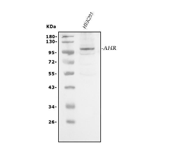
Western blot analysis of AHR using anti-AHR antibody (A00225-2).
Electrophoresis was performed on a 5-20% SDS-PAGE gel at 70V (Stacking gel) / 90V (Resolving gel) for 2-3 hours. The sample well of each lane was loaded with 30 ug of sample under reducing conditions.
Lane 1: human HEK293 whole cell lysates.
After electrophoresis, proteins were transferred to a nitrocellulose membrane at 150 mA for 50-90 minutes. Blocked the membrane with 5% non-fat milk/TBS for 1.5 hour at RT. The membrane was incubated with rabbit anti-AHR antigen affinity purified polyclonal antibody (Catalog # A00225-2) at 0.5 μg/mL overnight at 4°C, then washed with TBS-0.1%Tween 3 times with 5 minutes each and probed with a goat anti-rabbit IgG-HRP secondary antibody at a dilution of 1:5000 for 1.5 hour at RT. The signal is developed using an Enhanced Chemiluminescent detection (ECL) kit (Catalog # EK1002) with Tanon 5200 system. A specific band was detected for AHR at approximately 100 kDa. The expected band size for AHR is at 96 kDa.

Click image to see more details
IHC analysis of AHR using anti-AHR antibody (A00225-2).
AHR was detected in paraffin-embedded section of human placenta tissue. Heat mediated antigen retrieval was performed in citrate buffer (pH6, epitope retrieval solution) for 20 mins. The tissue section was blocked with 10% goat serum. The tissue section was then incubated with 2μg/ml rabbit anti-AHR Antibody (A00225-2) overnight at 4°C. Biotinylated goat anti-rabbit IgG was used as secondary antibody and incubated for 30 minutes at 37°C. The tissue section was developed using Strepavidin-Biotin-Complex (SABC)(Catalog # SA1022) with DAB as the chromogen.

Click image to see more details
IHC analysis of AHR using anti-AHR antibody (A00225-2).
AHR was detected in paraffin-embedded section of human cholangiocarcinoma tissue. Heat mediated antigen retrieval was performed in citrate buffer (pH6, epitope retrieval solution) for 20 mins. The tissue section was blocked with 10% goat serum. The tissue section was then incubated with 2μg/ml rabbit anti-AHR Antibody (A00225-2) overnight at 4°C. Biotinylated goat anti-rabbit IgG was used as secondary antibody and incubated for 30 minutes at 37°C. The tissue section was developed using Strepavidin-Biotin-Complex (SABC)(Catalog # SA1022) with DAB as the chromogen.

Click image to see more details
IHC analysis of AHR using anti-AHR antibody (A00225-2).
AHR was detected in paraffin-embedded section of human oesophagus squama cancer tissue. Heat mediated antigen retrieval was performed in citrate buffer (pH6, epitope retrieval solution) for 20 mins. The tissue section was blocked with 10% goat serum. The tissue section was then incubated with 2μg/ml rabbit anti-AHR Antibody (A00225-2) overnight at 4°C. Biotinylated goat anti-rabbit IgG was used as secondary antibody and incubated for 30 minutes at 37°C. The tissue section was developed using Strepavidin-Biotin-Complex (SABC)(Catalog # SA1022) with DAB as the chromogen.

Click image to see more details
IHC analysis of AHR using anti-AHR antibody (A00225-2).
AHR was detected in paraffin-embedded section of human tonsil tissue. Heat mediated antigen retrieval was performed in citrate buffer (pH6, epitope retrieval solution) for 20 mins. The tissue section was blocked with 10% goat serum. The tissue section was then incubated with 2μg/ml rabbit anti-AHR Antibody (A00225-2) overnight at 4°C. Biotinylated goat anti-rabbit IgG was used as secondary antibody and incubated for 30 minutes at 37°C. The tissue section was developed using Strepavidin-Biotin-Complex (SABC)(Catalog # SA1022) with DAB as the chromogen.

Click image to see more details
IHC analysis of AHR using anti-AHR antibody (A00225-2).
AHR was detected in paraffin-embedded section of mouse liver tissue. Heat mediated antigen retrieval was performed in citrate buffer (pH6, epitope retrieval solution) for 20 mins. The tissue section was blocked with 10% goat serum. The tissue section was then incubated with 2μg/ml rabbit anti-AHR Antibody (A00225-2) overnight at 4°C. Biotinylated goat anti-rabbit IgG was used as secondary antibody and incubated for 30 minutes at 37°C. The tissue section was developed using Strepavidin-Biotin-Complex (SABC)(Catalog # SA1022) with DAB as the chromogen.

Click image to see more details
IHC analysis of AHR using anti-AHR antibody (A00225-2).
AHR was detected in paraffin-embedded section of rat small intestine tissue. Heat mediated antigen retrieval was performed in citrate buffer (pH6, epitope retrieval solution) for 20 mins. The tissue section was blocked with 10% goat serum. The tissue section was then incubated with 2μg/ml rabbit anti-AHR Antibody (A00225-2) overnight at 4°C. Biotinylated goat anti-rabbit IgG was used as secondary antibody and incubated for 30 minutes at 37°C. The tissue section was developed using Strepavidin-Biotin-Complex (SABC)(Catalog # SA1022) with DAB as the chromogen.

Click image to see more details
IHC analysis of AHR using anti-AHR antibody (A00225-2).
AHR was detected in paraffin-embedded section of rat spleen tissue. Heat mediated antigen retrieval was performed in citrate buffer (pH6, epitope retrieval solution) for 20 mins. The tissue section was blocked with 10% goat serum. The tissue section was then incubated with 2μg/ml rabbit anti-AHR Antibody (A00225-2) overnight at 4°C. Biotinylated goat anti-rabbit IgG was used as secondary antibody and incubated for 30 minutes at 37°C. The tissue section was developed using Strepavidin-Biotin-Complex (SABC)(Catalog # SA1022) with DAB as the chromogen.

Click image to see more details
9. Flow Cytometry analysis of U87 cells using anti-AHR antibody (A00225-2).
Overlay histogram showing U87 cells stained with A00225-2 (Blue line). To facilitate intracellular staining, cells were fixed with 4% paraformaldehyde and permeabilized with permeabilization buffer. The cells were blocked with 10% normal goat serum. And then incubated with rabbit anti-AHR Antibody (A00225-2,1μg/1x106 cells) for 30 min at 20°C. DyLight®488 conjugated goat anti-rabbit IgG (BA1127, 5-10μg/1x106 cells) was used as secondary antibody for 30 minutes at 20°C. Isotype control antibody (Green line) was rabbit IgG (1μg/1x106) used under the same conditions. Unlabelled sample (Red line) was also used as a control.

Click image to see more details
10. Flow Cytometry analysis of U937 cells using anti-AHR antibody (A00225-2).
Overlay histogram showing U937 cells stained with A00225-2 (Blue line). To facilitate intracellular staining, cells were fixed with 4% paraformaldehyde and permeabilized with permeabilization buffer. The cells were blocked with 10% normal goat serum. And then incubated with rabbit anti-AHR Antibody (A00225-2,1μg/1x106 cells) for 30 min at 20°C. DyLight®488 conjugated goat anti-rabbit IgG (BA1127, 5-10μg/1x106 cells) was used as secondary antibody for 30 minutes at 20°C. Isotype control antibody (Green line) was rabbit IgG (1μg/1x106) used under the same conditions. Unlabelled sample (Red line) was also used as a control.
Specific Publications For Anti-AHR Antibody Picoband® (A00225-2)
Loading publications
Recommended Resources
Here are featured tools and databases that you might find useful.
- Boster's Pathways Library
- Protein Databases
- Bioscience Research Protocol Resources
- Data Processing & Analysis Software
- Photo Editing Software
- Scientific Literature Resources
- Research Paper Management Tools
- Molecular Biology Software
- Primer Design Tools
- Bioinformatics Tools
- Phylogenetic Tree Analysis
Customer Reviews
Have you used Anti-AHR Antibody Picoband®?
Share your experimental results or join a short interview to earn up to $1,000 in product credits or other rewards.
0 Reviews For Anti-AHR Antibody Picoband®
Customer Q&As
Have a question?
Find answers in Q&As, reviews.
Can't find your answer?
Submit your question
16 Customer Q&As for Anti-AHR Antibody Picoband®
Question
I have a question about product A00225-2, anti-AHR antibody. I was wondering if it would be possible to conjugate this antibody with biotin. I would need it to be without BSA or sodium azide. I am planning on using a buffer exchange of sodium azide with PBS only. Would there be problems for me to conjugate the antibody and store it in -20 degrees in small aliquots?
Verified Customer
Verified customer
Asked: 2020-04-27
Answer
It is not recommended storing this antibody with PBS buffer only in -20 degrees. If you want to store it in -20 degrees it is best to add some cryoprotectant like glycerol. If you want carrier free A00225-2 anti-AHR antibody, we can provide it to you in a special formula with trehalose and/or glycerol. These molecules will not interfere with conjugation chemistry and provide a good level of protection for the antibody from degradation. Please be sure to specify this in your purchase order.
Boster Scientific Support
Answered: 2020-04-27
Question
Would anti-AHR antibody A00225-2 work for IHC with placenta?
Verified Customer
Verified customer
Asked: 2020-04-08
Answer
According to the expression profile of placenta, AHR is highly expressed in placenta. So, it is likely that anti-AHR antibody A00225-2 will work for IHC with placenta.
Boster Scientific Support
Answered: 2020-04-08
Question
See below the WB image, lot number and protocol we used for placenta using anti-AHR antibody A00225-2. Please let me know if you require anything else.
Verified Customer
Verified customer
Asked: 2020-03-24
Answer
Thank you very much for the data. Our lab team are working to resolve this as quickly as possible, and we appreciate your patience and understanding! You have provided everything we needed. Please let me know if there is anything you need in the meantime.
Boster Scientific Support
Answered: 2020-03-24
Question
I was wanting to use your anti-AHR antibody for IHC for human placenta on frozen tissues, but I want to know if it has been validated for this particular application. Has this antibody been validated and is this antibody a good choice for human placenta identification?
Verified Customer
Verified customer
Asked: 2020-02-17
Answer
It shows on the product datasheet, A00225-2 anti-AHR antibody has been tested for Flow Cytometry, IHC, ICC, WB on human, mouse, rat tissues. We have an innovator award program that if you test this antibody and show it works in human placenta in IHC-frozen, you can get your next antibody for free.
Boster Scientific Support
Answered: 2020-02-17
Question
Our lab used your anti-AHR antibody for Flow Cytometry on placenta in the past. I am using human, and We intend to use the antibody for WB next. We are interested in examining placenta as well as liver in our next experiment. Could you please give me some suggestion on which antibody would work the best for WB?
Verified Customer
Verified customer
Asked: 2020-01-14
Answer
I viewed the website and datasheets of our anti-AHR antibody and I see that A00225-2 has been tested on human in both Flow Cytometry and WB. Thus A00225-2 should work for your application. Our Boster satisfaction guarantee will cover this product for WB in human even if the specific tissue type has not been validated. We do have a comprehensive range of products for WB detection and you can check out our website bosterbio.com to find out more information about them.
Boster Scientific Support
Answered: 2020-01-14
Question
We were well pleased with the WB result of your anti-AHR antibody. However we have observed positive staining in liver cytoplasm. using this antibody. Is that expected? Could you tell me where is AHR supposed to be expressed?
Verified Customer
Verified customer
Asked: 2019-11-25
Answer
Based on literature, liver does express AHR. Generally AHR expresses in cytoplasm. Regarding which tissues have AHR expression, here are a few articles citing expression in various tissues:
Liver, Pubmed ID: 8393992
Placenta, Pubmed ID: 15489334
Boster Scientific Support
Answered: 2019-11-25
Question
I see that the anti-AHR antibody A00225-2 works with IHC, what is the protocol used to produce the result images on the product page?
Verified Customer
Verified customer
Asked: 2019-11-12
Answer
You can find protocols for IHC on the "support/technical resources" section of our navigation menu. If you have any further questions, please send an email to support@bosterbio.com
Boster Scientific Support
Answered: 2019-11-12
Question
Is this A00225-2 anti-AHR antibody reactive to the isotypes of AHR?
Verified Customer
Verified customer
Asked: 2019-09-30
Answer
The immunogen of A00225-2 anti-AHR antibody is A synthetic peptide corresponding to a sequence of human AHR (AFLNKFQNGVLNETYPAELNNINNTQTTTHLQPLHH). Could you tell me which isotype you are interested in so I can help see if the immunogen is part of this isotype?
Boster Scientific Support
Answered: 2019-09-30
Question
Does A00225-2 anti-AHR antibody work on parafin embedded sections? If so, which fixation method do you recommend we use (PFA, paraformaldehyde, other)?
Verified Customer
Verified customer
Asked: 2019-09-24
Answer
You can see on the product datasheet, A00225-2 anti-AHR antibody as been tested on IHC. It is best to use PFA for fixation because it has better tissue penetration ability. PFA needs to be prepared fresh before use. Long term stored PFA turns into formalin, as the PFA molecules congregate and become formalin.
Boster Scientific Support
Answered: 2019-09-24
Question
We have observed staining in rat liver. Do you have any suggestions? Is anti-AHR antibody supposed to stain liver positively?
Verified Customer
Verified customer
Asked: 2019-07-10
Answer
Based on literature liver does express AHR. Based on Uniprot.org, AHR is expressed in placenta, liver, among other tissues. Regarding which tissues have AHR expression, here are a few articles citing expression in various tissues:
Liver, Pubmed ID: 8393992
Placenta, Pubmed ID: 15489334
Boster Scientific Support
Answered: 2019-07-10
Question
We are interested in using your anti-AHR antibody for cellular response to forskolin studies. Has this antibody been tested with western blotting on placenta tissue? We would like to see some validation images before ordering.
Verified Customer
Verified customer
Asked: 2019-03-15
Answer
I appreciate your inquiry. This A00225-2 anti-AHR antibody is validated on human cholangiocarcinoma tissue, placenta tissue, squama cancer tissue, tonsil tissue, mouse liver tissue, rat spleen tissue, small intestine tissue, u87 cells, u937 cells. It is guaranteed to work for Flow Cytometry, IHC, ICC, WB in human, mouse, rat. Our Boster guarantee will cover your intended experiment even if the sample type has not been be directly tested.
Boster Scientific Support
Answered: 2019-03-15
Question
We are currently using anti-AHR antibody A00225-2 for rat tissue, and we are happy with the ICC results. The species of reactivity given in the datasheet says human, mouse, rat. Is it true that the antibody can work on bovine tissues as well?
P. Evans
Verified customer
Asked: 2018-10-23
Answer
The anti-AHR antibody (A00225-2) has not been tested for cross reactivity specifically with bovine tissues, though there is a good chance of cross reactivity. We have an innovator award program that if you test this antibody and show it works in bovine you can get your next antibody for free. Please contact me if I can help you with anything.
Boster Scientific Support
Answered: 2018-10-23
Question
My question regards to test anti-AHR antibody A00225-2 on human placenta for research purposes, then I may be interested in using anti-AHR antibody A00225-2 for diagnostic purposes as well. Is the antibody suitable for diagnostic purposes?
Verified Customer
Verified customer
Asked: 2018-01-11
Answer
The products we sell, including anti-AHR antibody A00225-2, are only intended for research use. They would not be suitable for use in diagnostic work. If you have the means to develop a product into diagnostic use, and are interested in collaborating with us and develop our product into an IVD product, please contact us for more discussions.
Boster Scientific Support
Answered: 2018-01-11
Question
Thank you for helping with my inquiry over the phone. Here are the WB image, lot number and protocol we used for placenta using anti-AHR antibody A00225-2. Let me know if you need anything else.
K. Bhatt
Verified customer
Asked: 2014-05-27
Answer
Thanks for the data. You have provided everything we needed. Our lab team are working to resolve your inquiry as quickly as possible, and we appreciate your patience and understanding! Please let me know if there is anything you need in the meantime.
Boster Scientific Support
Answered: 2014-05-27
Question
Is there a BSA free version of anti-AHR antibody A00225-2 available?
D. Zhang
Verified customer
Asked: 2013-05-20
Answer
Thank you for your recent telephone inquiry. I can confirm that some lots of this anti-AHR antibody A00225-2 are BSA free. For now, these lots are available and we can make a BSA free formula for you free of charge. It will take 3 extra days to prepare. If you require this antibody BSA free again in future, please do not hesitate to contact me and I will be pleased to check which lots we have in stock that are BSA free.
Boster Scientific Support
Answered: 2013-05-20
Question
Is a blocking peptide available for product anti-AHR antibody (A00225-2)?
G. Evans
Verified customer
Asked: 2013-02-26
Answer
We do provide the blocking peptide for product anti-AHR antibody (A00225-2). If you would like to place an order for it please contact support@bosterbio.com and make a special request.
Boster Scientific Support
Answered: 2013-02-26
