Click image to see more details
-

-
-
-
-
+5
Product Info Summary
| SKU: | MP00024 |
|---|---|
| Size: | 100ug |
| Reactive Species: | Human, Monkey, Mouse, Rat |
| Host: | Mouse |
| Application: | ELISA, Flow Cytometry, IP, IF, IHC, WB |
Customers Who Bought This Also Bought

Product info
Product Name
Anti-Akt phospho S473 Monoclonal Antibody
SKU/Catalog Number
MP00024
Size
100ug
Form
Liquid (sterile filtered)
Description
Boster Bio Anti-Akt phospho S473 Monoclonal Antibody (Catalog # MP00024). Tested in ELISA, IF, IHC, WB applications. This antibody reacts with Human, Mouse, Rat, Monkey.
Storage & Handling
Store Anti-AKT pS473 (MOUSE) Monoclonal Antibody at -20°C prior to opening. Aliquot contents and freeze at -20°C or below for extended storage. Avoid cycles of freezing and thawing. Centrifuge product if not completely clear after standing at room temperature. This product is stable for several weeks at 4°C as an undiluted liquid. Dilute only prior to immediate use. Expiration date is one (1) year from date of opening. (Ship on dry ice.)
Cite This Product
Anti-Akt phospho S473 Monoclonal Antibody (Boster Biological Technology, Pleasanton CA, USA, Catalog # MP00024)
Host
Mouse
Contents
0.02 M Potassium Phosphate, 0.15 M Sodium Chloride, pH 7.2, 0.01% (w/v) Sodium Azide
Clonality
Monoclonal
Clone Number
Clone: 17F6.B11
Isotype
IgG1 kappa
Immunogen
Anti-AKT pS473 (MOUSE) Monoclonal Antibody was produced by repeated immunizations with a synthetic peptide corresponding to residues surrounding S473 of human AKT1 protein, followed by hybridoma development.
Cross-reactivity
No cross reactivity with other proteins.
Reactive Species
MP00024 is reactive to AKT1 in Human, Monkey, Mouse, Rat
Observed Molecular Weight
42 kDa
Calculated molecular weight
55.7 kDa
Background of AKT1
AKT phospho S473 is a component of the PI-3 kinase pathway and is activated by phosphorylation at Ser 473 and Thr 308. AKT is a cytoplasmic protein also known as AKT1, Protein Kinase B (PKB) and rac (related to A and C kinases). AKT is a key regulator of many signal transduction pathways. AKT Exhibits tight control over cell proliferation and cell viability. Overexpression or inappropriate activation of AKT is noted in many types of cancer. AKT mediates many of the downstream events of PI 3-kinase (a lipid kinase activated by growth factors, cytokines and insulin). PI 3-kinase recruits AKT to the membrane, where it is activated by PDK1 phosphorylation. Once phosphorylated, AKT dissociates from the membrane and phosphorylates targets in the cytoplasm and the cell nucleus. AKT has two main roles: (i) inhibition of apoptosis; (ii) promotion of proliferation. Anti-AKT pS473 (MOUSE) Monoclonal Antibody is ideal for investigators involved in Cell Signaling, Cancer, Neuroscience, Signal Transduction research.
Antibody Validation
Boster validates all antibodies on WB, IHC, ICC, Immunofluorescence, and ELISA with known positive control and negative samples to ensure specificity and high affinity, including thorough antibody incubations.
Application & Images
Applications
MP00024 is guaranteed for ELISA, Flow Cytometry, IP, IF, IHC, WB Boster Guarantee
Recommend Dilution
| Application | Dilution | Species |
|---|---|---|
| ELISA: 1:20 | 000 | |
| IF Microscopy: 1:500-1:3 | 000 | |
| WB: 1:500-1:3 | 000 | |
| Phospho AKT antibody is tested in ELISA | immunofluorescence | immunohistochemistry, flow cytometry, and western blotting. Expect a band approximately 56 kDa in size corresponding to phosphorylated AKT protein by western blotting in the appropriate cell lysate or extract. This phospho-specific monoclonal antibody reacts with human and mouse AKT pS473 and shows minimal reactivity by ELISA against the non-phosphorylated form of the immunizing peptide. Specific conditions for reactivity should be optimized by the end user. For immunohistochemistry use formalin-fixed paraffin-embedded sections. No pre-treatment of sample is required. |
Validation Images & Assay Conditions
Click image to see more details
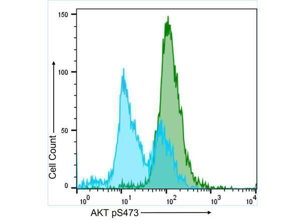
Flow Cytometry results of Anti-AKT pS473 (MOUSE) Monoclonal Antibody. The green histogram represents the A431 cells that were stimulated for 15 minutes with 100 ng/mL EGF. The blue histogram shows the untreated A431 cell population, which is bimodal. Both populations were stained with a 1:50 dilution of the Anti-AKT pS473 (MOUSE) Monoclonal Antibody for 30 mins at 4°C. The secondary antibody, Anti-MOUSE IgG (H&L) (GOAT) Antibody DyLight™ 488 Conjugated was used at a 1:200 dilution for 30 mins at 4°C.

Click image to see more details
Immunohistochemistry of Mouse anti-AKT pS473 antibody. Tissue: human prostate tissue. Fixation: formalin fixed paraffin embedded. Antigen retrieval: not required. Primary antibody: AKT pS473 antibody at 20 µg/mL for 1 h at RT. Secondary antibody: Dako's Techmate streptavidin-biotin reagents at 1:10,000 for 45 min at RT. Localization: AKT pS473 is nuclear and occasionally cytoplasmic. Staining: AKT pS473 as precipitated red signal with hematoxylin purple nuclear counterstain.

Click image to see more details
Western Blot of Mouse Anti-AKT pS473 antibody. Lane 1: non-phosphorylated AKT in untreated cells. Lane 2: phosphorylated AKT (indicated by arrowhead at ~56 kDa) on PDGF stimulated NIH/3T3 cell lysates. Load: 10 µg per lane. Primary antibody: AKT pS473 antibody at 1:10,000 in TBS with 0.05% Tween-20 with 1% BSA, for 1 h at 4° C. Secondary antibody: HRP conjugated Gt-a-Mouse IgG was used at a 1:20,000 dilution for 1 h at 4° C with FemtoMax™ enhanced chemiluminescent reagent . Other band(s): none.

Click image to see more details
Immunofluorescence Microscopy of Mouse Anti-AKTpS473 antibody using STED nanoscopy to evaluate AKT activation and migration. Tissue: A431 cells. Antigen retrieval: Panel A: serum starved,unstimulated cells. Panel B: serum starved, EGF stimulated for 15 mins. A massive increase in AKT-pS473 activation, as measured by intensity signal, peaked at 15 minutes and was associated with depolymerized tubulin. Staining: Panel A shows STED data (AKT-pS473, red channel) collected simultaneously with confocal signal (a-tubulin, green channel). Upon stimulation of cells with EGF, a rapid activation of AKT is observed (Panel B) along with a coincident change in the tubulin organization (yellow signal), as well as an extensive cell shape-change (cell membrane folding) and accumulation of AKTpS473 at the cell periphery.

Click image to see more details
High resolution STED immunofluorescence nanoscopy of Mouse anti-AKT pS473 antibody. Tissue: A431 cells. The merge images (A) and at high magnification (B) show phosphorylated AKT colocalized with the distal microtubules. Fixation: 4% paraformaldehyde for 5 min and after washes blocked with 10% NGS/0.2% Triton X-100 for 30 min. Antigen retrieval: serum deprivation for 12 h. Primary antibody: AKT pS473 antibody at 10 µg/mL and α-tubulin (cyan) at 1.4 µg/mL for 1 h at RT. Secondary antibody: Atto 647N anti-Mouse IgG (ATTO TEC GmbH), and DyLight™488 anti-Rabbit IgG were used at 1.0 µg/mL for 1h at RT for indirect detection. Localization: AKT pS473 is in the cytoplasm and also organized at the periphery of the cell. Staining: AKT pS473 as red signal with bis-benzimide (blue) nuclear counterstain.

Click image to see more details
Western Blot of Mouse Anti-Akt pS473 antibody. Lane 1: unstimulated NIH/3T3 lysates contain inactive unphosphorylated Akt1, green band. Lane 2: PDGF stimulated NIH/3T3 lysate contains both inactive (green band) and activated phosphorylated Akt1 (red band). Load: 10 µg per lane. Primary antibody: rabbit anti-Akt (pan) and mouse anti-Akt pS473 specific antibodies at 1:400 for overnight at 4°C. Secondary antibody: DyLight™ 549 conjugated anti-rabbit IgG (green) and DyLight™ 649 conjugated anti-mouse IgG (red) secondary antibodies at 1:10,000 for 45 min at RT. Block: 5% BLOTTO overnight at 4°C.

Click image to see more details
Western Blot of Mouse Anti-AKTpS473 antibody. Lane 1: A431 cells. Lane 2: A431 cells stimulated for 15 min with EGF. Load: 35 µg per lane. Primary antibody: AKTpS473 antibody at 1:400 for overnight at 4°C. Secondary antibody: DyLight™649 Conjugated Anti-AKT pS473 Monoclonal Antibody at 1:10,000 for 45 min at RT. Block: Blocking Buffer for Fluorescent Western Blotting overnight at 4°C. Predicted/Observed size: 56kDa. Other band(s): none.

Click image to see more details
Immunofluorescence confocal microscopy of Mouse Anti-AKT pS473 antibody. Tissue: EGF treated A431 cells. Fixation: 0.5% PFA. Antigen retrieval: EGF 15 min. Primary antibody: AKT pS473 antibody at 10 µg/mL for 1 h at RT. Secondary antibody: DyLight 488™ Goat anti-Rabbit IgG, MAb anti-AKT pS473, atto-647N anti-Mouse IgG (Active Motif). at 1:10,000 for 45 min at RT. Localization: AKT pS473 is nuclear and occasionally cytoplasmic. Staining: AKT pS473 as red signal with tubulin (cyan).

Click image to see more details
Western Blot of Mouse Anti-Akt pS473 antibody. A: Lane 1) PDGF stimulated NIH 3T3 cells [10µl]. Lane 2) NIH 3T3 cells [10µl]. Lane 3) Hela whole cell lysate [10µl] (weak signal). B: Lane 4) GST negative control protein [100ng]. Lane 5) GST negative control protein [25ng]. Lane 6) AKT 1 recombinant protein [100ng]. Lane 7) AKT 1 recombinant protein [25ng]. Block: 5% BSA overnight at 4°C. Primary antibody: Boster monoclonal anti-AKT antibody (200-301-268S, lot no. 27843) used at 1:1000 for overnight at 4°C. Secondary antibody: HRP Conjugated goat anti-mouse 1:25K for 45 min at RT. Detection: (TMBM-100) for 20 minutes, rinsed with deionized water, dried and scanned on conventional flatbed scanner.
Specific Publications For Anti-Akt phospho S473 Monoclonal Antibody (MP00024)
Loading publications
Recommended Resources
Here are featured tools and databases that you might find useful.
- Boster's Pathways Library
- Protein Databases
- Bioscience Research Protocol Resources
- Data Processing & Analysis Software
- Photo Editing Software
- Scientific Literature Resources
- Research Paper Management Tools
- Molecular Biology Software
- Primer Design Tools
- Bioinformatics Tools
- Phylogenetic Tree Analysis
Customer Reviews
Have you used Anti-Akt phospho S473 Monoclonal Antibody?
Share your experimental results or join a short interview to earn up to $1,000 in product credits or other rewards.
0 Reviews For Anti-Akt phospho S473 Monoclonal Antibody
Customer Q&As
Have a question?
Find answers in Q&As, reviews.
Can't find your answer?
Submit your question
15 Customer Q&As for Anti-Akt phospho S473 Monoclonal Antibody
Question
Does anti-Akt phospho S473 Monoclonal antibody MP00024 work for IHC with foreskin?
Verified Customer
Verified customer
Asked: 2020-04-30
Answer
According to the expression profile of foreskin, AKT1 is highly expressed in foreskin. So, it is likely that anti-Akt phospho S473 Monoclonal antibody MP00024 will work for IHC with foreskin.
Boster Scientific Support
Answered: 2020-04-30
Question
I was wanting to use your anti-Akt phospho S473 Monoclonal antibody for IHC for mouse foreskin on frozen tissues, but I want to know if it has been tested for this particular application. Has this antibody been tested and is this antibody a good choice for mouse foreskin identification?
D. Collins
Verified customer
Asked: 2020-02-07
Answer
You can see on the product datasheet, MP00024 anti-Akt phospho S473 Monoclonal antibody has been tested for IF, IHC, WB on human, mouse, rat tissues. We have an innovator award program that if you test this antibody and show it works in mouse foreskin in IHC-frozen, you can get your next antibody for free.
Boster Scientific Support
Answered: 2020-02-07
Question
I see that the anti-Akt phospho S473 Monoclonal antibody MP00024 works with IHC, what is the protocol used to produce the result images on the product page?
Verified Customer
Verified customer
Asked: 2019-11-06
Answer
You can find protocols for IHC on the "support/technical resources" section of our navigation menu. If you have any further questions, please send an email to support@bosterbio.com
Boster Scientific Support
Answered: 2019-11-06
Question
Is a blocking peptide available for product anti-Akt phospho S473 Monoclonal antibody (MP00024)?
Verified Customer
Verified customer
Asked: 2019-10-31
Answer
We do provide the blocking peptide for product anti-Akt phospho S473 Monoclonal antibody (MP00024). If you would like to place an order for it please contact support@bosterbio.com and make a special request.
Boster Scientific Support
Answered: 2019-10-31
Question
Do you have a BSA free version of anti-Akt phospho S473 Monoclonal antibody MP00024 available?
Verified Customer
Verified customer
Asked: 2019-08-16
Answer
We appreciate your recent telephone inquiry. I can confirm that some lots of this anti-Akt phospho S473 Monoclonal antibody MP00024 are BSA free. For now, these lots are available and we can make a BSA free formula for you free of charge. It will take 3 extra days to prepare. If you require this antibody BSA free again in future, please do not hesitate to contact me and I will be pleased to check which lots we have in stock that are BSA free.
Boster Scientific Support
Answered: 2019-08-16
Question
Is this MP00024 anti-Akt phospho S473 Monoclonal antibody reactive to the isotypes of AKT1?
Verified Customer
Verified customer
Asked: 2019-06-04
Answer
The immunogen of MP00024 anti-Akt phospho S473 Monoclonal antibody is anti-AKT pS473 (MOUSE) Monoclonal antibody was produced by repeated immunizations with a synthetic peptide corresponding to residues surrounding S473 of human AKT1 protein, followed by hybridoma development. Could you tell me which isotype you are interested in so I can help see if the immunogen is part of this isotype?
Boster Scientific Support
Answered: 2019-06-04
Question
We bought anti-Akt phospho S473 Monoclonal antibody for WB on adrenal gland in the past. I am using rat, and We are going to use the antibody for IHC next. My lab would like examining adrenal gland as well as cervix carcinoma in our next experiment. Could you please give me some suggestion on which antibody would work the best for IHC?
Verified Customer
Verified customer
Asked: 2019-05-22
Answer
I viewed the website and datasheets of our anti-Akt phospho S473 Monoclonal antibody and it appears that MP00024 has been tested on rat in both WB and IHC. Thus MP00024 should work for your application. Our Boster satisfaction guarantee will cover this product for IHC in rat even if the specific tissue type has not been validated. We do have a comprehensive range of products for IHC detection and you can check out our website bosterbio.com to find out more information about them.
Boster Scientific Support
Answered: 2019-05-22
Question
We appreciate helping with my inquiry over the phone. Here are the WB image, lot number and protocol we used for foreskin using anti-Akt phospho S473 Monoclonal antibody MP00024. Let me know if you need anything else.
Verified Customer
Verified customer
Asked: 2019-02-06
Answer
Thank you for the data. You have provided everything we needed. Our lab team are working to resolve your inquiry as quickly as possible, and we appreciate your patience and understanding! Please let me know if there is anything you need in the meantime.
Boster Scientific Support
Answered: 2019-02-06
Question
We are currently using anti-Akt phospho S473 Monoclonal antibody MP00024 for rat tissue, and we are content with the IF results. The species of reactivity given in the datasheet says human, mouse, rat. Is it likely that the antibody can work on monkey tissues as well?
Verified Customer
Verified customer
Asked: 2018-06-25
Answer
The anti-Akt phospho S473 Monoclonal antibody (MP00024) has not been tested for cross reactivity specifically with monkey tissues, though there is a good chance of cross reactivity. We have an innovator award program that if you test this antibody and show it works in monkey you can get your next antibody for free. Please contact me if I can help you with anything.
Boster Scientific Support
Answered: 2018-06-25
Question
I have a question about product MP00024, anti-Akt phospho S473 Monoclonal antibody. I was wondering if it would be possible to conjugate this antibody with biotin. I would need it to be without BSA or sodium azide. I am planning on using a buffer exchange of sodium azide with PBS only. Would there be problems for me to conjugate the antibody and store it in -20 degrees in small aliquots?
Verified Customer
Verified customer
Asked: 2018-02-16
Answer
It is not recommended storing this antibody with PBS buffer only in -20 degrees. If you want to store it in -20 degrees it is best to add some cryoprotectant like glycerol. If you want carrier free MP00024 anti-Akt phospho S473 Monoclonal antibody, we can provide it to you in a special formula with trehalose and/or glycerol. These molecules will not interfere with conjugation chemistry and provide a good level of protection for the antibody from degradation. Please be sure to specify this in your purchase order.
Boster Scientific Support
Answered: 2018-02-16
Question
Our lab were happy with the WB result of your anti-Akt phospho S473 Monoclonal antibody. However we have seen positive staining in liver cytoplasm. using this antibody. Is that expected? Could you tell me where is AKT1 supposed to be expressed?
J. Carter
Verified customer
Asked: 2018-01-31
Answer
Based on literature, liver does express AKT1. Generally AKT1 expresses in cytoplasm. Regarding which tissues have AKT1 expression, here are a few articles citing expression in various tissues:
Adrenal gland, Pubmed ID: 14702039
Cervix carcinoma, Pubmed ID: 17081983, 18669648
Cervix carcinoma, and Erythroleukemia, Pubmed ID: 23186163
Foreskin, Pubmed ID: 1718748
Liver, Pubmed ID: 24275569
Muscle, and Ovary, Pubmed ID: 15489334
Boster Scientific Support
Answered: 2018-01-31
Question
We have seen staining in rat muscle ovary. Any tips? Is anti-Akt phospho S473 Monoclonal antibody supposed to stain muscle ovary positively?
Verified Customer
Verified customer
Asked: 2017-06-12
Answer
According to literature muscle ovary does express AKT1. According to Uniprot.org, AKT1 is expressed in left adrenal gland, adrenal gland, muscle ovary, foreskin, cervix carcinoma, cervix carcinoma erythroleukemia, liver, among other tissues. Regarding which tissues have AKT1 expression, here are a few articles citing expression in various tissues:
Adrenal gland, Pubmed ID: 14702039
Cervix carcinoma, Pubmed ID: 17081983, 18669648
Cervix carcinoma, and Erythroleukemia, Pubmed ID: 23186163
Foreskin, Pubmed ID: 1718748
Liver, Pubmed ID: 24275569
Muscle, and Ovary, Pubmed ID: 15489334
Boster Scientific Support
Answered: 2017-06-12
Question
Please see the WB image, lot number and protocol we used for foreskin using anti-Akt phospho S473 Monoclonal antibody MP00024. Please let me know if you require anything else.
W. Gonzalez
Verified customer
Asked: 2016-04-05
Answer
Thank you very much for the data. Our lab team are working to resolve this as quickly as possible, and we appreciate your patience and understanding! You have provided everything we needed. Please let me know if there is anything you need in the meantime.
Boster Scientific Support
Answered: 2016-04-05
Question
Would MP00024 anti-Akt phospho S473 Monoclonal antibody work on parafin embedded sections? If so, which fixation method do you recommend we use (PFA, paraformaldehyde, other)?
R. Li
Verified customer
Asked: 2014-11-13
Answer
It shows on the product datasheet, MP00024 anti-Akt phospho S473 Monoclonal antibody as been validated on IHC. It is best to use PFA for fixation because it has better tissue penetration ability. PFA needs to be prepared fresh before use. Long term stored PFA turns into formalin, as the PFA molecules congregate and become formalin.
Boster Scientific Support
Answered: 2014-11-13
Question
We want to test anti-Akt phospho S473 Monoclonal antibody MP00024 on mouse foreskin for research purposes, then I may be interested in using anti-Akt phospho S473 Monoclonal antibody MP00024 for diagnostic purposes as well. Is the antibody suitable for diagnostic purposes?
G. Moore
Verified customer
Asked: 2013-01-31
Answer
The products we sell, including anti-Akt phospho S473 Monoclonal antibody MP00024, are only intended for research use. They would not be suitable for use in diagnostic work. If you have the means to develop a product into diagnostic use, and are interested in collaborating with us and develop our product into an IVD product, please contact us for more discussions.
Boster Scientific Support
Answered: 2013-01-31
