Click image to see more details
Product Info Summary
| SKU: | PB9685 |
|---|---|
| Size: | 100 μg/vial |
| Reactive Species: | Human, Mouse |
| Host: | Rabbit |
| Application: | Flow Cytometry, IF, ICC, WB |
Customers Who Bought This Also Bought

Product info
Product Name
Anti-ARID1A Antibody Picoband®
SKU/Catalog Number
PB9685
PB0724 is an alternative SKU for this antibody, used in previous lots.
Size
100 μg/vial
Form
Lyophilized
Description
Boster Bio Anti-ARID1A Antibody Picoband® catalog # PB9685. Tested in Flow Cytometry, IF, ICC, WB applications. This antibody reacts with Human, Mouse. The brand Picoband indicates this is a premium antibody that guarantees superior quality, high affinity, and strong signals with minimal background in Western blot applications. Only our best-performing antibodies are designated as Picoband, ensuring unmatched performance.
Storage & Handling
Store at -20˚C for one year from date of receipt. After reconstitution, at 4˚C for one month. It can also be aliquotted and stored frozen at -20˚C for six months. Avoid repeated freeze-thaw cycles.
Cite This Product
Anti-ARID1A Antibody Picoband® (Boster Biological Technology, Pleasanton CA, USA, Catalog # PB9685)
Host
Rabbit
Contents
Each vial contains 4 mg Trehalose, 0.9 mg NaCl and 0.2 mg Na2HPO4.
Clonality
Polyclonal
Isotype
Rabbit IgG
Immunogen
A synthetic peptide corresponding to a sequence in the middle region of human ARID1A, identical to the related mouse sequence.
Cross-reactivity
No cross-reactivity with other proteins
Reactive Species
PB9685 is reactive to ARID1A in Human, Mouse
Observed Molecular Weight
250-270 kDa
Calculated molecular weight
242.0 kDa
Background of ARID1A
AT-rich interactive domain-containing protein 1A, also known as p270, is a protein that in humans is encoded by the ARID1A gene. This gene encodes a member of the SWI/SNF families, whose members have helicase and ATPase activities and are thought to regulate transcription of certain genes by altering the chromatin structure around those genes. ARID1A is mapped to 1p36.11. It possesses at least two conserved domains that could be important for its function. First, it has a DNA-binding domain that can specifically bind an AT-rich DNA sequence known to be recognized by a SNF/SWI complex at the beta-globin locus. Second, the C-terminus of the protein can stimulate glucocorticoid receptor-dependent transcriptional activation.
Antibody Validation
Boster validates all antibodies on WB, IHC, ICC, Immunofluorescence, and ELISA with known positive control and negative samples to ensure specificity and high affinity, including thorough antibody incubations.
Application & Images
Applications
PB9685 is guaranteed for Flow Cytometry, IF, ICC, WB Boster Guarantee
Recommend Dilution
| Application | Dilution | Species |
|---|---|---|
| Western blot | 0.1-0.5μg/ml | Human, Mouse |
| Immunocytochemistry/Immunofluorescence | 5 μg/ml | Human |
| Flow Cytometry(Fixed) | 1-3 μg/1x106 cells | Human |
Tested application
Suggested blocking solution with 5% non-fat milk or BSA; (*)Recommended protein loading: 20-40 µg per lane
Validation Images & Assay Conditions
Click image to see more details
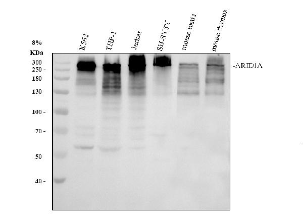
Western blot analysis of ARID1A using anti-ARID1A antibody (PB9685).
Electrophoresis was performed on a 8% SDS-PAGE gel at 80V (Stacking gel) / 120V (Resolving gel) for 2 hours. The sample well of each lane was loaded with 30 ug of sample under reducing conditions.
Lane 1: human K562 whole cell lysates,
Lane 2: human THP-1 whole cell lysates.
Lane 3: human Jurkat whole cell lysates.
Lane 4: human SH-SY5Y whole cell lysates.
Lane 5: mouse testis tissue lysates.
Lane 6: mouse thymus tissue lysates.
After electrophoresis, proteins were transferred to a nitrocellulose membrane at 150 mA for 50-90 minutes. Blocked the membrane with 5% non-fat milk/TBS for 1.5 hour at RT. The membrane was incubated with rabbit anti-ARID1A antigen affinity purified polyclonal antibody (PB9685) at 0.5 μg/mL overnight at 4°C, then washed with TBS-0.1%Tween 3 times with 5 minutes each and probed with a goat anti-rabbit IgG-HRP secondary antibody (Catalog # BA1054) at a dilution of 1:5000 for 1.5 hour at RT. The signal is developed using an ECL Plus Western Blotting Substrate (Catalog # AR1196-200) with Tanon 5200 system. A specific band was detected for ARID1A at approximately 250-270 kDa. The expected band size for ARID1A is at 242 kDa.

Click image to see more details
IF analysis of ARID1A using anti-ARID1A antibody (PB9685).
ARID1A was detected in an immunocytochemical section of A549 cells. Enzyme antigen retrieval was performed using IHC enzyme antigen retrieval reagent (AR0022) for 15 mins. The cells were blocked with 10% goat serum. And then incubated with 5 μg/mL rabbit anti-ARID1A Antibody (PB9685) overnight at 4°C. DyLight®488 Conjugated Goat Anti-Rabbit IgG (BA1127) was used as secondary antibody at 1:500 dilution and incubated for 30 minutes at 37°C. The section was counterstained with DAPI. Visualize using a fluorescence microscope and filter sets appropriate for the label used.

Click image to see more details
Flow Cytometry analysis of Hela cells using anti-ARID1A antibody (PB9685).
Overlay histogram showing Hela cells stained with PB9685 (Blue line). To facilitate intracellular staining, cells were fixed with 4% paraformaldehyde and permeabilized with permeabilization buffer. The cells were blocked with 10% normal goat serum. And then incubated with rabbit anti-ARID1A Antibody (PB9685, 1 μg/1x106 cells) for 30 min at 20°C. DyLight®488 conjugated goat anti-rabbit IgG (BA1127, 5-10 μg/1x106 cells) was used as secondary antibody for 30 minutes at 20°C. Isotype control antibody (Green line) was rabbit IgG (1 μg/1x106) used under the same conditions. Unlabelled sample without incubation with primary antibody and secondary antibody (Red line) was used as a blank control.
Specific Publications For Anti-ARID1A Antibody Picoband® (PB9685)
Loading publications
Recommended Resources
Here are featured tools and databases that you might find useful.
- Boster's Pathways Library
- Protein Databases
- Bioscience Research Protocol Resources
- Data Processing & Analysis Software
- Photo Editing Software
- Scientific Literature Resources
- Research Paper Management Tools
- Molecular Biology Software
- Primer Design Tools
- Bioinformatics Tools
- Phylogenetic Tree Analysis
Customer Reviews
Have you used Anti-ARID1A Antibody Picoband®?
Share your experimental results or join a short interview to earn up to $1,000 in product credits or other rewards.
0 Reviews For Anti-ARID1A Antibody Picoband®
Customer Q&As
Have a question?
Find answers in Q&As, reviews.
Can't find your answer?
Submit your question
15 Customer Q&As for Anti-ARID1A Antibody Picoband®
Question
I see that the anti-ARID1A antibody PB9685 works with WB, what is the protocol used to produce the result images on the product page?
G. Jones
Verified customer
Asked: 2020-04-07
Answer
You can find protocols for WB on the "support/technical resources" section of our navigation menu. If you have any further questions, please send an email to support@bosterbio.com
Boster Scientific Support
Answered: 2020-04-07
Question
I am looking for to test anti-ARID1A antibody PB9685 on human leukemic t-cell for research purposes, then I may be interested in using anti-ARID1A antibody PB9685 for diagnostic purposes as well. Is the antibody suitable for diagnostic purposes?
Verified Customer
Verified customer
Asked: 2020-01-20
Answer
The products we sell, including anti-ARID1A antibody PB9685, are only intended for research use. They would not be suitable for use in diagnostic work. If you have the means to develop a product into diagnostic use, and are interested in collaborating with us and develop our product into an IVD product, please contact us for more discussions.
Boster Scientific Support
Answered: 2020-01-20
Question
Our team were satisfied with the WB result of your anti-ARID1A antibody. However we have been able to see positive staining in cervix carcinoma erythroleukemia nucleus using this antibody. Is that expected? Could you tell me where is ARID1A supposed to be expressed?
Verified Customer
Verified customer
Asked: 2019-08-06
Answer
According to literature, cervix carcinoma erythroleukemia does express ARID1A. Generally ARID1A expresses in nucleus. Regarding which tissues have ARID1A expression, here are a few articles citing expression in various tissues:
Brain, Pubmed ID: 11734557
Cervix carcinoma, Pubmed ID: 16964243, 17081983, 18220336, 18669648, 20068231
Cervix carcinoma, and Erythroleukemia, Pubmed ID: 23186163
Colon carcinoma, Pubmed ID: 24129315
Embryonic kidney, Pubmed ID: 17525332
Gastric mucosa, Pubmed ID: 8804307
Leukemic T-cell, Pubmed ID: 19690332
Liver, Pubmed ID: 24275569
Boster Scientific Support
Answered: 2019-08-06
Question
Do you have a BSA free version of anti-ARID1A antibody PB9685 available?
Verified Customer
Verified customer
Asked: 2019-07-31
Answer
Thanks for your recent telephone inquiry. I can confirm that some lots of this anti-ARID1A antibody PB9685 are BSA free. For now, these lots are available and we can make a BSA free formula for you free of charge. It will take 3 extra days to prepare. If you require this antibody BSA free again in future, please do not hesitate to contact me and I will be pleased to check which lots we have in stock that are BSA free.
Boster Scientific Support
Answered: 2019-07-31
Question
We are currently using anti-ARID1A antibody PB9685 for human tissue, and we are content with the WB results. The species of reactivity given in the datasheet says human. Is it possible that the antibody can work on primate tissues as well?
Verified Customer
Verified customer
Asked: 2019-06-19
Answer
The anti-ARID1A antibody (PB9685) has not been tested for cross reactivity specifically with primate tissues, though there is a good chance of cross reactivity. We have an innovator award program that if you test this antibody and show it works in primate you can get your next antibody for free. Please contact me if I can help you with anything.
Boster Scientific Support
Answered: 2019-06-19
Question
We have been able to see staining in human cerebral cortex. Do you have any suggestions? Is anti-ARID1A antibody supposed to stain cerebral cortex positively?
Verified Customer
Verified customer
Asked: 2019-03-15
Answer
Based on literature cerebral cortex does express ARID1A. Based on Uniprot.org, ARID1A is expressed in cerebral cortex, brain, gastric mucosa, cervix carcinoma, embryonic kidney, leukemic t-cell, cervix carcinoma erythroleukemia, liver, colon carcinoma, among other tissues. Regarding which tissues have ARID1A expression, here are a few articles citing expression in various tissues:
Brain, Pubmed ID: 11734557
Cervix carcinoma, Pubmed ID: 16964243, 17081983, 18220336, 18669648, 20068231
Cervix carcinoma, and Erythroleukemia, Pubmed ID: 23186163
Colon carcinoma, Pubmed ID: 24129315
Embryonic kidney, Pubmed ID: 17525332
Gastric mucosa, Pubmed ID: 8804307
Leukemic T-cell, Pubmed ID: 19690332
Liver, Pubmed ID: 24275569
Boster Scientific Support
Answered: 2019-03-15
Question
Please see the WB image, lot number and protocol we used for leukemic t-cell using anti-ARID1A antibody PB9685. Please let me know if you require anything else.
Verified Customer
Verified customer
Asked: 2018-12-31
Answer
Thank you very much for the data. Our lab team are working to resolve this as quickly as possible, and we appreciate your patience and understanding! You have provided everything we needed. Please let me know if there is anything you need in the meantime.
Boster Scientific Support
Answered: 2018-12-31
Question
Is a blocking peptide available for product anti-ARID1A antibody (PB9685)?
Verified Customer
Verified customer
Asked: 2018-10-18
Answer
We do provide the blocking peptide for product anti-ARID1A antibody (PB9685). If you would like to place an order for it please contact support@bosterbio.com and make a special request.
Boster Scientific Support
Answered: 2018-10-18
Question
Would PB9685 anti-ARID1A antibody work on parafin embedded sections? If so, which fixation method do you recommend we use (PFA, paraformaldehyde, other)?
Verified Customer
Verified customer
Asked: 2018-10-01
Answer
As indicated on the product datasheet, PB9685 anti-ARID1A antibody as been validated on WB. It is best to use PFA for fixation because it has better tissue penetration ability. PFA needs to be prepared fresh before use. Long term stored PFA turns into formalin, as the PFA molecules congregate and become formalin.
Boster Scientific Support
Answered: 2018-10-01
Question
I am interested in using your anti-ARID1A antibody for coffin-siris syndrome 2 (css2) studies. Has this antibody been tested with western blotting on hepg2 whole cell lysate? We would like to see some validation images before ordering.
Verified Customer
Verified customer
Asked: 2018-08-27
Answer
Thanks for your inquiry. This PB9685 anti-ARID1A antibody is validated on sw620 whole cell lysate, hepg2 whole cell lysate. It is guaranteed to work for WB in human. Our Boster guarantee will cover your intended experiment even if the sample type has not been be directly tested.
Boster Scientific Support
Answered: 2018-08-27
Question
Thank you for helping with my inquiry over the phone. Here are the WB image, lot number and protocol we used for leukemic t-cell using anti-ARID1A antibody PB9685. Let me know if you need anything else.
Verified Customer
Verified customer
Asked: 2018-02-16
Answer
We appreciate the data. You have provided everything we needed. Our lab team are working to resolve your inquiry as quickly as possible, and we appreciate your patience and understanding! Please let me know if there is anything you need in the meantime.
Boster Scientific Support
Answered: 2018-02-16
Question
I have a question about product PB9685, anti-ARID1A antibody. I was wondering if it would be possible to conjugate this antibody with biotin. I would need it to be without BSA or sodium azide. I am planning on using a buffer exchange of sodium azide with PBS only. Would there be problems for me to conjugate the antibody and store it in -20 degrees in small aliquots?
Verified Customer
Verified customer
Asked: 2017-07-07
Answer
We do not advise storing this antibody with PBS buffer only in -20 degrees. If you want to store it in -20 degrees it is best to add some cryoprotectant like glycerol. If you want carrier free PB9685 anti-ARID1A antibody, we can provide it to you in a special formula with trehalose and/or glycerol. These molecules will not interfere with conjugation chemistry and provide a good level of protection for the antibody from degradation. Please be sure to specify this in your purchase order.
Boster Scientific Support
Answered: 2017-07-07
Question
I was wanting to use your anti-ARID1A antibody for WB for human leukemic t-cell on frozen tissues, but I want to know if it has been tested for this particular application. Has this antibody been tested and is this antibody a good choice for human leukemic t-cell identification?
A. Moore
Verified customer
Asked: 2017-07-06
Answer
It shows on the product datasheet, PB9685 anti-ARID1A antibody has been tested for WB on human tissues. We have an innovator award program that if you test this antibody and show it works in human leukemic t-cell in IHC-frozen, you can get your next antibody for free.
Boster Scientific Support
Answered: 2017-07-06
Question
Is this PB9685 anti-ARID1A antibody reactive to the isotypes of ARID1A?
B. Williams
Verified customer
Asked: 2016-11-15
Answer
The immunogen of PB9685 anti-ARID1A antibody is A synthetic peptide corresponding to a sequence in the middle region of human ARID1A (1021-1053aa KMWVDRYLAFTEEKAMGMTNLPAVGRKPLDLYR), identical to the related mouse sequence. Could you tell me which isotype you are interested in so I can help see if the immunogen is part of this isotype?
Boster Scientific Support
Answered: 2016-11-15
Question
Will anti-ARID1A antibody PB9685 work for WB with leukemic t-cell?
Z. Brown
Verified customer
Asked: 2015-11-03
Answer
According to the expression profile of leukemic t-cell, ARID1A is highly expressed in leukemic t-cell. So, it is likely that anti-ARID1A antibody PB9685 will work for WB with leukemic t-cell.
Boster Scientific Support
Answered: 2015-11-03
