Click image to see more details
-

-
-
-
-
+2
Product Info Summary
| SKU: | A05064 |
|---|---|
| Size: | 100 μg/vial |
| Reactive Species: | Human |
| Host: | Rabbit |
| Application: | IP, WB |
Customers Who Bought This Also Bought

Product info
Product Name
Anti-ARID2 Antibody Picoband®
SKU/Catalog Number
A05064
Size
100 μg/vial
Form
Lyophilized
Description
ARID2: subunit of the PBAF (SWI/SNF-B) chromatin remodeling complex; roles in transcriptional regulation and chromatin modification; page notes association with hepatocellular carcinoma. Assay context: IP/WB in human samples. In pathway profiling, can be contrasted with metabolic marker PLIN2/ADFP or membrane protease ADAM2 to relate chromatin state to cellular phenotype (putative).
Storage & Handling
Store at -20˚C for one year from date of receipt. After reconstitution, at 4˚C for one month. It can also be aliquotted and stored frozen at -20˚C for six months. Avoid repeated freeze-thaw cycles.
Cite This Product
Anti-ARID2 Antibody Picoband® (Boster Biological Technology, Pleasanton CA, USA, Catalog # A05064)
Host
Rabbit
Contents
Each vial contains 4 mg Trehalose, 0.9 mg NaCl and 0.2 mg Na2HPO4.
Clonality
Polyclonal
Isotype
Rabbit IgG
Immunogen
A synthetic peptide corresponding to a sequence at the N-terminus of human ARID2, identical to the related mouse and rat sequences.
Cross-reactivity
No cross-reactivity with other proteins.
Reactive Species
A05064 is reactive to ARID2 in Human
Observed Molecular Weight
245 kDa
Calculated molecular weight
197.4 kDa
Background of ARID2
AT-rich interactive domain-containing protein 2 (ARID2) is a protein that in humans is encoded by the ARID2 gene. It is mapped to 12q12. This gene encodes a member of the AT-rich interactive domain (ARID)-containing family of DNA-binding proteins. Members of the ARID family have roles in embryonic patterning, cell lineage gene regulation, cell cycle control, transcriptional regulation and chromatin structure modification. This protein functions as a subunit of the polybromo- and BRG1-associated factor or PBAF (SWI/SNF-B) chromatin remodeling complex which facilitates ligand-dependent transcriptional activation by nuclear receptors. Mutations in this gene are associated with hepatocellular carcinomas. A pseudogene of this gene is found on chromosome1.
Antibody Validation
Boster validates all antibodies on WB, IHC, ICC, Immunofluorescence, and ELISA with known positive control and negative samples to ensure specificity and high affinity, including thorough antibody incubations.
Application & Images
Applications
A05064 is guaranteed for IP, WB Boster Guarantee
Assay Dilutions Recommendation
The recommendations below provide a starting point for assay optimization. The actual working concentration varies and should be decided by the user.
Western blot, 0.1-0.5μg/ml
Immunoprecipitation, 0.5-2 μg/ml
Positive Control
WB: human THP-1 whole cell, human Jurkat whole cell, human HEL whole cell, human MCF-7 whole cell
IP: MCF-7 cell
Validation Images & Assay Conditions
Click image to see more details
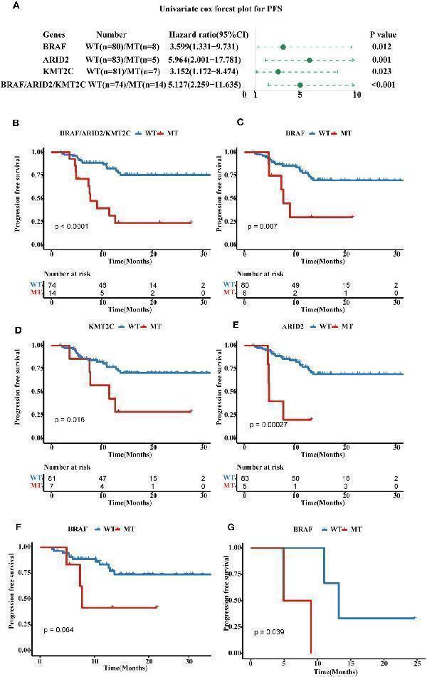
Prognostic associated somatic mutated genes in FPHYP CRC cohort. (A) Univariable analyses of PFS concerning somatic gene mutations in FPHYP CRC tumors. (B) Kaplan-Meier curves for PFS between three genes( BRAF , ARID2 , and KMT2C ) combined MT and WT groups. (C–E) Kaplan-Meier curves for PFS based on BRAF (C) , ARID2 (D) , and KMT2C (E) mutation status. (F, G) Kaplan-Meier plots of PFS for CRC patients undergoing exclusive first-line chemotherapy (F) and chemotherapy combined with bevacizumab (G) , stratified by BRAF mutation status. PFS, progression-free survival; MT, mutation type; WT, wiled type.
Index in PubMed under a CC BY license. PMID: 38023196

Click image to see more details
Prognostic associated somatic mutated genes in FPHYP CRC cohort. (A) Univariable analyses of OS concerning somatic gene mutations in FPHYP CRC tumors. (B) Kaplan-Meier curves for OS between three genes( BRAF , ARID2 , and KMT2C ) combined MT and WT groups. (C–E) Kaplan-Meier curves for PFS based on BRAF (C) , ARID2 (D) , and KMT2C (E) mutation status. OS, overall survival; MT, mutation type; WT, wiled type.
Index in PubMed under a CC BY license. PMID: 38023196

Click image to see more details
Construction of a four-gene mutation signature prediction disease progression and prognosis in FPHYP cohort. (A, B) Univariate and multivariate analyses were performed to assess the impact of clinicopathological features, individual somatic gene mutations, and the four-gene mutation signature on PFS (A) and OS (B) in CRC. (C) The Kaplan-Meier survival analysis for PFS in CRC patients between the four-gene combined MT and WT groups based on BRAF , ARID2 , KMT2C , and GNAQ mutation status. (D) The Kaplan-Meier survival analysis for OS in CRC cases between the four-gene combined MT and WT groups based on BRAF , ARID2 , KMT2C , and GNAQ mutation status. (E, F) ROC curves for PFS (E) and OS (F) that dependent on time were generated to evaluate the prognostic model’s performance, which is based on the gene mutation status within the FPHYP cohort. PFS, progression-free survival; OS, overall survival; MT, mutation type; WT, wiled type; ROC, receiver operating characteristic.
Index in PubMed under a CC BY license. PMID: 38023196

Click image to see more details
Immunohistochemical analysis of BRAF and ARID2 in CRC. (A, B) The BRAF expression original field was acquired from tissue sections (magnification, 200x) of the BRAF -MT (A) and BRAF -WT (B) groups. (C) Comparison of the IOD/Area value between BRAF -MT and BRAF -WT groups. (D, E) The BRAF expression original field was acquired from tissue sections (magnification, 200x) of stage I (D) and stage IV (E) groups. (F) Comparison of the IOD/Area value between stage I and IV groups. (G, H) The ARID2 expression original field was acquired from tissue sections (magnification, 200x) of the ARID2 -MT (G) and ARID2 -WT (H) groups. (I) Comparison of the IOD/Area value between ARID2 -MT and ARID2 -WT groups. (J, K) The ARID2 expression original field was acquired from tissue sections (magnification, 200x) of stage I (J) and stage IV (K) groups. (L) Comparison of the IOD/Area value between stage I and IV groups. MT, mutation type; WT, wiled type; IOD, cumulative optical density.
Index in PubMed under a CC BY license. PMID: 38023196

Click image to see more details
Western blot analysis of ARID2 using anti-ARID2 antibody (A05064).
Electrophoresis was performed on a 5-20% SDS-PAGE gel at 70V (Stacking gel) / 90V (Resolving gel) for 2-3 hours. The sample well of each lane was loaded with 30 ug of sample under reducing conditions.
Lane 1: human THP-1 whole cell lysates,
Lane 2: human Jurkat whole cell lysates,
Lane 3: human HEL whole cell lysates,
Lane 4: human MCF-7 whole cell lysates.
After electrophoresis, proteins were transferred to a nitrocellulose membrane at 150 mA for 50-90 minutes. Blocked the membrane with 5% non-fat milk/TBS for 1.5 hour at RT. The membrane was incubated with rabbit anti-ARID2 antigen affinity purified polyclonal antibody (A05064) at 0.5 μg/mL overnight at 4°C, then washed with TBS-0.1%Tween 3 times with 5 minutes each and probed with a goat anti-rabbit IgG-HRP secondary antibody at a dilution of 1:5000 for 1.5 hour at RT. The signal is developed using an Enhanced Chemiluminescent detection (ECL) kit (Catalog # EK1002) with Tanon 5200 system. A specific band was detected for ARID2 at approximately 245 kDa. The expected band size for ARID2 is at 197 kDa.

Click image to see more details
Immunoprecipitating ARID2 in MCF-7 whole cell lysate.
Western blot analysis of ARID2 using anti-ARID2 antibody (A05064).
Lane 1: MCF-7 whole cell lysates (30ug),
Lane 2: Rabbit control IgG instead of anti-ARID2 antibody in MCF-7 whole cell lysate,
Lane 3: anti-ARID2 antibody (2μg) + MCF-7 whole cell lysate (500μg).
After electrophoresis, proteins were transferred to a membrane. Then the membrane was incubated with rabbit anti-ARID2 antigen affinity purified polyclonal antibody (A05064) at a dilution of 0.5 μg/mL and probed with a goat anti-rabbit IgG-HRP secondary antibody (Catalog # BA1054). The signal is developed using ECL Plus Western Blotting Substrate (Catalog # AR1196-200). A specific band was detected for ARID2 at approximately 245 kDa. The expected band size for ARID2 is at 197 kDa.
Specific Publications For Anti-ARID2 Antibody Picoband® (A05064)
Loading publications
Recommended Resources
Here are featured tools and databases that you might find useful.
- Boster's Pathways Library
- Protein Databases
- Bioscience Research Protocol Resources
- Data Processing & Analysis Software
- Photo Editing Software
- Scientific Literature Resources
- Research Paper Management Tools
- Molecular Biology Software
- Primer Design Tools
- Bioinformatics Tools
- Phylogenetic Tree Analysis
Customer Reviews
Have you used Anti-ARID2 Antibody Picoband®?
Share your experimental results or join a short interview to earn up to $1,000 in product credits or other rewards.
0 Reviews For Anti-ARID2 Antibody Picoband®
Customer Q&As
Have a question?
Find answers in Q&As, reviews.
Can't find your answer?
Submit your question
10 Customer Q&As for Anti-ARID2 Antibody Picoband®
Question
Will anti-ARID2 antibody A05064 work on dog WB with kidney?
Verified Customer
Verified customer
Asked: 2020-04-17
Answer
Our lab technicians have not validated anti-ARID2 antibody A05064 on dog. You can run a BLAST between dog and the immunogen sequence of anti-ARID2 antibody A05064 to see if they may cross-react. If the sequence homology is close, then you can perform a pilot test. Keep in mind that since we have not validated dog samples, this use of the antibody is not covered by our guarantee. However we have an innovator award program that if you test this antibody and show it works in dog kidney in WB, you can get your next antibody for free.
Boster Scientific Support
Answered: 2020-04-17
Question
We are currently using anti-ARID2 antibody A05064 for human tissue, and we are happy with the IHC-P results. The species of reactivity given in the datasheet says human, mouse, rat. Is it likely that the antibody can work on pig tissues as well?
Verified Customer
Verified customer
Asked: 2020-02-05
Answer
The anti-ARID2 antibody (A05064) has not been validated for cross reactivity specifically with pig tissues, though there is a good chance of cross reactivity. We have an innovator award program that if you test this antibody and show it works in pig you can get your next antibody for free. Please contact me if I can help you with anything.
Boster Scientific Support
Answered: 2020-02-05
Question
Would A05064 anti-ARID2 antibody work on parafin embedded sections? If so, which fixation method do you recommend we use (PFA, paraformaldehyde, other)?
Verified Customer
Verified customer
Asked: 2020-02-03
Answer
You can see on the product datasheet, A05064 anti-ARID2 antibody as been tested on IHC-P. It is best to use PFA for fixation because it has better tissue penetration ability. PFA needs to be prepared fresh before use. Long term stored PFA turns into formalin, as the PFA molecules congregate and become formalin.
Boster Scientific Support
Answered: 2020-02-03
Question
Is this A05064 anti-ARID2 antibody reactive to the isotypes of ARID2?
Verified Customer
Verified customer
Asked: 2020-01-31
Answer
The immunogen of A05064 anti-ARID2 antibody is A synthetic peptide corresponding to a sequence of human ARID2 (DERRKGLAFLDELRQFHHSR). Could you tell me which isotype you are interested in so I can help see if the immunogen is part of this isotype?
Boster Scientific Support
Answered: 2020-01-31
Question
Will anti-ARID2 antibody A05064 work for IHC-P with heart?
Verified Customer
Verified customer
Asked: 2019-09-17
Answer
According to the expression profile of heart, ARID2 is highly expressed in heart. So, it is likely that anti-ARID2 antibody A05064 will work for IHC-P with heart.
Boster Scientific Support
Answered: 2019-09-17
Question
I appreciate helping with my inquiry over the phone. Here are the WB image, lot number and protocol we used for heart using anti-ARID2 antibody A05064. Let me know if you need anything else.
C. Gonzalez
Verified customer
Asked: 2019-07-23
Answer
I appreciate the data. You have provided everything we needed. Our lab team are working to resolve your inquiry as quickly as possible, and we appreciate your patience and understanding! Please let me know if there is anything you need in the meantime.
Boster Scientific Support
Answered: 2019-07-23
Question
Is a blocking peptide available for product anti-ARID2 antibody (A05064)?
Verified Customer
Verified customer
Asked: 2019-06-11
Answer
We do provide the blocking peptide for product anti-ARID2 antibody (A05064). If you would like to place an order for it please contact support@bosterbio.com and make a special request.
Boster Scientific Support
Answered: 2019-06-11
Question
you antibody to test anti-ARID2 antibody A05064 on mouse heart for research purposes, then I may be interested in using anti-ARID2 antibody A05064 for diagnostic purposes as well. Is the antibody suitable for diagnostic purposes?
Verified Customer
Verified customer
Asked: 2019-05-16
Answer
The products we sell, including anti-ARID2 antibody A05064, are only intended for research use. They would not be suitable for use in diagnostic work. If you have the means to develop a product into diagnostic use, and are interested in collaborating with us and develop our product into an IVD product, please contact us for more discussions.
Boster Scientific Support
Answered: 2019-05-16
Question
I see that the anti-ARID2 antibody A05064 works with IHC-P, what is the protocol used to produce the result images on the product page?
R. Li
Verified customer
Asked: 2018-10-17
Answer
You can find protocols for IHC-P on the "support/technical resources" section of our navigation menu. If you have any further questions, please send an email to support@bosterbio.com
Boster Scientific Support
Answered: 2018-10-17
Question
Can you help my question with product A05064, anti-ARID2 antibody. I was wondering if it would be possible to conjugate this antibody with biotin. I would need it to be without BSA or sodium azide. I am planning on using a buffer exchange of sodium azide with PBS only. Would there be problems for me to conjugate the antibody and store it in -20 degrees in small aliquots?
J. Taylor
Verified customer
Asked: 2014-03-13
Answer
We suggest not storing this antibody with PBS buffer only in -20 degrees. If you want to store it in -20 degrees it is best to add some cryoprotectant like glycerol. If you want carrier free A05064 anti-ARID2 antibody, we can provide it to you in a special formula with trehalose and/or glycerol. These molecules will not interfere with conjugation chemistry and provide a good level of protection for the antibody from degradation. Please be sure to specify this in your purchase order.
Boster Scientific Support
Answered: 2014-03-13
