Click image to see more details
Product Info Summary
| SKU: | M01146-1 |
|---|---|
| Size: | 100 μg/vial |
| Reactive Species: | Human |
| Host: | Mouse |
| Application: | Flow Cytometry, IHC, WB |
Customers Who Bought This Also Bought

Product info
Product Name
Anti-SLC4A1/CD233/Band 3 Antibody Picoband® (monoclonal, 5G2G7)
SKU/Catalog Number
M01146-1
Size
100 μg/vial
Form
Lyophilized
Description
Boster Bio Anti-SLC4A1 Antibody Picoband® (monoclonal, 5G2G7) catalog # M01146-1. Tested in Flow Cytometry, IHC, WB applications. This antibody reacts with Human. The brand Picoband indicates this is a premium antibody that guarantees superior quality, high affinity, and strong signals with minimal background in Western blot applications. Only our best-performing antibodies are designated as Picoband, ensuring unmatched performance.
Storage & Handling
At -20°C for one year from date of receipt. After reconstitution, at 4°C for one month. It can also be aliquotted and stored frozen at -20°C for six months. Avoid repeated freezing and thawing.
Cite This Product
Anti-SLC4A1/CD233/Band 3 Antibody Picoband® (monoclonal, 5G2G7) (Boster Biological Technology, Pleasanton CA, USA, Catalog # M01146-1)
Host
Mouse
Contents
Each vial contains 4 mg Trehalose, 0.9 mg NaCl and 0.2 mg Na2HPO4.
Clonality
Monoclonal
Clone Number
5G2G7
Isotype
Mouse IgG2b
Immunogen
E.coli-derived human SLC4A1 (Position: E28-N365). Human SLC4A1 shares 75.7% and 74.5% amino acid (aa) sequence identity with and rat SLC4A1, respectively.
Cross-reactivity
No cross-reactivity with other proteins.
Reactive Species
M01146-1 is reactive to SLC4A1 in Human
Observed Molecular Weight
102 kDa
Calculated molecular weight
101.8 kDa
Background of SLC4A1
Band 3 is also known as SLC4A1. The protein encoded by this gene is part of the anion exchanger (AE) family and is expressed in the erythrocyte plasma membrane, where it functions as a chloride/bicarbonate exchanger involved in carbon dioxide transport from tissues to lungs. The protein comprises two domains that are structurally and functionally distinct. The N-terminal 40kDa domain is located in the cytoplasm and acts as an attachment site for the red cell skeleton by binding ankyrin. The glycosylated C-terminal membrane-associated domain contains 12-14 membrane spanning segments and carries out the stilbene disulphonate-sensitive exchange transport of anions. The cytoplasmic tail at the extreme C-terminus of the membrane domain binds carbonic anhydrase II. The encoded protein associates with the red cell membrane protein glycophorin A and this association promotes the correct folding and translocation of the exchanger. This protein is predominantly dimeric but forms tetramers in the presence of ankyrin. Many mutations in this gene are known in man, and these mutations can lead to two types of disease: destabilization of red cell membrane leading to hereditary spherocytosis, and defective kidney acid secretion leading to distal renal tubular acidosis. Other mutations that do not give rise to disease result in novel blood group antigens, which form the Diego blood group system.
Antibody Validation
Boster validates all antibodies on WB, IHC, ICC, Immunofluorescence, and ELISA with known positive control and negative samples to ensure specificity and high affinity, including thorough antibody incubations.
Application & Images
Applications
M01146-1 is guaranteed for Flow Cytometry, IHC, WB Boster Guarantee
Recommend Dilution
| Application | Dilution | Species |
|---|---|---|
| Western blot | 0.25-0.5 μg/ml | Human |
| Immunohistochemistry(Paraffin-embedded Section) | 2-5 μg/ml | Human |
| Flow Cytometry (Fixed) | 1-3 μg/1x106 cells | Human |
Tested application
Suggested blocking solution with 5% non-fat milk or BSA; (*)Recommended protein loading: 20-40 µg per lane
Use TE buffer pH 9.0 for antigen retrieval; (*) citrate buffer pH 6.0 is an alternative.
Validation Images & Assay Conditions
Click image to see more details
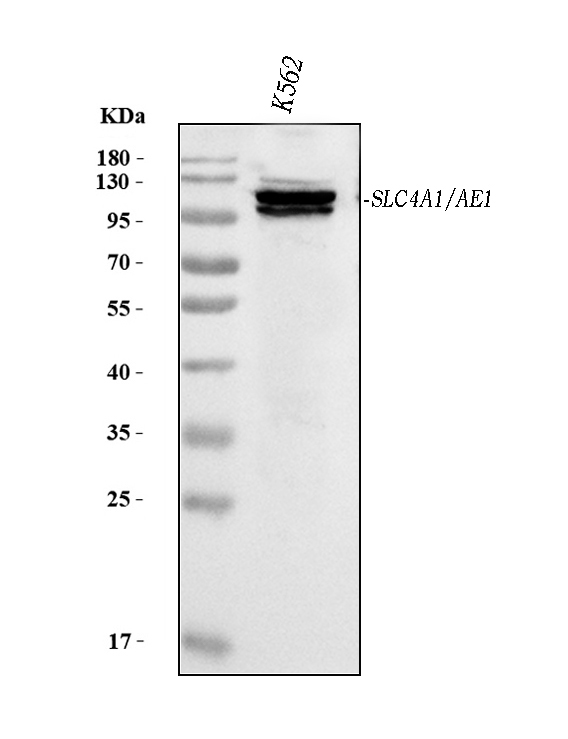
Western blot analysis of SLC4A1 using anti-SLC4A1 antibody (M01146-1).
Electrophoresis was performed on a 5-20% SDS-PAGE gel at 70V (Stacking gel) / 90V (Resolving gel) for 2-3 hours. The sample well of each lane was loaded with 30 ug of sample under reducing conditions.
Lane 1: human K562 whole cell lysates.
After electrophoresis, proteins were transferred to a nitrocellulose membrane at 150 mA for 50-90 minutes. Blocked the membrane with 5% non-fat milk/TBS for 1.5 hour at RT. The membrane was incubated with mouse anti-SLC4A1 antigen affinity purified monoclonal antibody (Catalog # M01146-1) at 0.5 μg/mL overnight at 4°C, then washed with TBS-0.1%Tween 3 times with 5 minutes each and probed with a goat anti-mouse IgG-HRP secondary antibody at a dilution of 1:10000 for 1.5 hour at RT. The signal is developed using an Enhanced Chemiluminescent detection (ECL) kit (Catalog # EK1001) with Tanon 5200 system. A specific band was detected for SLC4A1 at approximately 102 kDa. The expected band size for SLC4A1 is at 102 kDa.

Click image to see more details
IHC analysis of SLC4A1 using anti-SLC4A1 antibody (M01146-1).
SLC4A1 was detected in a paraffin-embedded section of human spleen tissue. Heat mediated antigen retrieval was performed in EDTA buffer (pH 8.0, epitope retrieval solution). The tissue section was blocked with 10% goat serum. The tissue section was then incubated with 2 μg/ml mouse anti-SLC4A1 Antibody (M01146-1) overnight at 4°C. Peroxidase Conjugated Goat Anti-mouse IgG was used as secondary antibody and incubated for 30 minutes at 37°C. The tissue section was developed using HRP Conjugated Mouse IgG Super Vision Assay Kit (Catalog # SV0001) with DAB as the chromogen.

Click image to see more details
Flow Cytometry analysis of HepG2 cells using anti-SLC4A1 antibody (M01146-1).
Overlay histogram showing HepG2 cells stained with M01146-1 (Blue line). The cells were fixed with 4% paraformaldehyde and blocked with 10% normal goat serum. And then incubated with mouse anti-SLC4A1 Antibody (M01146-1, 1 μg/1x106 cells) for 30 min at 20°C. DyLight®488 conjugated goat anti-mouse IgG (BA1126, 5-10 μg/1x106 cells) was used as secondary antibody for 30 minutes at 20°C. Isotype control antibody (Green line) was mouse IgG (1 μg/1x106) used under the same conditions. Unlabelled sample without incubation with primary antibody and secondary antibody (Red line) was used as a blank control.
Specific Publications For Anti-SLC4A1/CD233/Band 3 Antibody Picoband® (monoclonal, 5G2G7) (M01146-1)
Loading publications
Recommended Resources
Here are featured tools and databases that you might find useful.
- Boster's Pathways Library
- Protein Databases
- Bioscience Research Protocol Resources
- Data Processing & Analysis Software
- Photo Editing Software
- Scientific Literature Resources
- Research Paper Management Tools
- Molecular Biology Software
- Primer Design Tools
- Bioinformatics Tools
- Phylogenetic Tree Analysis
Customer Reviews
Have you used Anti-SLC4A1/CD233/Band 3 Antibody Picoband® (monoclonal, 5G2G7)?
Share your experimental results or join a short interview to earn up to $1,000 in product credits or other rewards.
0 Reviews For Anti-SLC4A1/CD233/Band 3 Antibody Picoband® (monoclonal, 5G2G7)
Customer Q&As
Have a question?
Find answers in Q&As, reviews.
Can't find your answer?
Submit your question
