Click image to see more details
-

-
-
-
-
+2
Product Info Summary
| SKU: | PA1816 |
|---|---|
| Size: | 100 μg/vial |
| Reactive Species: | Human, Mouse, Rat |
| Host: | Rabbit |
| Application: | Flow Cytometry, IF, IHC, ICC, WB |
Customers Who Bought This Also Bought

Product info
Product Name
Anti-Hsc70/HSPA8 Antibody Picoband®
SKU/Catalog Number
PA1816
BA2411 is an alternative SKU for this antibody, used in previous lots.
Size
100 μg/vial
Form
Lyophilized
Description
Boster Bio Anti-Hsc70/HSPA8 Antibody catalog # PA1816. Tested in Flow Cytometry, IF, IHC, ICC, WB applications. This antibody reacts with Human, Mouse, Rat. The brand Picoband indicates this is a premium antibody that guarantees superior quality, high affinity, and strong signals with minimal background in Western blot applications. Only our best-performing antibodies are designated as Picoband, ensuring unmatched performance.
Storage & Handling
Store at -20˚C for one year from date of receipt. After reconstitution, at 4˚C for one month. It can also be aliquotted and stored frozen at -20˚C for six months. Avoid repeated freeze-thaw cycles.
Cite This Product
Anti-Hsc70/HSPA8 Antibody Picoband® (Boster Biological Technology, Pleasanton CA, USA, Catalog # PA1816)
Host
Rabbit
Contents
Each vial contains antibody formulated with stabilizing components, 0.9mg NaCl, 0.2mg Na2HPO4, 0.05mg Thimerosal, 0.05mg NaN3.
*This antibody is supplied in a stabilized formulation.
Compatibility with conjugation reactions depends on the chemistry of the conjugation method used.
For conjugation methods that are not compatible with the stabilizing components present in this formulation, a carrier-free antibody format is required.
Clonality
Polyclonal
Isotype
Rabbit IgG
Immunogen
A synthetic peptide corresponding to a sequence at the C-terminus of human Hsc70, different from the related mouse and rat sequences by one amino acid.
Cross-reactivity
No cross-reactivity with other proteins
Reactive Species
PA1816 is reactive to HSPA8 in Human, Mouse, Rat
Observed Molecular Weight
71 kDa
Calculated molecular weight
70.9 kDa
Background of HSPA8
HSPA8 (heat shock 70kDa protein 8) also known as HSC70, HSC71, HSP73, HSPA10, FORMERLY, LAP1 or LPS-ASSOCIATED PROTEIN 1, is a heat shock protein that in humans is encoded by the HSPA8 gene. The HSPA8 gene contains 9 exons and spans 5 kb. The deduced HSPA8 protein has 646 amino acids and a predicted molecular mass of 70,899 Da. The HSPA8 gene is mapped on 11q24.1. HSPA8 plays an important role in cells by transiently associating with nascent polypeptides to facilitate correct folding. HSP73 also functions as an ATPase in the disassembly of clathrin-coated vesicles during transport of membrane components through the cell. Rapid decay involves AU-rich binding protein AUF1, which complexes with heat-shock proteins HSC70 and HSP70, translation initiation factor EIF4G, and poly (A)-binding protein. In the absence of Il3, Hsc70 formed a complex with Hsp40 and Hip, and this complex, in association with Eif4g and Pabp, formed a high-stability complex with Bim mRNA that protected it from ribonucleases.
Antibody Validation
Boster validates all antibodies on WB, IHC, ICC, Immunofluorescence, and ELISA with known positive control and negative samples to ensure specificity and high affinity, including thorough antibody incubations.
Application & Images
Applications
PA1816 is guaranteed for Flow Cytometry, IF, IHC, ICC, WB Boster Guarantee
Recommend Dilution
| Application | Dilution | Species |
|---|---|---|
| Western blot | 0.1-0.5μg/ml | Human, Mouse, Rat |
| Immunohistochemistry (Paraffin-embedded Section) | 0.5-1μg/ml | Human, Rat, Mouse |
| Immunocytochemistry/Immunofluorescence | 2μg/ml | Human |
| Flow Cytometry (Fixed) | 1-3μg/1x106 cells | Human |
Tested application
Suggested blocking solution with 5% non-fat milk or BSA; (*)Recommended protein loading: 20-40 µg per lane
Use TE buffer pH 9.0 for antigen retrieval; (*) citrate buffer pH 6.0 is an alternative.
Validation Images & Assay Conditions
Click image to see more details
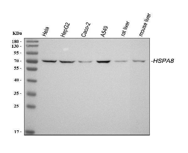
Western blot analysis of Hsc70 using anti-Hsc70 antibody (PA1816).
Electrophoresis was performed on a 5-20% SDS-PAGE gel at 70V (Stacking gel) / 90V (Resolving gel) for 2-3 hours. The sample well of each lane was loaded with 30 ug of sample under reducing conditions.
Lane 1: human Hela whole cell lysates,
Lane 2: human HepG2 whole cell lysates,
Lane 3: human Caco-2 whole cell lysates,
Lane 3: human A549 whole cell lysates,
Lane 3: rat liver tissue lysates,
Lane 3: mouse liver tissue lysates.
After electrophoresis, proteins were transferred to a nitrocellulose membrane at 150 mA for 50-90 minutes. Blocked the membrane with 5% non-fat milk/TBS for 1.5 hour at RT. The membrane was incubated with rabbit anti-Hsc70 antigen affinity purified polyclonal antibody (Catalog # PA1816) at 0.5 μg/mL overnight at 4°C, then washed with TBS-0.1%Tween 3 times with 5 minutes each and probed with a goat anti-rabbit IgG-HRP secondary antibody at a dilution of 1:5000 for 1.5 hour at RT. The signal is developed using an Enhanced Chemiluminescent detection (ECL) kit (Catalog # EK1002) with Tanon 5200 system. A specific band was detected for Hsc70 at approximately 71 kDa. The expected band size for Hsc70 is at 71 kDa.

Click image to see more details
IHC analysis of HSC70/HSPA8 using anti-HSC70/HSPA8 antibody (PA1816).
HSC70/HSPA8 was detected in a paraffin-embedded section of human ovarian cancer tissue. Heat mediated antigen retrieval was performed in EDTA buffer (pH 8.0, epitope retrieval solution). The tissue section was blocked with 10% goat serum. The tissue section was then incubated with 2 μg/ml rabbit anti-HSC70/HSPA8 Antibody (PA1816) overnight at 4°C. Peroxidase Conjugated Goat Anti-rabbit IgG was used as secondary antibody and incubated for 30 minutes at 37°C. The tissue section was developed using HRP Conjugated Rabbit IgG Super Vision Assay Kit (Catalog # SV0002) with DAB as the chromogen.

Click image to see more details
IHC analysis of Hsc70 using anti-Hsc70 antibody (PA1816).
Hsc70 was detected in paraffin-embedded section of Human Lung Cancer tissues. Heat mediated antigen retrieval was performed in citrate buffer (pH6, epitope retrieval solution) for 20 mins. The tissue section was blocked with 10% goat serum. The tissue section was then incubated with 1μg/ml rabbit anti-Hsc70 Antibody (PA1816) overnight at 4°C. Biotinylated goat anti-rabbit IgG was used as secondary antibody and incubated for 30 minutes at 37°C. The tissue section was developed using Strepavidin-Biotin-Complex (SABC)(Catalog # SA1022) with DAB as the chromogen.

Click image to see more details
IHC analysis of Hsc70 using anti-Hsc70 antibody (PA1816).
Hsc70 was detected in paraffin-embedded section of rat kidney tissues. Heat mediated antigen retrieval was performed in citrate buffer (pH6, epitope retrieval solution) for 20 mins. The tissue section was blocked with 10% goat serum. The tissue section was then incubated with 1μg/ml rabbit anti-Hsc70 Antibody (PA1816) overnight at 4°C. Biotinylated goat anti-rabbit IgG was used as secondary antibody and incubated for 30 minutes at 37°C. The tissue section was developed using Strepavidin-Biotin-Complex (SABC)(Catalog # SA1022) with DAB as the chromogen.

Click image to see more details
Flow Cytometry analysis of HL-60 cells using anti-Hsc70 antibody (PA1816).
Overlay histogram showing HL-60 cells stained with PA1816 (Blue line). To facilitate intracellular staining, cells were fixed with 4% paraformaldehyde and permeabilized with permeabilization buffer. The cells were blocked with 10% normal goat serum. And then incubated with rabbit anti-Hsc70 Antibody (PA1816, 1μg/1x106 cells) for 30 min at 20°C. DyLight®488 conjugated goat anti-rabbit IgG (BA1127, 5-10μg/1x106 cells) was used as secondary antibody for 30 minutes at 20°C. Isotype control antibody (Green line) was rabbit IgG (1μg/1x106) used under the same conditions. Unlabelled sample without incubation with primary antibody and secondary antibody (Red line) was used as a blank control.

Click image to see more details
IF analysis of Hsc70 using anti- Hsc70 antibody (PA1816).
Hsc70 was detected in immunocytochemical section of U20S cells. Enzyme antigen retrieval was performed using IHC enzyme antigen retrieval reagent (AR0022) for 15 mins. The cells were blocked with 10% goat serum. And then incubated with 2μg/mL rabbit anti-Hsc70 Antibody (PA1816) overnight at 4°C. DyLight®594 Conjugated Goat Anti-Rabbit IgG (BA1142) was used as secondary antibody at 1:100 dilution and incubated for 30 minutes at 37°C. The section was counterstained with DAPI. Visualize using a fluorescence microscope and filter sets appropriate for the label used.
Specific Publications For Anti-Hsc70/HSPA8 Antibody Picoband® (PA1816)
Loading publications
Recommended Resources
Here are featured tools and databases that you might find useful.
- Boster's Pathways Library
- Protein Databases
- Bioscience Research Protocol Resources
- Data Processing & Analysis Software
- Photo Editing Software
- Scientific Literature Resources
- Research Paper Management Tools
- Molecular Biology Software
- Primer Design Tools
- Bioinformatics Tools
- Phylogenetic Tree Analysis
Customer Reviews
Have you used Anti-Hsc70/HSPA8 Antibody Picoband®?
Share your experimental results or join a short interview to earn up to $1,000 in product credits or other rewards.
0 Reviews For Anti-Hsc70/HSPA8 Antibody Picoband®
Customer Q&As
Have a question?
Find answers in Q&As, reviews.
Can't find your answer?
Submit your question
16 Customer Q&As for Anti-Hsc70/HSPA8 Antibody Picoband®
Question
We are currently using anti-Hsc70/HSPA8 antibody PA1816 for rat tissue, and we are content with the WB results. The species of reactivity given in the datasheet says human, mouse, rat. Is it true that the antibody can work on primate tissues as well?
Verified Customer
Verified customer
Asked: 2020-03-26
Answer
The anti-Hsc70/HSPA8 antibody (PA1816) has not been validated for cross reactivity specifically with primate tissues, though there is a good chance of cross reactivity. We have an innovator award program that if you test this antibody and show it works in primate you can get your next antibody for free. Please contact me if I can help you with anything.
Boster Scientific Support
Answered: 2020-03-26
Question
Is a blocking peptide available for product anti-Hsc70/HSPA8 antibody (PA1816)?
Verified Customer
Verified customer
Asked: 2020-03-19
Answer
We do provide the blocking peptide for product anti-Hsc70/HSPA8 antibody (PA1816). If you would like to place an order for it please contact support@bosterbio.com and make a special request.
Boster Scientific Support
Answered: 2020-03-19
Question
I was wanting to use to test anti-Hsc70/HSPA8 antibody PA1816 on human cajal-retzius cell fetal brain cortex for research purposes, then I may be interested in using anti-Hsc70/HSPA8 antibody PA1816 for diagnostic purposes as well. Is the antibody suitable for diagnostic purposes?
D. Miller
Verified customer
Asked: 2019-11-18
Answer
The products we sell, including anti-Hsc70/HSPA8 antibody PA1816, are only intended for research use. They would not be suitable for use in diagnostic work. If you have the means to develop a product into diagnostic use, and are interested in collaborating with us and develop our product into an IVD product, please contact us for more discussions.
Boster Scientific Support
Answered: 2019-11-18
Question
We have observed staining in rat leukemic t-cell. What should we do? Is anti-Hsc70/HSPA8 antibody supposed to stain leukemic t-cell positively?
Verified Customer
Verified customer
Asked: 2019-09-24
Answer
According to literature leukemic t-cell does express HSPA8. According to Uniprot.org, HSPA8 is expressed in frontal cortex, placenta skin, embryonic kidney, colon carcinoma ovarian carcinoma, brain, cajal-retzius cell fetal brain cortex, lymphoblast, melanoma, leukemic t-cell, cervix carcinoma, cervix carcinoma erythroleukemia, liver, colon carcinoma, among other tissues. Regarding which tissues have HSPA8 expression, here are a few articles citing expression in various tissues:
Brain, Cajal-Retzius cell, and Fetal brain cortex, Pubmed ID: 1286667
Cervix carcinoma, Pubmed ID: 20068231
Cervix carcinoma, and Erythroleukemia, Pubmed ID: 23186163
Colon carcinoma, Pubmed ID: 24129315
Embryonic kidney, Pubmed ID: 17525332
Leukemic T-cell, Pubmed ID: 19690332
Liver, Pubmed ID: 24275569
Lymphoblast, Pubmed ID: 14654843
Melanoma, Pubmed ID: 17081065
Placenta, and Skin, Pubmed ID: 15489334
Boster Scientific Support
Answered: 2019-09-24
Question
Would PA1816 anti-Hsc70/HSPA8 antibody work on parafin embedded sections? If so, which fixation method do you recommend we use (PFA, paraformaldehyde, other)?
Verified Customer
Verified customer
Asked: 2019-08-20
Answer
You can see on the product datasheet, PA1816 anti-Hsc70/HSPA8 antibody as been tested on IHC. It is best to use PFA for fixation because it has better tissue penetration ability. PFA needs to be prepared fresh before use. Long term stored PFA turns into formalin, as the PFA molecules congregate and become formalin.
Boster Scientific Support
Answered: 2019-08-20
Question
We have tried in the past anti-Hsc70/HSPA8 antibody for IHC on brain in a previous experiment. I am using mouse, and We intend to use the antibody for WB next. My question regards examining brain as well as melanoma in our next experiment. Could you please give me some suggestion on which antibody would work the best for WB?
Verified Customer
Verified customer
Asked: 2019-05-21
Answer
I have checked the website and datasheets of our anti-Hsc70/HSPA8 antibody and I see that PA1816 has been tested on mouse in both IHC and WB. Thus PA1816 should work for your application. Our Boster satisfaction guarantee will cover this product for WB in mouse even if the specific tissue type has not been validated. We do have a comprehensive range of products for WB detection and you can check out our website bosterbio.com to find out more information about them.
Boster Scientific Support
Answered: 2019-05-21
Question
Thanks for helping with my inquiry over the phone. Here are the WB image, lot number and protocol we used for cajal-retzius cell fetal brain cortex using anti-Hsc70/HSPA8 antibody PA1816. Let me know if you need anything else.
Verified Customer
Verified customer
Asked: 2018-12-13
Answer
We appreciate the data. You have provided everything we needed. Our lab team are working to resolve your inquiry as quickly as possible, and we appreciate your patience and understanding! Please let me know if there is anything you need in the meantime.
Boster Scientific Support
Answered: 2018-12-13
Question
Our team were satisfied with the WB result of your anti-Hsc70/HSPA8 antibody. However we have observed positive staining in cajal-retzius cell fetal brain cortex cytoplasm. melanosome. nucleus using this antibody. Is that expected? Could you tell me where is HSPA8 supposed to be expressed?
Verified Customer
Verified customer
Asked: 2018-10-24
Answer
From literature, cajal-retzius cell fetal brain cortex does express HSPA8. Generally HSPA8 expresses in cytoplasm. melanosome. nucleus, nucleolus. Regarding which tissues have HSPA8 expression, here are a few articles citing expression in various tissues:
Brain, Cajal-Retzius cell, and Fetal brain cortex, Pubmed ID: 1286667
Cervix carcinoma, Pubmed ID: 20068231
Cervix carcinoma, and Erythroleukemia, Pubmed ID: 23186163
Colon carcinoma, Pubmed ID: 24129315
Embryonic kidney, Pubmed ID: 17525332
Leukemic T-cell, Pubmed ID: 19690332
Liver, Pubmed ID: 24275569
Lymphoblast, Pubmed ID: 14654843
Melanoma, Pubmed ID: 17081065
Placenta, and Skin, Pubmed ID: 15489334
Boster Scientific Support
Answered: 2018-10-24
Question
I was wanting to use your anti-Hsc70/HSPA8 antibody for IHC for human cajal-retzius cell fetal brain cortex on frozen tissues, but I want to know if it has been tested for this particular application. Has this antibody been tested and is this antibody a good choice for human cajal-retzius cell fetal brain cortex identification?
Verified Customer
Verified customer
Asked: 2018-07-27
Answer
You can see on the product datasheet, PA1816 anti-Hsc70/HSPA8 antibody has been tested for IHC, WB on human, mouse, rat tissues. We have an innovator award program that if you test this antibody and show it works in human cajal-retzius cell fetal brain cortex in IHC-frozen, you can get your next antibody for free.
Boster Scientific Support
Answered: 2018-07-27
Question
I am interested in using your anti-Hsc70/HSPA8 antibody for lipophagy studies. Has this antibody been tested with western blotting on mouse liver tissue? We would like to see some validation images before ordering.
Verified Customer
Verified customer
Asked: 2018-03-29
Answer
Thank you for your inquiry. This PA1816 anti-Hsc70/HSPA8 antibody is tested on human hela, a549 whole cell lysates, hela whole cell lysates, lung cancer tissue, rat liver tissue, spleen tissue, mouse liver tissue. It is guaranteed to work for IHC, WB in human, mouse, rat. Our Boster guarantee will cover your intended experiment even if the sample type has not been be directly tested.
Boster Scientific Support
Answered: 2018-03-29
Question
I see that the anti-Hsc70/HSPA8 antibody PA1816 works with IHC, what is the protocol used to produce the result images on the product page?
Verified Customer
Verified customer
Asked: 2017-10-16
Answer
You can find protocols for IHC on the "support/technical resources" section of our navigation menu. If you have any further questions, please send an email to support@bosterbio.com
Boster Scientific Support
Answered: 2017-10-16
Question
See below the WB image, lot number and protocol we used for cajal-retzius cell fetal brain cortex using anti-Hsc70/HSPA8 antibody PA1816. Please let me know if you require anything else.
J. Baker
Verified customer
Asked: 2017-03-22
Answer
Thank you very much for the data. Our lab team are working to resolve this as quickly as possible, and we appreciate your patience and understanding! You have provided everything we needed. Please let me know if there is anything you need in the meantime.
Boster Scientific Support
Answered: 2017-03-22
Question
Can you help my question with product PA1816, anti-Hsc70/HSPA8 antibody. I was wondering if it would be possible to conjugate this antibody with biotin. I would need it to be without BSA or sodium azide. I am planning on using a buffer exchange of sodium azide with PBS only. Would there be problems for me to conjugate the antibody and store it in -20 degrees in small aliquots?
B. Moore
Verified customer
Asked: 2017-02-15
Answer
We do not recommend storing this antibody with PBS buffer only in -20 degrees. If you want to store it in -20 degrees it is best to add some cryoprotectant like glycerol. If you want carrier free PA1816 anti-Hsc70/HSPA8 antibody, we can provide it to you in a special formula with trehalose and/or glycerol. These molecules will not interfere with conjugation chemistry and provide a good level of protection for the antibody from degradation. Please be sure to specify this in your purchase order.
Boster Scientific Support
Answered: 2017-02-15
Question
Does anti-Hsc70/HSPA8 antibody PA1816 work for IHC with cajal-retzius cell fetal brain cortex?
C. Anderson
Verified customer
Asked: 2016-07-27
Answer
According to the expression profile of cajal-retzius cell fetal brain cortex, HSPA8 is highly expressed in cajal-retzius cell fetal brain cortex. So, it is likely that anti-Hsc70/HSPA8 antibody PA1816 will work for IHC with cajal-retzius cell fetal brain cortex.
Boster Scientific Support
Answered: 2016-07-27
Question
Is this PA1816 anti-Hsc70/HSPA8 antibody reactive to the isotypes of HSPA8?
K. Roberts
Verified customer
Asked: 2013-03-01
Answer
The immunogen of PA1816 anti-Hsc70/HSPA8 antibody is A synthetic peptide corresponding to a sequence at the C-terminus of human Hsc70(563-582aa NDEDKQKILDKCNEIINWLD), different from the related mouse and rat sequences by one amino acid. Could you tell me which isotype you are interested in so I can help see if the immunogen is part of this isotype?
Boster Scientific Support
Answered: 2013-03-01
Question
Is there a BSA free version of anti-Hsc70/HSPA8 antibody PA1816 available?
D. Roberts
Verified customer
Asked: 2013-02-19
Answer
We appreciate your recent telephone inquiry. I can confirm that some lots of this anti-Hsc70/HSPA8 antibody PA1816 are BSA free. For now, these lots are available and we can make a BSA free formula for you free of charge. It will take 3 extra days to prepare. If you require this antibody BSA free again in future, please do not hesitate to contact me and I will be pleased to check which lots we have in stock that are BSA free.
Boster Scientific Support
Answered: 2013-02-19
