Click image to see more details
-

-
-
-
-
+26
Product Info Summary
| SKU: | A00183 |
|---|---|
| Size: | 100 μg/vial |
| Reactive Species: | Human, Mouse, Rat |
| Host: | Rabbit |
| Application: | Flow Cytometry, IF, IHC, ICC, WB |
Customers Who Bought This Also Bought

Product info
Product Name
Anti-Bax Antibody Picoband®
SKU/Catalog Number
A00183
Size
100 μg/vial
Form
Lyophilized
Description
Boster Bio Anti-Bax Antibody Picoband® catalog # A00183. Tested in Flow Cytometry, IF, IHC, ICC, WB applications. This antibody reacts with Human, Mouse, Rat. The brand Picoband indicates this is a premium antibody that guarantees superior quality, high affinity, and strong signals with minimal background in Western blot applications. Only our best-performing antibodies are designated as Picoband, ensuring unmatched performance.
Storage & Handling
Store at -20˚C for one year from date of receipt. After reconstitution, at 4˚C for one month. It can also be aliquotted and stored frozen at -20˚C for six months. Avoid repeated freeze-thaw cycles.
Cite This Product
Anti-Bax Antibody Picoband® (Boster Biological Technology, Pleasanton CA, USA, Catalog # A00183)
Host
Rabbit
Contents
Each vial contains 4 mg Trehalose, 0.9 mg NaCl and 0.2 mg Na2HPO4.
Clonality
Polyclonal
Isotype
Rabbit IgG
Immunogen
A synthetic peptide corresponding to a sequence at the N-terminus of human Bax, different from the related mouse and rat sequences by five amino acids.
Cross-reactivity
No cross-reactivity with other proteins
Reactive Species
A00183 is reactive to BAX in Human, Mouse, Rat
Observed Molecular Weight
21 kDa
Calculated molecular weight
21.2 kDa
Background of BAX
Apoptosis regulator BAX, also known as bcl-2-like protein 4, is a protein that in humans is encoded by the BAX gene. The protein encoded by this gene belongs to the BCL2 protein family. BCL2 family members form hetero- or homodimers and act as anti- or pro-apoptotic regulators that are involved in a wide variety of cellular activities. This protein forms a heterodimer with BCL2, and functions as an apoptotic activator. Additionally, this protein is reported to interact with, and increase the opening of, the mitochondrial voltage-dependent anion channel (VDAC), which leads to the loss in membrane potential and the release of cytochrome c. The expression of this gene is regulated by the tumor suppressor P53 and has been shown to be involved in P53-mediated apoptosis. Multiple alternatively spliced transcript variants, which encode different isoforms, have been reported for this gene.
Antibody Validation
Boster validates all antibodies on WB, IHC, ICC, Immunofluorescence, and ELISA with known positive control and negative samples to ensure specificity and high affinity, including thorough antibody incubations.
Application & Images
Applications
A00183 is guaranteed for Flow Cytometry, IF, IHC, ICC, WB Boster Guarantee
Recommend Dilution
| Application | Dilution | Species |
|---|---|---|
| Western blot | 0.1-0.5μg/ml | Human, Rat |
| Immunohistochemistry (Paraffin-embedded Section) | 2-5μg/ml | Human, Mouse, Rat |
| Immunocytochemistry/Immunofluorescence | 5 μg/ml | Human |
| Immunofluorescence | 10 μg/ml | Human |
| Flow Cytometry (Fixed) | 1-3μg/1x106 cells | Human |
Tested application
Suggested blocking solution with 5% non-fat milk or BSA; (*)Recommended protein loading: 20-40 µg per lane
Use TE buffer pH 9.0 for antigen retrieval; (*) citrate buffer pH 6.0 is an alternative.
Validation Images & Assay Conditions
Click image to see more details
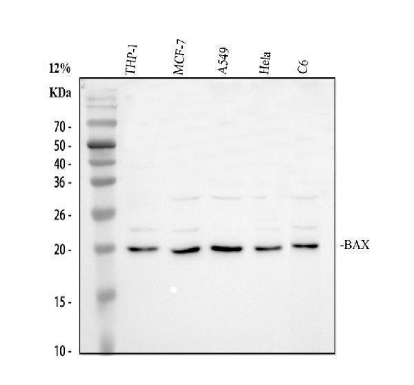
Western blot analysis of Bax using anti-Bax antibody (A00183).
Electrophoresis was performed on a 12% SDS-PAGE gel at 80V (Stacking gel) / 120V (Resolving gel) for 2 hours. The sample well of each lane was loaded with 30 ug of sample under reducing conditions.
Lane 1: human THP-1 whole cell lysates,
Lane 2: human MCF-7 whole cell lysates,
Lane 3: human A549 whole cell lysates,
Lane 4: human Hela whole cell lysates,
Lane 5: rat C6 whole cell lysates.
After electrophoresis, proteins were transferred to a nitrocellulose membrane at 150 mA for 50-90 minutes. Blocked the membrane with 5% non-fat milk/TBS for 1.5 hour at RT. The membrane was incubated with rabbit anti-Bax antigen affinity purified polyclonal antibody (A00183) at 0.5 μg/mL overnight at 4°C, then washed with TBS-0.1%Tween 3 times with 5 minutes each and probed with a goat anti-rabbit IgG-HRP secondary antibody (Catalog # BA1054) at a dilution of 1:5000 for 1.5 hour at RT. The signal is developed using an ECL Plus Western Blotting Substrate (Catalog # AR1196-200) with Tanon 5200 system. A specific band was detected for Bax at approximately 21 kDa. The expected band size for Bax is at 21 kDa.

Click image to see more details
IHC analysis of Bax using anti-Bax antibody (A00183).
Bax was detected in a paraffin-embedded section of human lung cancer tissue. Heat mediated antigen retrieval was performed in EDTA buffer (pH 8.0, epitope retrieval solution). The tissue section was blocked with 10% goat serum. The tissue section was then incubated with 2 μg/ml rabbit anti-Bax Antibody (A00183) overnight at 4°C. Peroxidase Conjugated Goat Anti-rabbit IgG was used as secondary antibody and incubated for 30 minutes at 37°C. The tissue section was developed using HRP Conjugated Rabbit IgG Super Vision Assay Kit (Catalog # SV0002) with DAB as the chromogen.

Click image to see more details
Western blot analysis of BAX using anti-BAX antibody (A00183).
Electrophoresis was performed on a 12% SDS-PAGE gel at 80V (Stacking gel) / 120V (Resolving gel) for 2 hours. The sample well of each lane was loaded with 30 ug of sample under reducing conditions.
Lane 1: human Hela- WT whole cell lysates,
Lane 2: human Hela-BAX KO whole cell lysates.
After electrophoresis, proteins were transferred to a nitrocellulose membrane at 150 mA for 50-90 minutes. Blocked the membrane with 5% non-fat milk/TBS for 1.5 hour at RT. The membrane was incubated with rabbit anti-BAX antigen affinity purified polyclonal antibody (A00183) at 0.5 μg/mL overnight at 4°C, then washed with TBS-0.1%Tween 3 times with 5 minutes each and probed with a goat anti-rabbit IgG-HRP secondary antibody at a dilution of 1:5000 for 1.5 hour at RT. The signal is developed using an ECL Plus Western Blotting Substrate (Catalog # AR1196-200) with Tanon 5200 system. A specific band was detected for BAX at approximately 21 kDa. The expected band size for BAX is at 21 kDa.

Click image to see more details
IHC analysis of Bax using anti-Bax antibody (A00183).
Bax was detected in a paraffin-embedded section of mouse kidney tissue. Heat mediated antigen retrieval was performed in EDTA buffer (pH 8.0, epitope retrieval solution). The tissue section was blocked with 10% goat serum. The tissue section was then incubated with 2 μg/ml rabbit anti-Bax Antibody (A00183) overnight at 4°C. Peroxidase Conjugated Goat Anti-rabbit IgG was used as secondary antibody and incubated for 30 minutes at 37°C. The tissue section was developed using HRP Conjugated Rabbit IgG Super Vision Assay Kit (Catalog # SV0002) with DAB as the chromogen.

Click image to see more details
IHC analysis of Bax using anti-Bax antibody (A00183).
Bax was detected in a paraffin-embedded section of rat kidney tissue. Heat mediated antigen retrieval was performed in EDTA buffer (pH 8.0, epitope retrieval solution). The tissue section was blocked with 10% goat serum. The tissue section was then incubated with 2 μg/ml rabbit anti-Bax Antibody (A00183) overnight at 4°C. Peroxidase Conjugated Goat Anti-rabbit IgG was used as secondary antibody and incubated for 30 minutes at 37°C. The tissue section was developed using HRP Conjugated Rabbit IgG Super Vision Assay Kit (Catalog # SV0002) with DAB as the chromogen.

Click image to see more details
IF analysis of Bax using anti-Bax antibody (A00183).
Bax was detected in an immunocytochemical section of A549 cells. Enzyme antigen retrieval was performed using IHC enzyme antigen retrieval reagent (AR0022) for 15 mins. The cells were blocked with 10% goat serum. And then incubated with 5 μg/mL rabbit anti-Bax Antibody (A00183) overnight at 4°C. DyLight®488 Conjugated Goat Anti-Rabbit IgG (BA1127) was used as secondary antibody at 1:500 dilution and incubated for 30 minutes at 37°C. The section was counterstained with DAPI. Visualize using a fluorescence microscope and filter sets appropriate for the label used.

Click image to see more details
IF analysis of BAX using anti-BAX antibody (A00183).
BAX was detected in a paraffin-embedded section of FFPE mouse uterus tissue. Heat mediated antigen retrieval was performed in EDTA buffer (pH 8.0, epitope retrieval solution). The tissue section was blocked with 10% goat serum. The tissue section was then incubated with rabbit anti-BAX Antibody (A00183) at 10 μg/ml overnight at 4°C. DyLight®488 Conjugated Goat Anti-Rabbit IgG (BA1127) was used as secondary antibody at 1:500 dilution and incubated for 30 minutes at 37°C. The section was counterstained with DAPI. Visualize using a fluorescence microscope and filter sets appropriate for the label used.

Click image to see more details
Flow Cytometry analysis of THP-1 cells using anti-Bax antibody (A00183).
Overlay histogram showing THP-1 cells stained with A00183 (Blue line). To facilitate intracellular staining, cells were fixed with 4% paraformaldehyde and permeabilized with permeabilization buffer. The cells were blocked with 10% normal goat serum. And then incubated with rabbit anti-Bax Antibody (A00183, 1 μg/1x106 cells) for 30 min at 20°C. DyLight®488 conjugated goat anti-rabbit IgG (BA1127, 5-10 μg/1x106 cells) was used as secondary antibody for 30 minutes at 20°C. Isotype control antibody (Green line) was rabbit IgG (1 μg/1x106) used under the same conditions. Unlabelled sample without incubation with primary antibody and secondary antibody (Red line) was used as a blank control.

Click image to see more details
a Western blotting was used to examine mitochondrial apoptotic pathway-related proteins after treatment with control (i), PANI-PEG-CS (ii), Ir(III) complex (iii), and Ir(III)@PANI-PEG-CS (iv). Key proteins involved in apoptosis such as Bax, Bcl-2, cytochrome c, and cleaved caspase-3 were examined to better understand how each treatment affects cell death at the mitochondrial level. b To further investigate the underlying molecular mechanisms, western blotting was used to investigate the PI3K/AKT/mTOR pathway (i), PANI-PEG-CS (ii), Ir(III) complex (iii), and Ir(III)@PANI-PEG-CS (iv). Expression levels of PI3K, AKT (total and phosphorylated), and mTOR were evaluated to assess whether this survival pathway was activated or suppressed. Protein levels were quantified and compared to the control group to determine statistical significance. * p < 0.05, ** p < 0.01, *** p < 0.001 in comparison to the respective control
Index in Springer Nature under a CC BY license. DOI: 10.1186/s12645-025-00336-z

Click image to see more details
( A, B ) Protein expression levels of p21, p53, Bax, and Bcl-2 were detected by Western blot assays and compared by quantitative analysis of the gray value. * p <0.05 and ** p <0.01 vs. siR-NC group. siR-NC – control siRNA; siR-HMGB3 – HMGB3 siRNA; HMGB3 – high mobility group-box 3.
Index in PubMed under a CC BY license. PMID: 27774979

Click image to see more details
( A ) Expression of Bcl-2 (A1–A3), ( B ) Bax (B1–B3), ( C ) Caspase-3 (C1–C3) in ovaries of HEV inoculation rabbits. A1, B1 and C1 are control group. A2, B2, C2 are HEV RNA positive ovaries in 28 dpi. A3, B3, C3 are HEV RNA positive ovaries in 49 dpi. (Original magnification:40×).
Index in PubMed under a CC BY license. PMID: 29435117

Click image to see more details
Celastrol attenuates ganglion cells apoptosis in the retina of EAE rats. Treatment of celastrol decreased the number of TUNEL-positive cells (A) , upregulated expression of Bcl-2 (B) and downregulated expression of Bax, cleaved-caspase 3 and cleaved-PARP. Scale bar: 100 μm. Data were shown as mean ± SD, n = 5. ∗∗ P < 0.01 versus control group, ## P < 0.01 versus EAE group, †† P < 0.01 versus low dosage of celastrol group.
Index in PubMed under a CC BY license. PMID: 28239352

Click image to see more details
Effects of maltol on the levels of inflammation cytokines in cisplatin-induced renal toxicity. ( A ) Effects of maltol on the positive expressions of Bax, Bcl-2, iNOS and COX-2 in renal tissues were examined by IHC in renal tissues (magnification × 200), And the column chart shows stained area, semiquantitative analysis of Bax, Bcl-2, iNOS and COX-2 expression in kidneys to IHC. ( B ) Inflammation cytokines level of TNF-α, IL-1β, iNOS and NF-κB in serum of mice were measured by ELISA kits. All values were expressed as mean ± S.D. * p < 0.05, ** p < 0.01 vs . normal group; # p < 0.05, ## p < 0.01 vs . cisplatin group.
Index in PubMed under a CC BY license. PMID: 30374107

Click image to see more details
V protein overexpression in DF-1 cells inhibited apoptosis through the Bcl-2\Bax-Caspase-3 pathway. A V and control (pCAGEN-flag) plasmids were transfected into DF-1 cells for 48 h before the cells were harvested. The RNA was extracted according to the previously described method. The mRNA levels of some apoptosis-related genes (proapoptosis genes Caspase-3, Caspase-9, and FasLG, and the antiapoptosis gene Bcl-2) were detected using Q-PCR. B DF-1 cells were transfected with V and control (pCAGEN-flag) plasmids for 48 h. Whole-cell extracts were prepared for Western blot analysis that was specific for the indicated proteins.
Index in PubMed under a CC BY license. PMID: 30290847

Click image to see more details
Anti-SEMA4D antibody can inhibit the survival of AML cell lines in vivo and in vitro. A CCK-8 analysis of U937 and Molm-13 cells treated with VX15/2503 or not. B Cell apoptosis rate of U937 and Molm-13 cells treated with VX15/2503 or not was detected by flow cytometry using Annexin V-APC/PI staining. C Western blotting analysis was used to determine the expression of apoptosis-related proteins (Bcl-2, Bax, and cleaved-caspase3) in U937 and Molm-13 cells treated with VX15/2503 or not. Results of densitometry analysis of relative expression levels after normalization to loading control β-actin are presented. D Western blotting analysis was used to determine the expression of p-PI3K, PI3K, p-Akt, Akt in U937 and Molm-13 cells treated with VX15/2503 or not. Results of densitometry analysis of relative expression levels after normalization to loading control β-actin are presented. E Schematic outline of the mouse model delineating this experiment. F Nude mice were subcutaneously inoculated with U937 cells to establish AML xenograft tumors and treated with VX15/2503. Volumes of tumors were monitored by direct measurement. G Tumor size of xenograft mice in two groups. H Weights of tumors of xenograft mice in two groups. I Immunohistochemistry stain was used to measure the expression of Bcl-2, Bax, cleaved-caspase3, p-PI3K, p-Akt in xenograft tumors. J Western blotting analysis was used to determine the expression of Bcl-2, Bax, cleaved-caspase3, p-PI3K, PI3K, p-Akt, Akt in xenograft tumors. Results of densitometry analysis of relative expression levels after normalization to loading control β-actin are presented. Data with statistical significance are as indicated, *P < 0.05, **P < 0.01, ***P < 0.001, ns not significant
Index in PubMed under a CC BY license. PMID: 35794581

Click image to see more details
(A) Proliferation ability of MDA-MB-231 and MCF-7 induced by TGF-β1 by plate cloning experiment. The effect of TGF-β1 on the proliferation of MDA-MB-231 cells (B) and MCF-7 (C) cells were analyzed by CCK-8 (*p < 0.05, **p < 0.01). Analysis of the effect of TGF-β1 on the apoptosis of MDA-MB-231 cells (D) and MCF-7 (E) cells by Annexin V-FITC/PI stain flow cytometry (*p < 0.05, **p < 0.01). The expression level of the apoptosis and TP63 proteins in MDR-MB-231 cells (F) and MCF-7 (G) cells with TGF-β1 induced. GAPDH was used as an internal control. Quantitative analysis of TP63, Bcl-2 and Bax are expressed as the mean ± SD. *, **p < 0.01 vs. control group.
Index in PubMed under a CC BY license. PMID: 35480110

Click image to see more details
SEMA4D promotes proliferation and inhibits apoptosis of AML cells. A Western blot was used to detect SEMA4D protein level when U937 and Molm-13 cells were transfected with stably knocking down or overexpressing SEMA4D lentivirus. B CCK-8 analysis of U937 and Molm-13 cells transfected with lentivirus targeting SEMA4D or control. C Colony formation assay of U937 and Molm-13 cells transfected with lentivirus targeting SEMA4D or control. D Cell apoptosis rate of U937 and Molm-13 cells transfected with lentivirus targeting SEMA4D or control was detected by flow cytometry using Annexin V-APC/PI staining. E Western blotting analysis was used to determine the expression of apoptosis-related proteins (Bcl-2, Bax, and cleaved-caspase3) in U937 and Molm-13 cells transfected with lentivirus targeting SEMA4D or control. Results of densitometry analysis of relative expression levels after normalization to loading control β-actin are presented. Data with statistical significance are as indicated, *P < 0.05, **P < 0.01, ***P < 0.001, ns not significant
Index in PubMed under a CC BY license. PMID: 35794581

Click image to see more details
SEMA4D functions through its receptor PlexinB1. A Western blot was used to detect PlexinB1 protein level when U937 and Molm-13 cells were transfected with siRNA-PlexinB1 or siRNA-control. B CCK-8 analysis of U937 and Molm-13 cells transfected with lentivirus targeting SEMA4D when PlexinB1 was knocked down or not. C Western blotting analysis was used to determine the expression of apoptosis-related proteins (Bcl-2, Bax, and cleaved-caspase3) in U937 and Molm-13 cells transfected with lentivirus targeting SEMA4D when PlexinB1 was knocked down or not. Results of densitometry analysis of relative expression levels after normalization to loading control β-actin are presented. D Cell apoptosis rate of U937 and Molm-13 cells transfected with lentivirus targeting SEMA4D when PlexinB1 was knocked down or not was detected by flow cytometry using Annexin V-APC/PI staining. E Western blotting analysis was used to determine the expression of p-PI3K, PI3K, p-Akt, Akt in U937 and Molm-13 cells transfected with lentivirus targeting SEMA4D when PlexinB1 was knocked down or not. Results of densitometry analysis of relative expression levels after normalization to loading control β-actin are presented. Data with statistical significance are as indicated, *P < 0.05, **P < 0.01, ***P < 0.001, ns not significant
Index in PubMed under a CC BY license. PMID: 35794581

Click image to see more details
Key proteins of apoptosis are upregulated in ACLF rats. ( A ) Western blot analysis for the BAX, CASP3, C-CASP3, CASP7, C-CASP7, CASP8, C-CASP8, and GAPDH proteins. Normal group: Lanes 1 to 3, ACLF group: Lanes 4 to 6. Relative expression level of ( B ) CASP3/GAPDH ( C ) C-CASP3/GAPDH ( D ) CASP7/GAPDH ( E ) C-CASP7/GAPDH ( F ) BAX/GAPDH ( G ) CASP8/GAPDH ( H ) C-CASP8/GAPDH. * P < 0.05 and ** P < 0.01. n = 3 per group.
Index in PubMed under a CC BY license. PMID: 38172209

Click image to see more details
CircHIPK3 promotes H/R-induced cardiomyocyte apoptosis. A The intracellular ROS level was detected by flow cytometry. n = 3. B Annexin V-FITC/PI flow cytometry was used to evaluate the effect of circHIPK3 on cardiomyocyte apoptosis. n = 3. C Apoptosis-related proteins, including procaspase-3, cleaved caspase-3, Bax, and Bcl-2, were detected by western blotting. n = 3. * P < 0.05 compared with the normal group. # P < 0.05 compared with the H/R group.
Index in PubMed under a CC BY license. PMID: 33824287

Click image to see more details
MiR-20b-5p inhibits autophagy and apoptosis of cardiomyocytes under H/R conditions. A Transfection efficacy of miR-20b-5p mimics and miR-20b-5p inhibitors in cardiomyocytes. B Western blot showed the effect of transfection of miR-20b-5p mimics and miR-20b-5p inhibitors on the expression of LC3II and P62 in normal cardiomyocytes. n = 3. C Western blot showed the effect of transfection of miR-20b-5p mimics and miR-20b-5p inhibitors on the expression of apoptosis-related proteins, including procaspase-3, cleaved caspase-3, Bax, and Bcl-2 in normal cardiomyocytes. n = 3. D Western blot showed the effects of miR-20b-5p mimics and miR-20b-5p inhibitor transfection on the expression of LC3II and P62 in cardiomyocytes under H/R conditions. n = 3. E Annexin V-FITC/PI flow cytometry was used to evaluate the effect of miR-20b-5p mimics and miR-20b-5p inhibitors on cardiomyocyte apoptosis under H/R conditions. n = 3. F Western blot analyzed the expression of apoptosis-related proteins, including procaspase-3, cleaved caspase-3, Bax, and Bcl-2 in cardiomyocytes under H/R conditions by miR-20b-5p mimics and miR-20b-5p inhibitors transfection. n = 3. * P < 0.05 compared with the mimics-NC (MNC) group. # P < 0.05 compared with the inhibitors-NC (INC) group.
Index in PubMed under a CC BY license. PMID: 33824287

Click image to see more details
Protective effects of maltol on cisplatin-induced injury in HEK293 cells. ( A ) The cytotoxic effects of cisplatin on HEK293 cells. ( B ) Effect of maltol on the activity of normal cells. ( C ) The viability of HEK293 cells incubated with maltol after cisplatin exposure. Effects of maltol on the protein expression levels of Bcl-2, Bax and caspase 3, 8, 9 as well as GAPDH protein was used as a loading control. ( D ) Cells were used for western blot analysis of indicated proteins (upper panel). Column chart represents relative protein levels compared with the control group after normalization to GAPDH levels (lower panel) Values are expressed as mean ± S.D. n = 8. ** p < 0.01 vs . normal group; # p < 0.05, ## p < 0.01 vs . cisplatin group.
Index in PubMed under a CC BY license. PMID: 30374107

Click image to see more details
CircHIPK3 regulates autophagy and apoptosis via the CircHIPK3/miR-20b-5p/ATG7 axis. A Luciferase reporter assay showed that miR-20b-5p mimics directly binds to the 3′-UTR of ATG7 and inhibits luciferase activity. * P < 0.05 compared with the NC-mimics group. B ATG7 protein expression was detected by western blotting. n = 3. * P < 0.05 compared with the MNC group. # P < 0.05 compared with the INC group. C The protein expression levels of LC3-II, P62, and ATG7 were measured by western blotting. n = 3. D The intracellular ROS level was detected by flow cytometry. n = 3. E Annexin V-FITC/PI flow cytometry was used to evaluate apoptosis under different treatment conditions. n = 3. F Apoptosis-related proteins, including procaspase-3, cleaved caspase-3, Bax, and Bcl-2, were detected by western blotting. n = 3. * P < 0.05 compared with the H/R group. # P < 0.05 compared with the H/R + sicircHIPK3 group.
Index in PubMed under a CC BY license. PMID: 33824287

Click image to see more details
TXNL1 induces apoptosis in DF-1 cells through the Bcl-2\Bax-Caspase-3 pathway. A Dot plot showing the flow cytometric analysis of phosphatidylserine (PS) translocation after staining with annexin V and PI in mock, control (pCMV-HA), and TXNL1-transfected DF-1 cells at 48 h. A representative of three independent experiments is shown. The lower right quadrant represents the early apoptotic cells (annexin v-positive), while the upper right quadrant shows the late apoptotic and necrotic cell populations (annexin v and PI-positive). B Dot plot showing the flow cytometric analysis of PS translocation after staining with annexin V and PI in mock, NC, and si290-transfected DF-1 cells at 36 h. C pCMV-HA-TXNL1 and control plasmids were transfected into DF-1 cells for 48 h before the cells were harvested. The RNA was extracted according to the previously described method. The mRNA level of some apoptosis-related genes (proapoptosis genes Caspase-3, Caspase-9, and FasLG and the antiapoptosis gene Bcl-2) were detected using Q-PCR. D DF-1 cells were transfected with pCMV-HA-TXNL1 and control plasmids for 48 h. Whole-cell extracts were prepared for Western blot analysis that was specific for the proteins indicated. E TXNL1 and control were transfected into DF-1 cells for 48 h before the cells were harvested. The mRNA levels of some interferon-related genes (IFN-α, IFN-β, IFN-γ, IRF1, IRF3) were detected using Q-PCR. F Si290 and NC were transfected into DF-1 cells for 36 h before the cells were harvested. The mRNA level of some apoptosis-related genes were detected using Q-PCR. G DF-1 cells were transfected with si290 and NC for 36 h. Whole-cell extracts were prepared for Western blot analysis that was specific for the indicated proteins. H si290 and NC were transfected into DF-1 cells for 36 h before the cells were harvested. The mRNA levels of some interferon-related genes were detected using Q-PCR.
Index in PubMed under a CC BY license. PMID: 30290847

Click image to see more details
The influences of circPOSTN silencing on proliferation, apoptosis and aerobic glycolysis of glioma cells. a – l LN229 and U251 cells were transfected with si-circPOSTN or si-NC. a The interference efficiency of si-circPOSTN was analyzed with RT-qPCR assay in LN229 and U251 cells. b , c Effect of circPOSTN silencing on the cell viability of LN229 and U251 cells was assessed with MTT assay. d The apoptosis rate was computed with flow cytometry assay in transfected LN229 and U251 cells. e The western blot assay showed the expression levels of Bcl-2 and Bax in LN229 and U251 cells. f The caspase-3 activity was measured with a caspase-3 assay kit. g – i The concentration of glucose and lactate in the culture medium, as well as ATP production level were measured with a series of kits, respectively. j The protein expression levels of HK2 and LDHA were determined with western blot assay in transfected LN229 and U251 cells. k – l LDHA enzyme activity and ROS accumulation were evaluated in LN229 and U251 cells post-transfection with lactate dehydrogenase activity detection kit and reactive oxygen species assay kit, respectively. * P < 0.05
Index in PubMed under a CC BY license. PMID: 32774168

Click image to see more details
Expression level of protein of Bcl-2, Bax and Caspase-3 in ovary tissue of different groups at 28 dpi and 49 dpi. ( A ) intensity of expression of Bcl-2 ( B ) Bax, ( C ) Caspase-3 in ovaries of HEV inoculation rabbits. ( D ) Western blot analysis of Bcl-2, Bax and Caspase-3 in ovaries in different groups.
Index in PubMed under a CC BY license. PMID: 29435117

Click image to see more details
Knockdown of circPOSTN mediated-effects on proliferation and apoptosis of glioma cells could be eliminated by silencing miR-361-5p. a – j LN229 and U251 cells were transfected with si-NC, si-circPOSTN, si-circPOSTN + anti-miR-NC, or si-circPOSTN + anti-miR-361-5p. a , b The relativity expression level of miR-361-5p was analyzed with RT-qPCR assay in LN229 and U251 cells. c , d MTT assay was administrated to assess cell viability of LN229 and U251 cells after transfection. e , f The apoptosis of transfected LN229 and U251 cells was monitored by flow cytometry. g , h The western blot assay was employed to show the expression levels of Bcl-2 and Bax in LN229 and U251 cells. i , j The caspase-3 activity was examined by caspase-3 assay kit. * P < 0.05
Index in PubMed under a CC BY license. PMID: 32774168

Click image to see more details
TGF-β1 suppresses the HCC cells proliferative capacity but does not promote apoptosis. SMMC-7721 and BEL-7402 cells were treated with 0, 5ng/mL and 10ng/mL TGF-β1 for 48 hours. A . apoptosis B . western blotting examined the expression levels of Bcl-2 and Bax. Data are expressed as the mean ± SEM of three independent experiments, #P>0.05.
Index in PubMed under a CC BY license. PMID: 28076850

Click image to see more details
TPX2 regulated proliferation, apoptosis, and aerobic glycolysis in glioma cells. a – l LN229 and U251 cells were introduced with si-NC or si-TPX2. a The transfection efficiency of si-TPX2 was checked with RT-qPCR assay in LN229 and U251 cells. b , c The cell viability of LN229 and U251 cells was determined with MTT assay. d The apoptosis rate of transfected LN229 and U251 cells was represented by flow cytometry assay. e The western blot assay was used to assay the expression levels of Bcl-2 and Bax in LN229 and U251 cells. f The activity of caspase-3 was detected with a caspase-3 assay kit. g – i The glucose, lactate, and ATP production levels were shown. j The protein expression levels of HK2 and LDHA were estimated by western blot assay in LN229 and U251 cells. k , l LDHA enzyme activity and ROS content were evaluated in LN229 and U251 cells post-transfection. * P < 0.05
Index in PubMed under a CC BY license. PMID: 32774168

Click image to see more details
The effect of the combination of alteronol and ADM on protein levels of apoptosis-related molecules in 4T1 cells. (A) The protein levels of Bax and Bcl-2 were measured by western blot. (B) Quantitative analysis of Bax and Bcl-2 protein levels in 4T1 cells after treatment with alteronol and/or ADM. (C) The protein levels of cleaved PARP, cleaved caspase-9, and cleaved caspase-3 were examined by western blot. (D) Quantitative analysis of cleaved PARP, cleaved caspase-9, and cleaved caspase-3 protein levels after the indicated treatments. ∗ P < 0.05, ∗∗ P < 0.01 vs. control group. # P < 0.05, ## P < 0.01 vs. alteronol group. & P < 0.05, && P < 0.01 vs. ADM group. All data are expressed as mean ± SD of three independent experiments.
Index in PubMed under a CC BY license. PMID: 31001113
Specific Publications For Anti-Bax Antibody Picoband® (A00183)
Loading publications
Recommended Resources
Here are featured tools and databases that you might find useful.
- Boster's Pathways Library
- Protein Databases
- Bioscience Research Protocol Resources
- Data Processing & Analysis Software
- Photo Editing Software
- Scientific Literature Resources
- Research Paper Management Tools
- Molecular Biology Software
- Primer Design Tools
- Bioinformatics Tools
- Phylogenetic Tree Analysis
Customer Reviews
Have you used Anti-Bax Antibody Picoband®?
Share your experimental results or join a short interview to earn up to $1,000 in product credits or other rewards.
0 Reviews For Anti-Bax Antibody Picoband®
Customer Q&As
Have a question?
Find answers in Q&As, reviews.
Can't find your answer?
Submit your question
16 Customer Q&As for Anti-Bax Antibody Picoband®
Question
Can you help my question with product A00183, anti-Bax antibody. I was wondering if it would be possible to conjugate this antibody with biotin. I would need it to be without BSA or sodium azide. I am planning on using a buffer exchange of sodium azide with PBS only. Would there be problems for me to conjugate the antibody and store it in -20 degrees in small aliquots?
Verified Customer
Verified customer
Asked: 2020-03-11
Answer
We suggest not storing this antibody with PBS buffer only in -20 degrees. If you want to store it in -20 degrees it is best to add some cryoprotectant like glycerol. If you want carrier free A00183 anti-Bax antibody, we can provide it to you in a special formula with trehalose and/or glycerol. These molecules will not interfere with conjugation chemistry and provide a good level of protection for the antibody from degradation. Please be sure to specify this in your purchase order.
Boster Scientific Support
Answered: 2020-03-11
Question
Our team were happy with the WB result of your anti-Bax antibody. However we have observed positive staining in ovarian carcinoma isoform delta: cytoplasm. using this antibody. Is that expected? Could you tell me where is BAX supposed to be expressed?
Verified Customer
Verified customer
Asked: 2020-02-28
Answer
From literature, ovarian carcinoma does express BAX. Generally BAX expresses in isoform alpha: mitochondrion outer membrane, isoform beta: cytoplasm., isoform gamma: cytoplasm., isoform delta: cytoplasm. Regarding which tissues have BAX expression, here are a few articles citing expression in various tissues:
B-cell, Pubmed ID: 8358790
Brain, Pubmed ID: 9920818
Ovarian carcinoma, Pubmed ID: 14702039
Skin, Pubmed ID: 15489334
Boster Scientific Support
Answered: 2020-02-28
Question
Does anti-Bax antibody A00183 work for WB with b-cell?
Verified Customer
Verified customer
Asked: 2020-02-12
Answer
According to the expression profile of b-cell, BAX is highly expressed in b-cell. So, it is likely that anti-Bax antibody A00183 will work for WB with b-cell.
Boster Scientific Support
Answered: 2020-02-12
Question
We ordered your anti-Bax antibody for ICC on b-cell in the past. I am using mouse, and We are going to use the antibody for WB next. you antibody examining b-cell as well as brain in our next experiment. Could give a recommendation on which antibody would work the best for WB?
Verified Customer
Verified customer
Asked: 2020-01-30
Answer
I have checked the website and datasheets of our anti-Bax antibody and it appears that A00183 has been tested on mouse in both ICC and WB. Thus A00183 should work for your application. Our Boster satisfaction guarantee will cover this product for WB in mouse even if the specific tissue type has not been validated. We do have a comprehensive range of products for WB detection and you can check out our website bosterbio.com to find out more information about them.
Boster Scientific Support
Answered: 2020-01-30
Question
I was wanting to use your anti-Bax antibody for WB for mouse b-cell on frozen tissues, but I want to know if it has been validated for this particular application. Has this antibody been validated and is this antibody a good choice for mouse b-cell identification?
Verified Customer
Verified customer
Asked: 2019-12-16
Answer
You can see on the product datasheet, A00183 anti-Bax antibody has been tested for Flow Cytometry, IHC, ICC, WB on human, mouse, rat tissues. We have an innovator award program that if you test this antibody and show it works in mouse b-cell in IHC-frozen, you can get your next antibody for free.
Boster Scientific Support
Answered: 2019-12-16
Question
Is there a BSA free version of anti-Bax antibody A00183 available?
Verified Customer
Verified customer
Asked: 2019-08-08
Answer
Thanks for your recent telephone inquiry. I can confirm that some lots of this anti-Bax antibody A00183 are BSA free. For now, these lots are available and we can make a BSA free formula for you free of charge. It will take 3 extra days to prepare. If you require this antibody BSA free again in future, please do not hesitate to contact me and I will be pleased to check which lots we have in stock that are BSA free.
Boster Scientific Support
Answered: 2019-08-08
Question
I have attached the WB image, lot number and protocol we used for b-cell using anti-Bax antibody A00183. Please let me know if you require anything else.
Verified Customer
Verified customer
Asked: 2019-07-04
Answer
Thank you very much for the data. Our lab team are working to resolve this as quickly as possible, and we appreciate your patience and understanding! You have provided everything we needed. Please let me know if there is anything you need in the meantime.
Boster Scientific Support
Answered: 2019-07-04
Question
Does A00183 anti-Bax antibody work on parafin embedded sections? If so, which fixation method do you recommend we use (PFA, paraformaldehyde, other)?
Verified Customer
Verified customer
Asked: 2019-06-24
Answer
As indicated on the product datasheet, A00183 anti-Bax antibody as been validated on WB. It is best to use PFA for fixation because it has better tissue penetration ability. PFA needs to be prepared fresh before use. Long term stored PFA turns into formalin, as the PFA molecules congregate and become formalin.
Boster Scientific Support
Answered: 2019-06-24
Question
Is this A00183 anti-Bax antibody reactive to the isotypes of BAX?
Verified Customer
Verified customer
Asked: 2019-06-11
Answer
The immunogen of A00183 anti-Bax antibody is A synthetic peptide corresponding to a sequence at the N-terminus of human Bax (17-48aa EQIMKTGALLLQGFIQDRAGRMGGEAPELALD), different from the related mouse and rat sequences by five amino acids. Could you tell me which isotype you are interested in so I can help see if the immunogen is part of this isotype?
Boster Scientific Support
Answered: 2019-06-11
Question
We are currently using anti-Bax antibody A00183 for human tissue, and we are satisfied with the ICC results. The species of reactivity given in the datasheet says human, mouse, rat. Is it possible that the antibody can work on horse tissues as well?
D. Krishna
Verified customer
Asked: 2018-12-31
Answer
The anti-Bax antibody (A00183) has not been tested for cross reactivity specifically with horse tissues, though there is a good chance of cross reactivity. We have an innovator award program that if you test this antibody and show it works in horse you can get your next antibody for free. Please contact me if I can help you with anything.
Boster Scientific Support
Answered: 2018-12-31
Question
We appreciate helping with my inquiry over the phone. Here are the WB image, lot number and protocol we used for b-cell using anti-Bax antibody A00183. Let me know if you need anything else.
L. Banerjee
Verified customer
Asked: 2017-01-09
Answer
We appreciate the data. You have provided everything we needed. Our lab team are working to resolve your inquiry as quickly as possible, and we appreciate your patience and understanding! Please let me know if there is anything you need in the meantime.
Boster Scientific Support
Answered: 2017-01-09
Question
My lab would like to test anti-Bax antibody A00183 on mouse b-cell for research purposes, then I may be interested in using anti-Bax antibody A00183 for diagnostic purposes as well. Is the antibody suitable for diagnostic purposes?
N. Parker
Verified customer
Asked: 2016-12-08
Answer
The products we sell, including anti-Bax antibody A00183, are only intended for research use. They would not be suitable for use in diagnostic work. If you have the means to develop a product into diagnostic use, and are interested in collaborating with us and develop our product into an IVD product, please contact us for more discussions.
Boster Scientific Support
Answered: 2016-12-08
Question
We have seen staining in rat skin. Any tips? Is anti-Bax antibody supposed to stain skin positively?
D. Brown
Verified customer
Asked: 2016-04-21
Answer
From literature skin does express BAX. From Uniprot.org, BAX is expressed in mucosa of transverse colon, b-cell, brain, ovarian carcinoma, skin, among other tissues. Regarding which tissues have BAX expression, here are a few articles citing expression in various tissues:
B-cell, Pubmed ID: 8358790
Brain, Pubmed ID: 9920818
Ovarian carcinoma, Pubmed ID: 14702039
Skin, Pubmed ID: 15489334
Boster Scientific Support
Answered: 2016-04-21
Question
We are interested in using your anti-Bax antibody for hypothalamus development studies. Has this antibody been tested with western blotting on a549 cells? We would like to see some validation images before ordering.
Z. Mitchell
Verified customer
Asked: 2015-12-11
Answer
I appreciate your inquiry. This A00183 anti-Bax antibody is validated on rat thymus tissue, mouse thymus tissue, hela whole cell lysates, a549 cells. It is guaranteed to work for Flow Cytometry, IHC, ICC, WB in human, mouse, rat. Our Boster guarantee will cover your intended experiment even if the sample type has not been be directly tested.
Boster Scientific Support
Answered: 2015-12-11
Question
Is a blocking peptide available for product anti-Bax antibody (A00183)?
G. Carter
Verified customer
Asked: 2014-09-10
Answer
We do provide the blocking peptide for product anti-Bax antibody (A00183). If you would like to place an order for it please contact support@bosterbio.com and make a special request.
Boster Scientific Support
Answered: 2014-09-10
Question
I see that the anti-Bax antibody A00183 works with WB, what is the protocol used to produce the result images on the product page?
C. Baker
Verified customer
Asked: 2013-12-11
Answer
You can find protocols for WB on the "support/technical resources" section of our navigation menu. If you have any further questions, please send an email to support@bosterbio.com
Boster Scientific Support
Answered: 2013-12-11
