Click image to see more details
-

-
-
-
-
+1
Product Info Summary
| SKU: | A00181 |
|---|---|
| Size: | 100 μg/vial |
| Reactive Species: | Human, Mouse, Rat |
| Host: | Rabbit |
| Application: | Flow Cytometry, IP, IF, IHC, ICC, WB |
Customers Who Bought This Also Bought

Product info
Product Name
Anti-Bcl-XL/BCL2L1 Antibody Picoband®
SKU/Catalog Number
A00181
Size
100 μg/vial
Form
Lyophilized
Description
Boster Bio Anti-Bcl-XL/BCL2L1 Antibody Picoband® catalog # A00181. Tested in Flow Cytometry, IP, ICC/IF, IHC, WB applications. This antibody reacts with Human, Mouse, Rat. The brand Picoband indicates this is a premium antibody that guarantees superior quality, high affinity, and strong signals with minimal background in Western blot applications. Only our best-performing antibodies are designated as Picoband, ensuring unmatched performance.
Storage & Handling
Store at -20˚C for one year from date of receipt. After reconstitution, at 4˚C for one month. It can also be aliquotted and stored frozen at -20˚C for six months. Avoid repeated freeze-thaw cycles.
Cite This Product
Anti-Bcl-XL/BCL2L1 Antibody Picoband® (Boster Biological Technology, Pleasanton CA, USA, Catalog # A00181)
Host
Rabbit
Contents
Each vial contains 4 mg Trehalose, 0.9 mg NaCl and 0.2 mg Na2HPO4.
Clonality
Polyclonal
Isotype
Rabbit IgG
Immunogen
E.coli-derived human Bcl-XL recombinant protein (Position: M1-T219). Human Bcl-XL shares 97.9% amino acid (aa) sequence identity with both mouse and rat Bcl-XL.
Cross-reactivity
No cross-reactivity with other proteins
Reactive Species
A00181 is reactive to BCL2L1 in Human, Mouse, Rat
Observed Molecular Weight
26-30 kDa
Calculated molecular weight
26.0 kDa
Background of BCL2L1
Bcl-2-like protein 1, also known as Bcl-XL, is a protein that in humans is encoded by the BCL2L1 gene. The protein encoded by this gene belongs to the BCL-2 protein family. BCL-2 family members form hetero- or homodimers and act as anti- or pro-apoptotic regulators that are involved in a wide variety of cellular activities. The proteins encoded by this gene are located at the outer mitochondrial membrane, and have been shown to regulate outer mitochondrial membrane channel (VDAC) opening. VDAC regulates mitochondrial membrane potential, and thus controls the production of reactive oxygen species and release of cytochrome C by mitochondria, both of which are the potent inducers of cell apoptosis. Alternative splicing results in multiple transcript variants encoding two different isoforms. The longer isoform (Bcl-xL) acts as an apoptotic inhibitor and the shorter form (Bcl-xS) acts as an apoptotic activator.
Antibody Validation
Boster validates all antibodies on WB, IHC, ICC, Immunofluorescence, and ELISA with known positive control and negative samples to ensure specificity and high affinity, including thorough antibody incubations.
Application & Images
Applications
A00181 is guaranteed for Flow Cytometry, IP, IF, IHC, ICC, WB Boster Guarantee
Recommend Dilution
| Application | Dilution | Species |
|---|---|---|
| Western blot | 0.1-0.5μg/ml | Human, Mouse, Rat |
| Immunohistochemistry (Paraffin-embedded Section) | 2-5μg/ml | Human |
| Immunocytochemistry/Immunofluorescence | 5 μg/ml | Human |
| Immunoprecipitation | 0.5-2 μg/ml | Human |
| Flow Cytometry(Fixed) | 1-3μg/1x106 cells | Human |
Tested application
Suggested blocking solution with 5% non-fat milk or BSA; (*)Recommended protein loading: 20-40 µg per lane
Use TE buffer pH 9.0 for antigen retrieval; (*) citrate buffer pH 6.0 is an alternative.
Validation Images & Assay Conditions
Click image to see more details
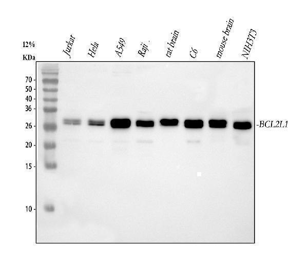
Western blot analysis of Bcl-XL using anti-Bcl-XL antibody (A00181).
Electrophoresis was performed on a 5-20% SDS-PAGE gel at 70V (Stacking gel) / 90V (Resolving gel) for 2-3 hours. The sample well of each lane was loaded with 30 ug of sample under reducing conditions.
Lane 1: human Jurkat whole cell lysates,
Lane 2: human Hela whole cell lysates,
Lane 3: human A549 whole cell lysates,
Lane 4: human Raji whole cell lysates,
Lane 5: rat brain tissue lysates,
Lane 6: rat C6 whole cell lysates,
Lane 7: mouse brain tissue lysates,
Lane 8: mouse NIH/3T3 whole cell lysates.
After electrophoresis, proteins were transferred to a nitrocellulose membrane at 150 mA for 50-90 minutes. Blocked the membrane with 5% non-fat milk/TBS for 1.5 hour at RT. The membrane was incubated with rabbit anti-Bcl-XL antigen affinity purified polyclonal antibody (Catalog # A00181) at 0.5 μg/mL overnight at 4°C, then washed with TBS-0.1%Tween 3 times with 5 minutes each and probed with a goat anti-rabbit IgG-HRP secondary antibody at a dilution of 1:5000 for 1.5 hour at RT. The signal is developed using an Enhanced Chemiluminescent detection (ECL) kit (Catalog # EK1002) with Tanon 5200 system. A specific band was detected for Bcl-XL at approximately 26-30 kDa. The expected band size for Bcl-XL is at 26 kDa.

Click image to see more details
IHC analysis of Bcl-XL using anti-Bcl-XL antibody (A00181).
Bcl-XL was detected in a paraffin-embedded section of human pancrease cancer tissue. Heat mediated antigen retrieval was performed in EDTA buffer (pH 8.0, epitope retrieval solution). The tissue section was blocked with 10% goat serum. The tissue section was then incubated with 2 μg/ml rabbit anti-Bcl-XL Antibody (A00181) overnight at 4°C. Peroxidase Conjugated Goat Anti-rabbit IgG was used as secondary antibody and incubated for 30 minutes at 37°C. The tissue section was developed using HRP Conjugated Rabbit IgG Super Vision Assay Kit (Catalog # SV0002) with DAB as the chromogen.

Click image to see more details
IF analysis of Bcl-XL using anti-Bcl-XL antibody (A00181).
Bcl-XL was detected in an immunocytochemical section of TPC-1 cells. Enzyme antigen retrieval was performed using IHC enzyme antigen retrieval reagent (AR0022) for 15 mins. The cells were blocked with 10% goat serum. And then incubated with 5 μg/mL rabbit anti-Bcl-XL Antibody (A00181) overnight at 4°C. DyLight®488 Conjugated Goat Anti-Rabbit IgG (BA1127) was used as secondary antibody at 1:500 dilution and incubated for 30 minutes at 37°C. The section was counterstained with DAPI. Visualize using a fluorescence microscope and filter sets appropriate for the label used.

Click image to see more details
Immunoprecipitating (IP) Bcl-XL in A549 whole cell lysate.
Western blot analysis of Bcl-XL using anti-Bcl-XL antibody (A00181);
Lane 1: A549 whole cell lysates (30ug);
Lane 2: Rabbit control IgG instead of anti-Bcl-XL antibody in A549 whole cell lysate;
Lane 3: anti-Bcl-XL antibody (2μg) + A549 whole cell lysate (500μg).
After electrophoresis, proteins were transferred to a membrane. Then the membrane was incubated with rabbit anti-Bcl-XL antigen affinity purified polyclonal antibody (A00181) at a dilution of 0.5 μg/mL and probed with a goat anti-rabbit IgG-HRP secondary antibody (Catalog # BA1054). The signal is developed using ECL Plus Western Blotting Substrate (Catalog # AR1196-200). A specific band was detected for Bcl-XL at approximately 26-30 kDa. The expected band size for Bcl-XL is at 26 kDa.

Click image to see more details
Flow Cytometry analysis of Jurkat cells using anti-Bcl-XL antibody (A00181).
Overlay histogram showing Jurkat cells stained with A00181 (Blue line). To facilitate intracellular staining, cells were fixed with 4% paraformaldehyde and permeabilized with permeabilization buffer. The cells were blocked with 10% normal goat serum. And then incubated with rabbit anti-Bcl-XL Antibody (A00181, 1 μg/1x106 cells) for 30 min at 20°C. DyLight®488 conjugated goat anti-rabbit IgG (BA1127, 5-10 μg/1x106 cells) was used as secondary antibody for 30 minutes at 20°C. Isotype control antibody (Green line) was rabbit IgG (1 μg/1x106) used under the same conditions. Unlabelled sample (Red line) was also used as a control.
Specific Publications For Anti-Bcl-XL/BCL2L1 Antibody Picoband® (A00181)
Loading publications
Recommended Resources
Here are featured tools and databases that you might find useful.
- Boster's Pathways Library
- Protein Databases
- Bioscience Research Protocol Resources
- Data Processing & Analysis Software
- Photo Editing Software
- Scientific Literature Resources
- Research Paper Management Tools
- Molecular Biology Software
- Primer Design Tools
- Bioinformatics Tools
- Phylogenetic Tree Analysis
Customer Reviews
Have you used Anti-Bcl-XL/BCL2L1 Antibody Picoband®?
Share your experimental results or join a short interview to earn up to $1,000 in product credits or other rewards.
0 Reviews For Anti-Bcl-XL/BCL2L1 Antibody Picoband®
Customer Q&As
Have a question?
Find answers in Q&As, reviews.
Can't find your answer?
Submit your question
16 Customer Q&As for Anti-Bcl-XL/BCL2L1 Antibody Picoband®
Question
We need using your anti-Bcl-XL/BCL2L1 antibody for negative regulation of neuron apoptotic process studies. Has this antibody been tested with western blotting on rat thymus tissue? We would like to see some validation images before ordering.
Verified Customer
Verified customer
Asked: 2020-02-19
Answer
Thank you for your inquiry. This A00181 anti-Bcl-XL/BCL2L1 antibody is validated on rat thymus tissue, nih3t3 whole cell lysates, hepg2 whole cell lysates, sgc7901 whole cell lysates. It is guaranteed to work for Flow Cytometry, IHC, ICC, WB in human, mouse, rat. Our Boster guarantee will cover your intended experiment even if the sample type has not been be directly tested.
Boster Scientific Support
Answered: 2020-02-19
Question
Will anti-Bcl-XL/BCL2L1 antibody A00181 work for WB with upper lobe of left lung?
Verified Customer
Verified customer
Asked: 2020-01-09
Answer
According to the expression profile of upper lobe of left lung, BCL2L1 is highly expressed in upper lobe of left lung. So, it is likely that anti-Bcl-XL/BCL2L1 antibody A00181 will work for WB with upper lobe of left lung.
Boster Scientific Support
Answered: 2020-01-09
Question
I was wanting to use your anti-Bcl-XL/BCL2L1 antibody for WB for human upper lobe of left lung on frozen tissues, but I want to know if it has been validated for this particular application. Has this antibody been validated and is this antibody a good choice for human upper lobe of left lung identification?
Verified Customer
Verified customer
Asked: 2019-12-13
Answer
It shows on the product datasheet, A00181 anti-Bcl-XL/BCL2L1 antibody has been validated for Flow Cytometry, IHC, ICC, WB on human, mouse, rat tissues. We have an innovator award program that if you test this antibody and show it works in human upper lobe of left lung in IHC-frozen, you can get your next antibody for free.
Boster Scientific Support
Answered: 2019-12-13
Question
We have seen staining in mouse upper lobe of left lung. Any tips? Is anti-Bcl-XL/BCL2L1 antibody supposed to stain upper lobe of left lung positively?
Verified Customer
Verified customer
Asked: 2019-09-13
Answer
According to literature upper lobe of left lung does express BCL2L1. According to Uniprot.org, BCL2L1 is expressed in upper lobe of left lung, colon carcinoma, lung, among other tissues. Regarding which tissues have BCL2L1 expression, here are a few articles citing expression in various tissues:
Colon carcinoma, Pubmed ID: 17974005
Lung, Pubmed ID: 15489334
Boster Scientific Support
Answered: 2019-09-13
Question
I appreciate helping with my inquiry over the phone. Here are the WB image, lot number and protocol we used for upper lobe of left lung using anti-Bcl-XL/BCL2L1 antibody A00181. Let me know if you need anything else.
Verified Customer
Verified customer
Asked: 2019-07-01
Answer
Thank you for the data. You have provided everything we needed. Our lab team are working to resolve your inquiry as quickly as possible, and we appreciate your patience and understanding! Please let me know if there is anything you need in the meantime.
Boster Scientific Support
Answered: 2019-07-01
Question
Would A00181 anti-Bcl-XL/BCL2L1 antibody work on parafin embedded sections? If so, which fixation method do you recommend we use (PFA, paraformaldehyde, other)?
Verified Customer
Verified customer
Asked: 2019-06-25
Answer
As indicated on the product datasheet, A00181 anti-Bcl-XL/BCL2L1 antibody as been tested on WB. It is best to use PFA for fixation because it has better tissue penetration ability. PFA needs to be prepared fresh before use. Long term stored PFA turns into formalin, as the PFA molecules congregate and become formalin.
Boster Scientific Support
Answered: 2019-06-25
Question
My colleagues were satisfied with the WB result of your anti-Bcl-XL/BCL2L1 antibody. However we have been able to see positive staining in lung isoform bcl-x(l): mitochondrion inner using this antibody. Is that expected? Could you tell me where is BCL2L1 supposed to be expressed?
Verified Customer
Verified customer
Asked: 2018-02-05
Answer
According to literature, lung does express BCL2L1. Generally BCL2L1 expresses in isoform bcl-x(l): mitochondrion inner. Regarding which tissues have BCL2L1 expression, here are a few articles citing expression in various tissues:
Colon carcinoma, Pubmed ID: 17974005
Lung, Pubmed ID: 15489334
Boster Scientific Support
Answered: 2018-02-05
Question
Is this A00181 anti-Bcl-XL/BCL2L1 antibody reactive to the isotypes of BCL2L1?
Verified Customer
Verified customer
Asked: 2017-12-06
Answer
The immunogen of A00181 anti-Bcl-XL/BCL2L1 antibody is E.coli-derived human Bcl-XL recombinant protein (Position: M1-T219). Human Bcl-XL shares 97.9% amino acid (aa) sequence identity with both mouse and rat Bcl-XL. Could you tell me which isotype you are interested in so I can help see if the immunogen is part of this isotype?
Boster Scientific Support
Answered: 2017-12-06
Question
See below the WB image, lot number and protocol we used for upper lobe of left lung using anti-Bcl-XL/BCL2L1 antibody A00181. Please let me know if you require anything else.
G. Singh
Verified customer
Asked: 2017-09-05
Answer
Thank you very much for the data. Our lab team are working to resolve this as quickly as possible, and we appreciate your patience and understanding! You have provided everything we needed. Please let me know if there is anything you need in the meantime.
Boster Scientific Support
Answered: 2017-09-05
Question
I would like to test anti-Bcl-XL/BCL2L1 antibody A00181 on human upper lobe of left lung for research purposes, then I may be interested in using anti-Bcl-XL/BCL2L1 antibody A00181 for diagnostic purposes as well. Is the antibody suitable for diagnostic purposes?
Verified Customer
Verified customer
Asked: 2017-08-07
Answer
The products we sell, including anti-Bcl-XL/BCL2L1 antibody A00181, are only intended for research use. They would not be suitable for use in diagnostic work. If you have the means to develop a product into diagnostic use, and are interested in collaborating with us and develop our product into an IVD product, please contact us for more discussions.
Boster Scientific Support
Answered: 2017-08-07
Question
We are currently using anti-Bcl-XL/BCL2L1 antibody A00181 for mouse tissue, and we are content with the IHC results. The species of reactivity given in the datasheet says human, mouse, rat. Is it likely that the antibody can work on zebrafish tissues as well?
Verified Customer
Verified customer
Asked: 2017-07-31
Answer
The anti-Bcl-XL/BCL2L1 antibody (A00181) has not been tested for cross reactivity specifically with zebrafish tissues, but there is a good chance of cross reactivity. We have an innovator award program that if you test this antibody and show it works in zebrafish you can get your next antibody for free. Please contact me if I can help you with anything.
Boster Scientific Support
Answered: 2017-07-31
Question
We ordered your anti-Bcl-XL/BCL2L1 antibody for Flow Cytometry on upper lobe of left lung in the past. I am using mouse, and We want to use the antibody for WB next. My question regards examining upper lobe of left lung as well as colon carcinoma in our next experiment. Could you please give me some suggestion on which antibody would work the best for WB?
K. Wu
Verified customer
Asked: 2017-05-09
Answer
I looked at the website and datasheets of our anti-Bcl-XL/BCL2L1 antibody and it appears that A00181 has been validated on mouse in both Flow Cytometry and WB. Thus A00181 should work for your application. Our Boster satisfaction guarantee will cover this product for WB in mouse even if the specific tissue type has not been validated. We do have a comprehensive range of products for WB detection and you can check out our website bosterbio.com to find out more information about them.
Boster Scientific Support
Answered: 2017-05-09
Question
Is a blocking peptide available for product anti-Bcl-XL/BCL2L1 antibody (A00181)?
K. Jackson
Verified customer
Asked: 2015-05-08
Answer
We do provide the blocking peptide for product anti-Bcl-XL/BCL2L1 antibody (A00181). If you would like to place an order for it please contact support@bosterbio.com and make a special request.
Boster Scientific Support
Answered: 2015-05-08
Question
Can you help my question with product A00181, anti-Bcl-XL/BCL2L1 antibody. I was wondering if it would be possible to conjugate this antibody with biotin. I would need it to be without BSA or sodium azide. I am planning on using a buffer exchange of sodium azide with PBS only. Would there be problems for me to conjugate the antibody and store it in -20 degrees in small aliquots?
M. Carter
Verified customer
Asked: 2014-12-17
Answer
We suggest not storing this antibody with PBS buffer only in -20 degrees. If you want to store it in -20 degrees it is best to add some cryoprotectant like glycerol. If you want carrier free A00181 anti-Bcl-XL/BCL2L1 antibody, we can provide it to you in a special formula with trehalose and/or glycerol. These molecules will not interfere with conjugation chemistry and provide a good level of protection for the antibody from degradation. Please be sure to specify this in your purchase order.
Boster Scientific Support
Answered: 2014-12-17
Question
I see that the anti-Bcl-XL/BCL2L1 antibody A00181 works with WB, what is the protocol used to produce the result images on the product page?
J. Patel
Verified customer
Asked: 2014-05-12
Answer
You can find protocols for WB on the "support/technical resources" section of our navigation menu. If you have any further questions, please send an email to support@bosterbio.com
Boster Scientific Support
Answered: 2014-05-12
Question
Is there a BSA free version of anti-Bcl-XL/BCL2L1 antibody A00181 available?
T. Parker
Verified customer
Asked: 2013-01-08
Answer
We appreciate your recent telephone inquiry. I can confirm that some lots of this anti-Bcl-XL/BCL2L1 antibody A00181 are BSA free. For now, these lots are available and we can make a BSA free formula for you free of charge. It will take 3 extra days to prepare. If you require this antibody BSA free again in future, please do not hesitate to contact me and I will be pleased to check which lots we have in stock that are BSA free.
Boster Scientific Support
Answered: 2013-01-08
