This website uses cookies to ensure you get the best experience on our website.
- Table of Contents
74 Citations 17 Q&As
312 Citations 16 Q&As
42 Citations 16 Q&As


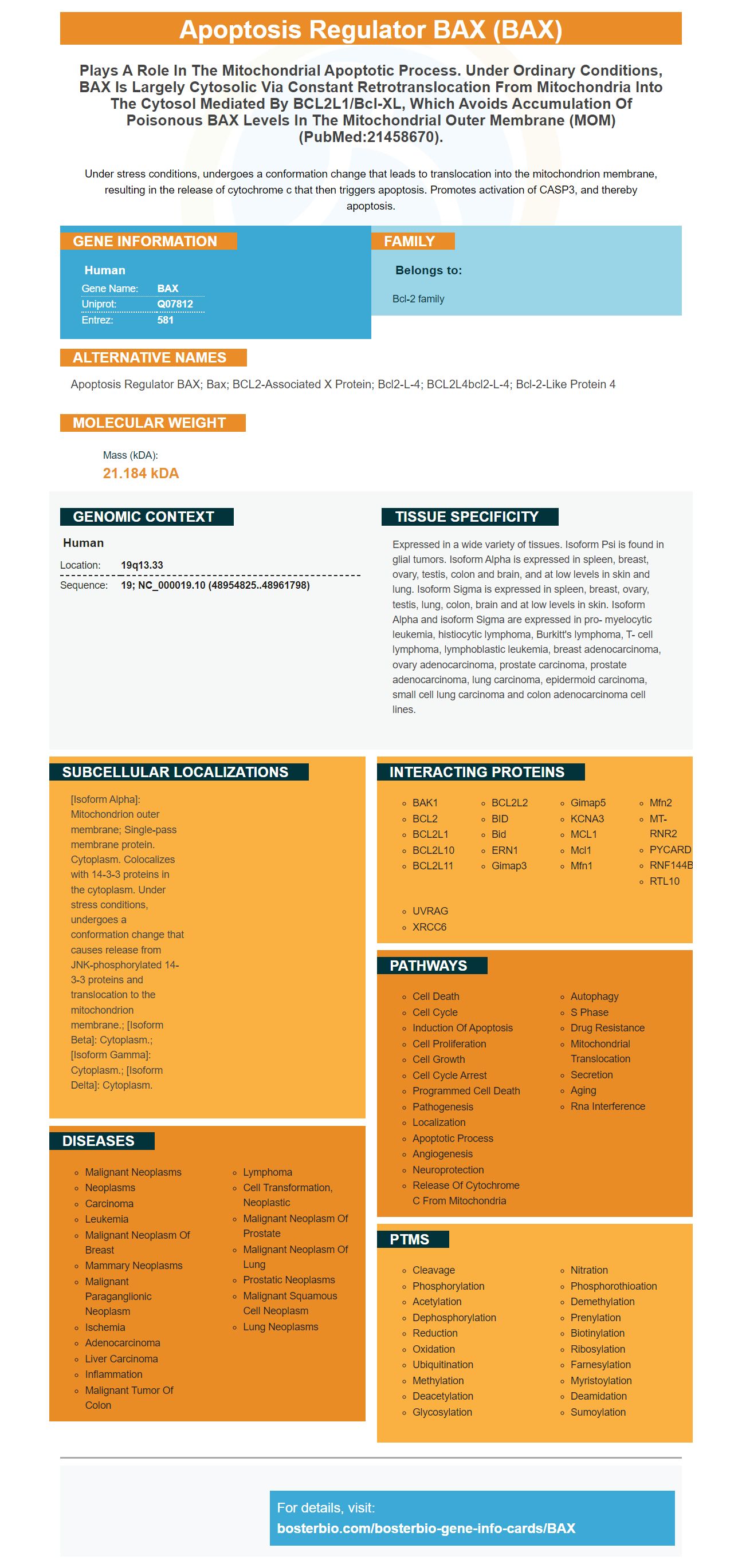
Facts about Apoptosis regulator BAX.

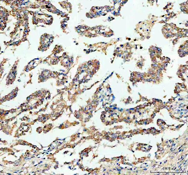
Under stress conditions, undergoes a conformation change that leads to translocation into the mitochondrion membrane, resulting in the release of cytochrome c that then triggers apoptosis. Promotes activation of CASP3, and thereby apoptosis.
| Human | |
|---|---|
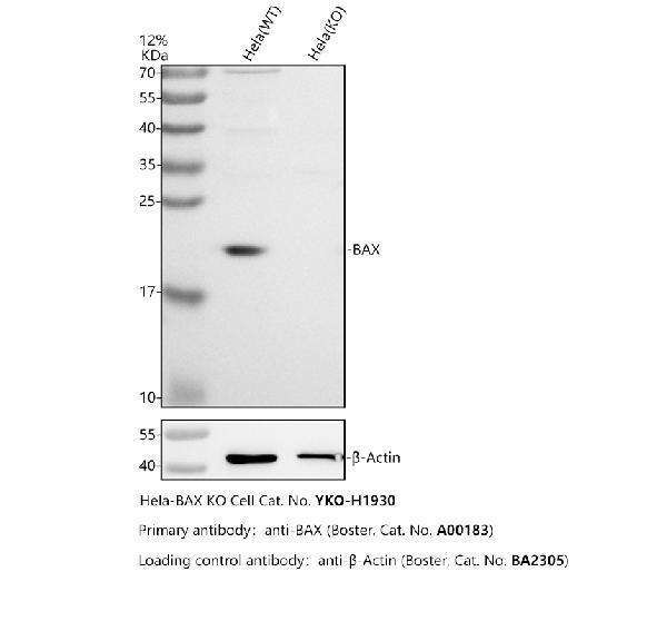
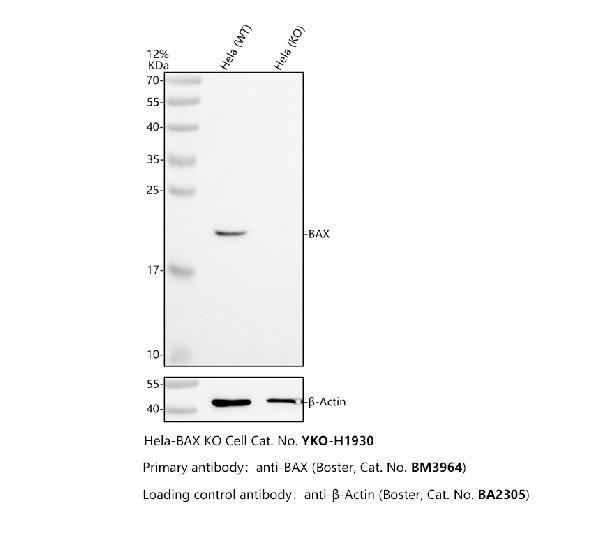
| Gene Name: | BAX |
| Uniprot: | Q07812 |
| Entrez: | 581 |

| Belongs to: |
|---|
| Bcl-2 family |

apoptosis regulator BAX; Bax; BCL2-associated X protein; Bcl2-L-4; BCL2L4bcl2-L-4; Bcl-2-like protein 4
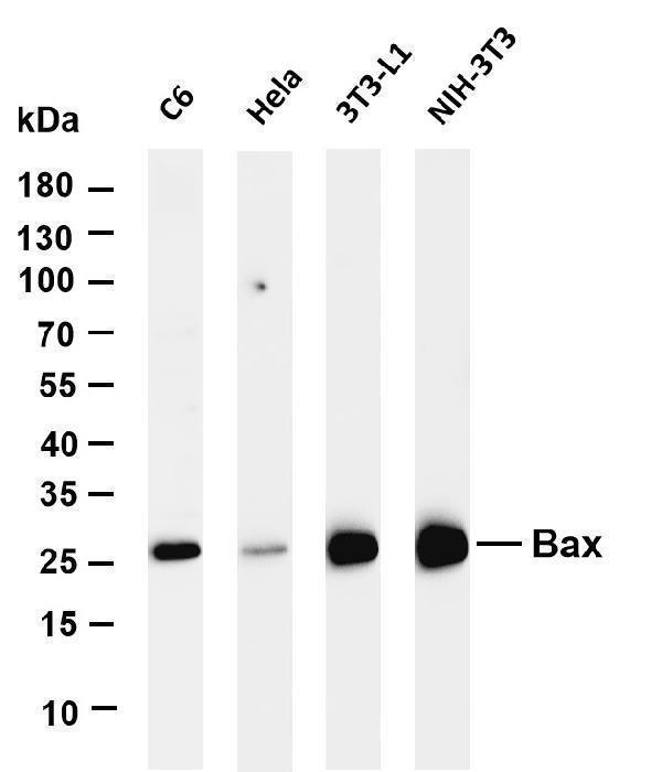
Mass (kDA):
21.184 kDA

| Human | |
|---|---|
| Location: | 19q13.33 |
| Sequence: | 19; NC_000019.10 (48954825..48961798) |
Expressed in a wide variety of tissues. Isoform Psi is found in glial tumors. Isoform Alpha is expressed in spleen, breast, ovary, testis, colon and brain, and at low levels in skin and lung. Isoform Sigma is expressed in spleen, breast, ovary, testis, lung, colon, brain and at low levels in skin. Isoform Alpha and isoform Sigma are expressed in pro- myelocytic leukemia, histiocytic lymphoma, Burkitt's lymphoma, T- cell lymphoma, lymphoblastic leukemia, breast adenocarcinoma, ovary adenocarcinoma, prostate carcinoma, prostate adenocarcinoma, lung carcinoma, epidermoid carcinoma, small cell lung carcinoma and colon adenocarcinoma cell lines.
[Isoform Alpha]: Mitochondrion outer membrane; Single-pass membrane protein. Cytoplasm. Colocalizes with 14-3-3 proteins in the cytoplasm. Under stress conditions, undergoes a conformation change that causes release from JNK-phosphorylated 14-3-3 proteins and translocation to the mitochondrion membrane.; [Isoform Beta]: Cytoplasm.; [Isoform Gamma]: Cytoplasm.; [Isoform Delta]: Cytoplasm.


PMID: 8358790 by Oltvai Z.N., et al. Bcl-2 heterodimerizes in vivo with a conserved homolog, Bax, that accelerates programmed cell death.
PMID: 7607685 by Apte S.S., et al. Mapping of the human BAX gene to chromosome 19q13.3-q13.4 and isolation of a novel alternatively spliced transcript, BAX delta.
*Showing only the more recent 20. More publications can be found for each product on its corresponding product page