Click image to see more details
-

-
-
-
-
+6
Product Info Summary
| SKU: | PB9075 |
|---|---|
| Size: | 100 μg/vial |
| Reactive Species: | Human, Mouse, Rat |
| Host: | Rabbit |
| Application: | IHC, WB |
Customers Who Bought This Also Bought

Product info
Product Name
Anti-BDNF Antibody Picoband®
SKU/Catalog Number
PB9075
Size
100 μg/vial
Form
Lyophilized
Description
Boster Bio Anti-BDNF Antibody Picoband® catalog # PB9075. Tested in IHC, WB applications. This antibody reacts with Human, Mouse, Rat. The brand Picoband indicates this is a premium antibody that guarantees superior quality, high affinity, and strong signals with minimal background in Western blot applications. Only our best-performing antibodies are designated as Picoband, ensuring unmatched performance.
Storage & Handling
Store at -20˚C for one year from date of receipt. After reconstitution, at 4˚C for one month. It can also be aliquotted and stored frozen at -20˚C for six months. Avoid repeated freeze-thaw cycles.
Cite This Product
Anti-BDNF Antibody Picoband® (Boster Biological Technology, Pleasanton CA, USA, Catalog # PB9075)
Host
Rabbit
Contents
Each vial contains 4 mg Trehalose, 0.9 mg NaCl and 0.2 mg Na2HPO4.
Clonality
Polyclonal
Isotype
Rabbit IgG
Immunogen
Anti-BDNF Picoband™ Antibody (PB9075) was raised against E.coli-derived human BDNF recombinant protein (Position: H129-R247). Human BDNF shares 100% amino acid (aa) sequence identity with both mouse and rat BDNF.
Cross-reactivity
No cross-reactivity with other proteins
Reactive Species
PB9075 is reactive to Bdnf in Human, Mouse, Rat
Observed Molecular Weight
14 kDa
Calculated molecular weight
28.1 kDa
Background of Bdnf
Brain-derived neurotrophic factor, also known as BDNF, is a secreted protein that, in humans, is encoded by the BDNF gene. BDNF is a member of the neurotrophin family of growth factors, which are related to the canonical nerve growth factor. It is mapped to 11p14.1. BDNF is a prosurvival factor induced by cortical neurons that is necessary for survival of striatal neurons in the brain. It is expressed within peripheral ganglia and is not restricted to neuronal target fields. BDNF has been purified and shown to reduce the amount of naturally occurring neuronal cell death in portions of the peripheral nervous system.
Antibody Validation
Boster validates all antibodies on WB, IHC, ICC, Immunofluorescence, and ELISA with known positive control and negative samples to ensure specificity and high affinity, including thorough antibody incubations.
Application & Images
Applications
PB9075 is guaranteed for IHC, WB Boster Guarantee
Recommend Dilution
| Application | Dilution | Species |
|---|---|---|
| Western blot | 0.1-0.5μg/ml | Human, Mouse, Rat |
| Immunohistochemistry (Paraffin-embedded Section) | 2-5μg/ml | Mouse, Rat |
Tested application
Suggested blocking solution with 5% non-fat milk or BSA; (*)Recommended protein loading: 20-40 µg per lane
Use TE buffer pH 9.0 for antigen retrieval; (*) citrate buffer pH 6.0 is an alternative.
Validation Images & Assay Conditions
Click image to see more details
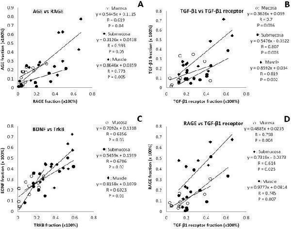
( A ) Correlation between AGE and RAGE in different layers; ( B ) Correlation between TGF-β1 and TGF-β1receptor in different layers; ( C ) Correlation between BDNF and TrkB in different layers; ( D ) Correlation between RAGE and TGF-β1receptor in different layers.
Index in PubMed under a CC BY license. PMID: 29930382

Click image to see more details
( A ) Correlation between AGE and RAGE in muscle layer and submucosa layer with circumferential constant a; ( B ) Correlation between AGE and RAGE in muscle layer and submucosa layer with longitudinal constant a; ( C ) Correlation between TGF-β1 and TGF-β1 receptor in mucosa layer and TGF-β1 muscle layer with circumferential and longitudinal material constant a; ( D ) Correlation between BDNF in muscle and submucosa layers with longitudinal constant a.
Index in PubMed under a CC BY license. PMID: 29930382

Click image to see more details
The fraction of AGE, RAGE, TGF-β1, TGF- β1 receptor, BDNF and TrkB in the different layers of the colon between two groups. In the different layers, the fraction of AGE, RAGE, TGF-β1 and TGF- β1 receptor was bigger whereas the fraction of BDNF and TrkB was smaller in the Diabetes group than in Control group. Compared with Control group: *P < 0.05, **P < 0.01.
Index in PubMed under a CC BY license. PMID: 29930382

Click image to see more details
The representative samples of immunohistochemical staining for AGE, RAGE, TGF-β1, TGF- β1 receptor, BDNF and TrkB in the colon wall of two groups. The microscopy with high magnification have been inserted in each single histological photo (arrow) in order to display the localization of markers. The staining of all proteins was stronger in the muscle layer than other layers. In the different layers, the staining of AGE, RAGE, TGF-β1 and TGF- β1 receptor was stronger whereas the staining of BDNF and TrkB was weaker in the Diabetes group than in Control group. Bar = 100 um.
Index in PubMed under a CC BY license. PMID: 29930382

Click image to see more details
SPION-mediated magnetic actuation upregulates the expression of neurotrophic factors associated with repair phenotypes in Schwann cells. The protein expression of repair phenotype-related neurotrophic factors BDNF, GDNF, Olig1 and VEGF in different experimental groups was detected by immunohistochemical staining at 3 (a) , 7 (d) , 14 (g) and 21 days (j) after crush injury, and the protein expression levels were quantitatively analyzed ( b , e , h , k ). c , f , i , l The protein expression levels of such neurotrophic factors at the above time points were detected by ELISA, and the results were consistent with the immunohistochemical analysis. Each experiment was carried out in triplicate. The values are represented as the mean ± SD. Scale bar = 50 µm in panels a , d , g , j . * P < 0.05, ** P < 0.01
Index in PubMed under a CC BY license. PMID: 35351151

Click image to see more details
IHC analysis of BDNF using anti-BDNF antibody (PB9075).
BDNF was detected in a paraffin-embedded section of human brain tissue. Heat mediated antigen retrieval was performed in EDTA buffer (pH 8.0, epitope retrieval solution). The tissue section was blocked with 10% goat serum. The tissue section was then incubated with 2 μg/ml rabbit anti-BDNF Antibody (PB9075) overnight at 4°C. Peroxidase Conjugated Goat Anti-rabbit IgG was used as secondary antibody and incubated for 30 minutes at 37°C. The tissue section was developed using HRP Conjugated Rabbit IgG Super Vision Assay Kit (Catalog # SV0002) with DAB as the chromogen.

Click image to see more details
Western blot analysis of BDNF using anti-BDNF antibody (PB9075).
Electrophoresis was performed on a 12% SDS-PAGE gel at 80V (Stacking gel) / 120V (Resolving gel) for 2 hours.
Lane 1: recombinant human BDNF protein 5 ng,
Lane 2: recombinant human BDNF protein 2.5 ng.
After electrophoresis, proteins were transferred to a nitrocellulose membrane at 150 mA for 50-90 minutes. Blocked the membrane with 5% non-fat milk/TBS for 1.5 hour at RT. The membrane was incubated with rabbit anti-BDNF antigen affinity purified polyclonal antibody (PB9075) at 0.5 μg/mL overnight at 4°C, then washed with TBS-0.1%Tween 3 times with 5 minutes each and probed with a goat anti-rabbit IgG-HRP secondary antibody (Catalog # BA1054) at a dilution of 1:5000 for 1.5 hour at RT. The signal is developed using an ECL Plus Western Blotting Substrate (Catalog # AR1196-200) with Tanon 5200 system. A specific band was detected for BDNF at approximately 14 kDa.

Click image to see more details
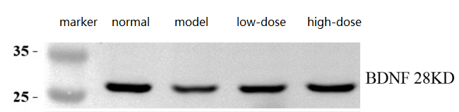
Western blot analysis of BDNF using anti-BDNF antibody (PB9075).
Electrophoresis was performed on a 12% SDS-PAGE gel at 80V (Stacking gel) / 120V (Resolving gel) for 2 hours.
Lane 1: normal group-Hippocampal tissue lysates from normal mouse tissue lysates,
Lane 2: model group-Hippocampal tissue lysates from depressed mouse,
Lane 3: low-dose group-Hippocampal tissue lysates from depressed mouse treated with an in-house drug,
Lane 4: high-dose group-Hippocampal tissue lysates from depressed mouse treated with an in-house drug.
After electrophoresis, proteins were transferred to a nitrocellulose membrane at 150 mA for 50-90 minutes. Blocked the membrane with 5% non-fat milk/TBS for 1.5 hour at RT. The membrane was incubated with rabbit anti-BDNF antigen affinity purified polyclonal antibody (PB9075) at 1:2000 overnight at 4°C, then washed with TBS-0.1%Tween 3 times with 5 minutes each and probed with a goat anti-rabbit IgG-HRP secondary antibody (Catalog # BA1054) at a dilution of 1:10000 for 1 hour at RT. The signal is developed using an ECL Plus Western Blotting Substrate (Catalog # AR1196-200) with Tanon 5200 system. A specific band was detected for BDNF at approximately 28 kDa.

Click image to see more details
IHC analysis of BDNF using anti-BDNF antibody (PB9075).
BDNF was detected in a paraffin-embedded section of mouse brain tissue. Heat mediated antigen retrieval was performed in EDTA buffer (pH 8.0, epitope retrieval solution). The tissue section was blocked with 10% goat serum. The tissue section was then incubated with 2 μg/ml rabbit anti-BDNF Antibody (PB9075) overnight at 4°C. Peroxidase Conjugated Goat Anti-rabbit IgG was used as secondary antibody and incubated for 30 minutes at 37°C. The tissue section was developed using HRP Conjugated Rabbit IgG Super Vision Assay Kit (Catalog # SV0002) with DAB as the chromogen.

Click image to see more details
IHC analysis of BDNF using anti-BDNF antibody (PB9075).
BDNF was detected in a paraffin-embedded section of rat brain tissue. Heat mediated antigen retrieval was performed in EDTA buffer (pH 8.0, epitope retrieval solution). The tissue section was blocked with 10% goat serum. The tissue section was then incubated with 2 μg/ml rabbit anti-BDNF Antibody (PB9075) overnight at 4°C. Peroxidase Conjugated Goat Anti-rabbit IgG was used as secondary antibody and incubated for 30 minutes at 37°C. The tissue section was developed using HRP Conjugated Rabbit IgG Super Vision Assay Kit (Catalog # SV0002) with DAB as the chromogen.
Specific Publications For Anti-BDNF Antibody Picoband® (PB9075)
Loading publications
Recommended Resources
Here are featured tools and databases that you might find useful.
- Boster's Pathways Library
- Protein Databases
- Bioscience Research Protocol Resources
- Data Processing & Analysis Software
- Photo Editing Software
- Scientific Literature Resources
- Research Paper Management Tools
- Molecular Biology Software
- Primer Design Tools
- Bioinformatics Tools
- Phylogenetic Tree Analysis
Customer Reviews
Have you used Anti-BDNF Antibody Picoband®?
Share your experimental results or join a short interview to earn up to $1,000 in product credits or other rewards.
1 Reviews For Anti-BDNF Antibody Picoband®
Anti-BDNF (PB9075) shows clear, specific bands in mouse brain tissues by WB, with BDNF levels markedly reduced in depressed mice and restored after treatment, demonstrating excellent antibody performance.
Excellent
| SKU | PB9075 |
|---|---|
| Application | Western Blot |
| Sample | mouse brain tissue |
| Sample Processing Description | ① Hippocampal tissue from normal mice, ② Hippocampal tissue from depressed mice, ③ Hippocampal tissue from depressed mice treated with our in-house drug, ④ Hippocampal tissue from depressed mice treated with a positive control drug. Total protein was extracted from all samples. |
| Other Reagents | RIPA lysis buffer, Protease inhibitor, Running buffer, Transfer buffer, Blocking buffer |
| Primary Antibody | BDNF Antibody Picoband® |
| Primary Incubation | 1:2000, overnight at 4 ℃ |
| Secondary Antibody | HRP Conjugated AffiniPure Goat Anti-Rabbit IgG (H+L) (BA1054) |
| Secondary Incubation | 1:10000, 1 h in RT |
| Detection | Substrate: ECL substrate, Image system: ChemiDoc MP |
| Results Summary | BDNF belongs to the neurotrophin family and functions by binding to the TrkB receptor on the surface of neurons, activating intracellular signaling pathways to maintain, protect, and enhance neuronal function. In simple terms, it helps brain cells stay healthier, more active, and better connected. Experimental results show that BDNF levels are significantly decreased in the brains of depressed mice, but are restored after treatment. |
Qi Ding, Shandong First Medical University
Verified customer
Submitted 2026-03-26
Customer Q&As
Have a question?
Find answers in Q&As, reviews.
Can't find your answer?
Submit your question
1 Customer Q&As for Anti-BDNF Antibody Picoband®
Question
We are currently using anti-BDNF antibody PB9075 for human tissue, and we are satisfied with the ICC results. The species of reactivity given in the datasheet says human, mouse, rat. Is it possible that the antibody can work on goat tissues as well?
Verified Customer
Verified customer
Asked: 2019-07-18
Answer
The anti-BDNF antibody (PB9075) has not been tested for cross reactivity specifically with goat tissues, though there is a good chance of cross reactivity. We have an innovator award program that if you test this antibody and show it works in goat you can get your next antibody for free. Please contact me if I can help you with anything.
Boster Scientific Support
Answered: 2019-07-18
