Click image to see more details
-

-
-
-
-
+3
Product Info Summary
| SKU: | M00327-2 |
|---|---|
| Size: | 100 μg/vial |
| Reactive Species: | Human, Mouse, Rat |
| Host: | Mouse |
| Application: | Flow Cytometry, IF, IHC, ICC, WB |
Customers Who Bought This Also Bought

Product info
Product Name
Anti-Beclin 1 Antibody Picoband® (monoclonal, 2D12A3)
SKU/Catalog Number
M00327-2
Size
100 μg/vial
Form
Lyophilized
Description
Boster Bio Anti-Beclin 1 Antibody Picoband® (monoclonal, 2D12A3) catalog # M00327-2. Tested in Flow Cytometry, IF, IHC, ICC, WB applications. This antibody reacts with Human, Mouse, Rat. The brand Picoband indicates this is a premium antibody that guarantees superior quality, high affinity, and strong signals with minimal background in Western blot applications. Only our best-performing antibodies are designated as Picoband, ensuring unmatched performance.
Storage & Handling
At -20°C for one year from date of receipt. After reconstitution, at 4°C for one month. It can also be aliquotted and stored frozen at -20°C for six months. Avoid repeated freezing and thawing.
Cite This Product
Anti-Beclin 1 Antibody Picoband® (monoclonal, 2D12A3) (Boster Biological Technology, Pleasanton CA, USA, Catalog # M00327-2)
Host
Mouse
Contents
Each vial contains 4 mg Trehalose, 0.9 mg NaCl and 0.2 mg Na2HPO4.
Clonality
Monoclonal
Clone Number
2D12A3
Isotype
Mouse IgG1
Immunogen
E.coli-derived human Beclin 1 recombinant protein (Position: M1-S354). Human Beclin 1 shares 97% amino acid (aa) sequence identity with both mouse and rat Beclin 1.
Cross-reactivity
No cross-reactivity with other proteins.
Reactive Species
M00327-2 is reactive to BECN1 in Human, Mouse, Rat
Observed Molecular Weight
52-60 kDa
Calculated molecular weight
51.9 kDa
Background of BECN1
Beclin-1, also known as also known as ATG6 or VPS30 is a protein that in humans is encoded by the BECN1 gene. Beclin-1 and its binding partner class III phosphoinositide 3-kinase (PI3K), also named Vps34, are required for the initiation of the formation of the autophagosome in autophagy. This gene participates in the regulation of autophagy and has an important role in development, tumorigenesis, and neurodegeneration. Schizophrenia is associated with low levels of Beclin-1 in the hippocampus of the affected which causes diminished autophagywhich in turn results in increased neuronal cell death. It has been found that beclin-1 can promote autophagy in autophagy-defective yeast with a targeted disruption of apg6/vps30, and in human MCF7 breast carcinoma cells.
Antibody Validation
Boster validates all antibodies on WB, IHC, ICC, Immunofluorescence, and ELISA with known positive control and negative samples to ensure specificity and high affinity, including thorough antibody incubations.
Application & Images
Applications
M00327-2 is guaranteed for Flow Cytometry, IF, IHC, ICC, WB Boster Guarantee
Recommend Dilution
| Application | Dilution | Species |
|---|---|---|
| Western Blot (WB) | 0.25-0.5 μg/ml | Human, Mouse, Rat |
| Immunohistochemistry (IHC) | 2-5 μg/ml | Human |
| Immunofluorescence (IF) | 5 μg/ml | Human |
| Flow Cytometry (FC) | 1-3 μg/1x106 cells | Human |
Tested application
Suggested blocking solution with 5% non-fat milk or BSA; (*)Recommended protein loading: 20-40 µg per lane
Use TE buffer pH 9.0 for antigen retrieval; (*) citrate buffer pH 6.0 is an alternative.
Validation Images & Assay Conditions
Click image to see more details
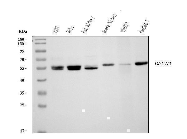
Western blot analysis of Beclin 1 using anti-Beclin 1 antibody (M00327-2).
Electrophoresis was performed on a 5-20% SDS-PAGE gel at 70V (Stacking gel) / 90V (Resolving gel) for 2-3 hours. The sample well of each lane was loaded with 30 ug of sample under reducing conditions.
Lane 1: human 293T whole cell lysates,
Lane 2: human Hela whole cell lysates,
Lane 3: rat kidney tissue lysates,
Lane 4: mouse kidney tissue lysates,
Lane 5: mouse NIH/3T3 whole cell lysates,
Lane 6: mouse RAW264.7 whole cell lysates.
After electrophoresis, proteins were transferred to a nitrocellulose membrane at 150 mA for 50-90 minutes. Blocked the membrane with 5% non-fat milk/TBS for 1.5 hour at RT. The membrane was incubated with mouse anti-Beclin 1 antigen affinity purified monoclonal antibody (Catalog # M00327-2) at 0.5 μg/mL overnight at 4°C, then washed with TBS-0.1%Tween 3 times with 5 minutes each and probed with a goat anti-mouse IgG-HRP secondary antibody at a dilution of 1:10000 for 1.5 hour at RT. The signal is developed using an Enhanced Chemiluminescent detection (ECL) kit (Catalog # EK1001) with Tanon 5200 system. A specific band was detected for Beclin 1 at approximately 52-60 kDa. The expected band size for Beclin 1 is at 52 kDa.

Click image to see more details
IHC analysis of Beclin 1 using anti-Beclin 1 antibody (M00327-2).
Beclin 1 was detected in a paraffin-embedded section of human breast cancer tissue. Heat mediated antigen retrieval was performed in EDTA buffer (pH 8.0, epitope retrieval solution). The tissue section was blocked with 10% goat serum. The tissue section was then incubated with 2 μg/ml mouse anti-Beclin 1 Antibody (M00327-2) overnight at 4°C. Peroxidase Conjugated Goat Anti-mouse IgG was used as secondary antibody and incubated for 30 minutes at 37°C. The tissue section was developed using HRP Conjugated Mouse IgG Super Vision Assay Kit (Catalog # SV0001) with DAB as the chromogen.

Click image to see more details
IHC analysis of Beclin 1 using anti-Beclin 1 antibody (M00327-2).
Beclin 1 was detected in a paraffin-embedded section of human hepatocellular carcinoma tissue. Heat mediated antigen retrieval was performed in EDTA buffer (pH 8.0, epitope retrieval solution). The tissue section was blocked with 10% goat serum. The tissue section was then incubated with 2 μg/ml mouse anti-Beclin 1 Antibody (M00327-2) overnight at 4°C. Peroxidase Conjugated Goat Anti-mouse IgG was used as secondary antibody and incubated for 30 minutes at 37°C. The tissue section was developed using HRP Conjugated Mouse IgG Super Vision Assay Kit (Catalog # SV0001) with DAB as the chromogen.

Click image to see more details
IHC analysis of Beclin 1 using anti-Beclin 1 antibody (M00327-2).
Beclin 1 was detected in a paraffin-embedded section of human lung cancer tissue. Heat mediated antigen retrieval was performed in EDTA buffer (pH 8.0, epitope retrieval solution). The tissue section was blocked with 10% goat serum. The tissue section was then incubated with 2 μg/ml mouse anti-Beclin 1 Antibody (M00327-2) overnight at 4°C. Peroxidase Conjugated Goat Anti-mouse IgG was used as secondary antibody and incubated for 30 minutes at 37°C. The tissue section was developed using HRP Conjugated Mouse IgG Super Vision Assay Kit (Catalog # SV0001) with DAB as the chromogen.

Click image to see more details
IHC analysis of Beclin 1 using anti-Beclin 1 antibody (M00327-2).
Beclin 1 was detected in a paraffin-embedded section of human colonic adenoma tissue. Heat mediated antigen retrieval was performed in EDTA buffer (pH 8.0, epitope retrieval solution). The tissue section was blocked with 10% goat serum. The tissue section was then incubated with 2 μg/ml mouse anti-Beclin 1 Antibody (M00327-2) overnight at 4°C. Peroxidase Conjugated Goat Anti-mouse IgG was used as secondary antibody and incubated for 30 minutes at 37°C. The tissue section was developed using HRP Conjugated Mouse IgG Super Vision Assay Kit (Catalog # SV0001) with DAB as the chromogen.

Click image to see more details
IF analysis of Beclin 1 using anti-Beclin 1 antibody (M00327-2).
Beclin 1 was detected in an immunocytochemical section of HepG2 cells. Enzyme antigen retrieval was performed using IHC enzyme antigen retrieval reagent (AR0022) for 15 mins. The cells were blocked with 10% goat serum. And then incubated with 5 μg/mL mouse anti-Beclin 1 Antibody (M00327-2) overnight at 4°C. DyLight®488 Conjugated Goat Anti-Mouse IgG (BA1126) was used as secondary antibody at 1:100 dilution and incubated for 30 minutes at 37°C. The section was counterstained with DAPI. Visualize using a fluorescence microscope and filter sets appropriate for the label used.

Click image to see more details
Flow Cytometry analysis of PC-3 cells using anti-Beclin 1 antibody (M00327-2).
Overlay histogram showing PC-3 cells stained with M00327-2 (Blue line). To facilitate intracellular staining, cells were fixed with 4% paraformaldehyde and permeabilized with permeabilization buffer. The cells were blocked with 10% normal goat serum. And then incubated with mouse anti-Beclin 1 Antibody (M00327-2, 1 μg/1x106 cells) for 30 min at 20°C. DyLight®488 conjugated goat anti-mouse IgG (BA1126, 5-10 μg/1x106 cells) was used as secondary antibody for 30 minutes at 20°C. Isotype control antibody (Green line) was mouse IgG (1 μg/1x106) used under the same conditions. Unlabelled sample without incubation with primary antibody and secondary antibody (Red line) was used as a blank control.
Specific Publications For Anti-Beclin 1 Antibody Picoband® (monoclonal, 2D12A3) (M00327-2)
Loading publications
Recommended Resources
Here are featured tools and databases that you might find useful.
- Boster's Pathways Library
- Protein Databases
- Bioscience Research Protocol Resources
- Data Processing & Analysis Software
- Photo Editing Software
- Scientific Literature Resources
- Research Paper Management Tools
- Molecular Biology Software
- Primer Design Tools
- Bioinformatics Tools
- Phylogenetic Tree Analysis
Customer Reviews
Have you used Anti-Beclin 1 Antibody Picoband® (monoclonal, 2D12A3)?
Share your experimental results or join a short interview to earn up to $1,000 in product credits or other rewards.
0 Reviews For Anti-Beclin 1 Antibody Picoband® (monoclonal, 2D12A3)
Customer Q&As
Have a question?
Find answers in Q&As, reviews.
Can't find your answer?
Submit your question
