Click image to see more details
Product Info Summary
| SKU: | A01263 |
|---|---|
| Size: | 200ug |
| Reactive Species: | Human, Mouse, Leopard frog |
| Host: | Rabbit |
| Application: | ELISA, IF, WB |
Customers Who Bought This Also Bought

Product info
Product Name
Anti-beta Actin ACTB Antibody
SKU/Catalog Number
A01263
Size
200ug
Form
Liquid (sterile filtered)
Description
Boster Bio Anti-beta Actin ACTB Antibody (Catalog # A01263). Tested in ELISA, IF, WB applications. This antibody reacts with Human, Mouse, Leopard Frog.
Storage & Handling
Store beta-Actin Loading Control Antibody at -20°C prior to opening. Aliquot contents and freeze at -20°C or below for extended storage. Avoid cycles of freezing and thawing. Centrifuge product if not completely clear after standing at room temperature. This product is stable for several weeks at 4°C as an undiluted liquid. Dilute only prior to immediate use. Expiration date is one (1) year from date of opening. (Ship on dry ice.)
Cite This Product
Anti-beta Actin ACTB Antibody (Boster Biological Technology, Pleasanton CA, USA, Catalog # A01263)
Host
Rabbit
Contents
0.02 M Potassium Phosphate, 0.15 M Sodium Chloride, pH 7.2, 0.01% (w/v) Sodium Azide
Clonality
Polyclonal
Isotype
IgG
Immunogen
beta-Actin Loading Control Antibody was prepared from whole rabbit serum produced by repeated immunizations with a synthetic peptide corresponding to C-Terminal region near amino acids 350-375 of Human beta Actin.
Cross-reactivity
No cross reactivity with other proteins.
Reactive Species
A01263 is reactive to ACTB in Human, Mouse, Leopard frog
Observed Molecular Weight
68 kDa
Calculated molecular weight
41.7 kDa
Background of ACTB
In vertebrates 3 main groups of actin isoforms, alpha, beta and gamma have been identified. Alpha actins are found in muscle tissues and are a major constituent of the contractile apparatus. Beta and gamma actins coexist in most cell types as components of the cyto-skeleton and as mediators of internal cell motility. Beta actins are highly conserved proteins that are involved in cell motility, structure and integrity. Beta actins are cytoplasmic proteins. Anti-Actin is a loading control antibody and is critical for the correct interpretation of your western blot. Beta-Actin Loading Control Antibody is used to normalize the levels of protein detected by confirming that protein loading is uniform across the gel.
Antibody Validation
Boster validates all antibodies on WB, IHC, ICC, Immunofluorescence, and ELISA with known positive control and negative samples to ensure specificity and high affinity, including thorough antibody incubations.
Application & Images
Applications
A01263 is guaranteed for ELISA, IF, WB Boster Guarantee
Recommend Dilution
| Application | Dilution | Species |
|---|---|---|
| ELISA: 1:10 | 000 - 1:40 | 000 |
| IF Microscopy: 1:500 - 1:2 | 000 | |
| WB: 1:1 | 000 - 1:4 | 000 |
| Anti-beta Actin Antibody has been tested for use in ELISA | immunofluorescence | and western blot. Specific conditions for reactivity should be optimized by the end user. Beta actin present in fibroblast connective tissue stains very brightly. Beta actin present in neuromuscular junctions also stains. Paraformaldehyde fixation yields brighter staining than formalin or methanol fixation. Expect a band at ~42 kDa in size corresponding to beta actin by western blotting in the appropriate cell lysate or extract. |
Tested application
Suggested blocking solution with 5% non-fat milk or BSA; (*)Recommended protein loading: 20-40 µg per lane
Use TE buffer pH 9.0 for antigen retrieval; (*) citrate buffer pH 6.0 is an alternative.
Validation Images & Assay Conditions
Click image to see more details
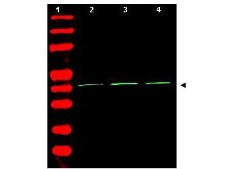
Western Blot of Rabbit Anti-Beta Actin Antibody. Lane 1: molecular weight. Lane 2: human embryonic kidney 293 . Lane 3: human lung carcinoma A549 . Lane 4: mouse brain . Load: 35 µg per lane. Primary antibody: Beta Actin antibody at 1:1,500 for overnight at 4°C. Secondary antibody: IRDye800™ rabbit secondary antibody at 1:10,000 for 45 min at RT. Block: 5% BLOTTO overnight at 4°C. Predicted/Observed size: ~42 kDa corresponding to beta Actin (arrowhead). Other band(s): none.

Click image to see more details
Immunohistochemistry of Rabbit Anti-Beta Actin Antibody. Tissue: sections 4.2 mm thick of rana pipiens tissue. Fixation: formalin fixed paraffin embedded. Antigen retrieval: not required. Primary antibody: Beta Actin antibody at 1:200 for 1 h at RT. Secondary antibody: Peroxidase rabbit secondary antibody at 1:10,000 for 45 min at RT. Localization: Beta Actin is at the neuromuscular junction. Staining: Beta Actin as precipitated green signal with red and blue counterstain.

Click image to see more details
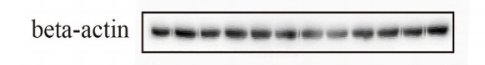
Western blot analysis of Beta Actin using anti-Beta Actin antibody (A01263).
Electrophoresis was performed on a 5-20% SDS-PAGE gel at 80V (Stacking gel) / 120V (Resolving gel) for 2 hours. The sample well of each lane was loaded with 30 ug of sample under reducing conditions.
Lane 1-12: human HEK293T whole cell lysates,
After electrophoresis, proteins were transferred to a nitrocellulose membrane at 150 mA for 50-90 minutes. Blocked the membrane with 5% non-fat milk/TBS for 1.5 hour at RT. The membrane was incubated with rabbit anti-Beta Actin antigen affinity purified polyclonal antibody (A01263) at 1:1000 overnight at 4°C, then washed with TBS-0.1%Tween 3 times with 5 minutes each and probed with a goat anti-rabbit IgG-HRP secondary antibody (Catalog # BA1054) at a dilution of 1:5000 for 1 hour at RT. The signal is developed using an ECL Plus Western Blotting Substrate with Azure Biosystems c600 system. A specific band was detected for Beta Actin at approximately 42 kDa. The expected band size for Beta Actin is at 42 kDa.

Click image to see more details
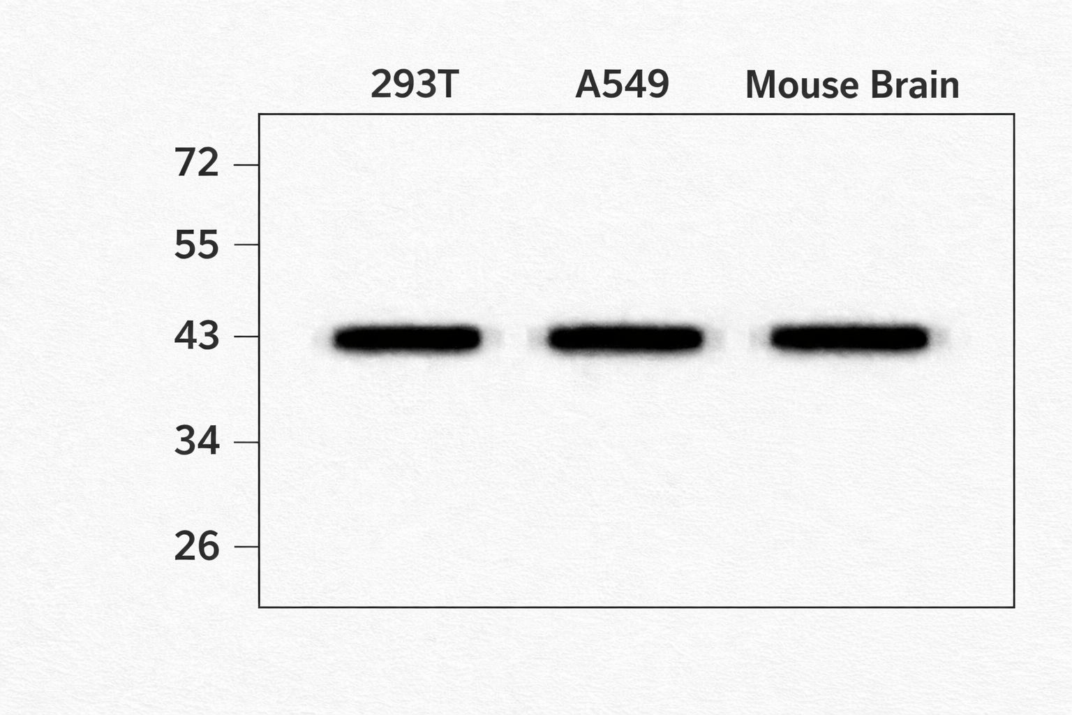
Western blot analysis of beta Actin using anti-beta Actin antibody (A01263).
Electrophoresis was performed on a 5-20% SDS-PAGE gel at 80V (Stacking gel) / 120V (Resolving gel) for 2 hours. The sample well of each lane was loaded with 30-35 ug of sample under reducing conditions.
Lane 1: human 293T whole cell lysates,
Lane 2: human A549 whole cell lysates,
Lane 3: mouse brain tissue lysates.
After electrophoresis, proteins were transferred to a nitrocellulose membrane at 150 mA for 50-90 minutes. Blocked the membrane with 5% non-fat milk/TBS for 1.5 hour at RT. The membrane was incubated with rabbit anti-beta Actin antigen affinity purified polyclonal antibody (A01263) at 1: 1000 overnight at 4°C, then washed with TBS-0.1%Tween 3 times with 5 minutes each and probed with a goat anti-rabbit IgG-HRP secondary antibody at a dilution of 1:5000 for 1.5 hour at RT. The signal is developed using an ECL Plus Western Blotting Substrate (Catalog # AR1196-200) with Tanon 5200 system. A specific band was detected for beta Actin at approximately 42 kDa. The expected band size for beta Actin is at 42 kDa.
Specific Publications For Anti-beta Actin ACTB Antibody (A01263)
Loading publications
Recommended Resources
Here are featured tools and databases that you might find useful.
- Boster's Pathways Library
- Protein Databases
- Bioscience Research Protocol Resources
- Data Processing & Analysis Software
- Photo Editing Software
- Scientific Literature Resources
- Research Paper Management Tools
- Molecular Biology Software
- Primer Design Tools
- Bioinformatics Tools
- Phylogenetic Tree Analysis
Customer Reviews
Have you used Anti-beta Actin ACTB Antibody?
Share your experimental results or join a short interview to earn up to $1,000 in product credits or other rewards.
2 Reviews For Anti-beta Actin ACTB Antibody
Produced clear, sharp bands with no background. Excellent specificity and consistency across replicates. Ideal as a loading control for WB.
Excellent
| SKU | A01263 |
|---|---|
| Application | Western Blot |
| Sample | Human 293T and A549 cell lysates, Mouse brain tissue |
| Sample Processing Description | Cells were lysed in RIPA buffer containing protease inhibitors. Lysates were clarified by centrifugation and quantified using a BCA assay. 30–35 µg of protein was loaded per lane on a 5–20% SDS-PAGE gel and transferred to nitrocellulose membranes. |
| Primary Antibody | Anti-beta Actin ACTB Antibody |
| Primary Incubation | 1:1000, incubated overnight at 4°C. |
| Secondary Antibody | Goat anti-rabbit IgG-HRP (Catalog # BA1054). |
| Secondary Incubation | 1:5000 dilution, 1 hour at room temperature. |
| Other Reagents used | 5% non-fat milk/TBST blocking buffer, ECL Plus substrate, Azure Biosystems c600 imaging system. |
| Detection | Chemiluminescent detection. Strong single band observed at ~42 kDa, corresponding to beta actin. |
| Results Summary | Produced clear, sharp bands with no background. Excellent specificity and consistency across replicates. Ideal as a loading control for WB. |
David Lee
Verified customer
Submitted 2026-01-08
The antibody is highly efficient and specific, showing a clear target band with no non-specific bands.
Excellent
| SKU | A01263 |
|---|---|
| Application | Western Blot |
| Sample | HEK293T |
| Blocking Agent | 5% Non-fat milk |
| Primary Incubation | 1:1000, overnight at 4 ℃ |
| Secondary Antibody | Anti-Rabbit IgG Secondary Antibody, HRP-conjugated |
| Secondary Incubation | Incubate at room temperature for 1 hour |
| Detection | Azure Biosystems c600, ECL substrate |
| Results Summary | I will definitely purchase BosterBio products again and recommend them to my classmates and colleagues. |
Ziru Wang, Jilin University
Verified customer
Submitted 2025-10-10
Customer Q&As
Have a question?
Find answers in Q&As, reviews.
Can't find your answer?
Submit your question
