Click image to see more details
Product Info Summary
| SKU: | A00334-2 |
|---|---|
| Size: | 100 μg/vial |
| Reactive Species: | Human |
| Host: | Rabbit |
| Application: | Flow Cytometry, IF, ICC, WB |
Customers Who Bought This Also Bought

Product info
Product Name
Anti-Caspase-3(p17)/CASP3 Antibody Picoband®
SKU/Catalog Number
A00334-2
Size
100 μg/vial
Form
Lyophilized
Description
Boster Bio Anti-Caspase-3(p17)/CASP3 Antibody Picoband® catalog # A00334-2. Tested in Flow Cytometry, IF, ICC, WB applications. This antibody reacts with Human. The brand Picoband indicates this is a premium antibody that guarantees superior quality, high affinity, and strong signals with minimal background in Western blot applications. Only our best-performing antibodies are designated as Picoband, ensuring unmatched performance.
Storage & Handling
At -20°C for one year from date of receipt. After reconstitution, at 4°C for one month. It can also be aliquotted and stored frozen at -20°C for six months. Avoid repeated freezing and thawing.
Cite This Product
Anti-Caspase-3(p17)/CASP3 Antibody Picoband® (Boster Biological Technology, Pleasanton CA, USA, Catalog # A00334-2)
Host
Rabbit
Contents
Each vial contains 4 mg Trehalose, 0.9 mg NaCl, 0.2 mg Na2HPO4.
Clonality
Polyclonal
Isotype
Rabbit IgG
Immunogen
A synthetic peptide corresponding to a sequence in the middle region of human Caspase-3(p17)/CASP3, which shares 78.3% and 87% amino acid (aa) sequence identity with mouse and rat CASP3.
Cross-reactivity
No cross-reactivity with other proteins.
Reactive Species
A00334-2 is reactive to CASP3 in Human
Observed Molecular Weight
32 kDa
Calculated molecular weight
31.6 kDa
Background of CASP3
Caspase 3 is a caspase protein which interacts with Survivin, XIAP, CFLAR, Caspase 8, HCLS1, Deleted in Colorectal Cancer, TRAF3 and GroEL. This gene which is located on 4q35 encodes a protein that is a member of the cysteine-aspartic acid protease (caspase) family. Sequential activation of caspases plays a central role in the execution-phase of cell apoptosis. Caspases exist as inactive proenzymes that undergo proteolytic processing at conserved aspartic residues to produce two subunits, large and small, that dimerize to form the active enzyme. It is the predominant caspase involved in the cleavage of amyloid-beta 4A precursor protein, which is associated with neuronal death in Alzheimer's disease. And the caspase-3 activation in heart failure sequentially cleaves SRF and generates a truncated SRF that appears to function as a dominant-negative transcription factor. Additionally, the caspase-3 influence on bone mineral absorbance should be considered in any in vivo application of caspase-3 inhibitors to the treatment of human disease. In erythroid precursors undergoing terminal differentiation, Hsp70 prevents active CASP3 from cleaving GATA1 and inducing apoptosis.
Antibody Validation
Boster validates all antibodies on WB, IHC, ICC, Immunofluorescence, and ELISA with known positive control and negative samples to ensure specificity and high affinity, including thorough antibody incubations.
Application & Images
Applications
A00334-2 is guaranteed for Flow Cytometry, IF, ICC, WB Boster Guarantee
Recommend Dilution
| Application | Dilution | Species |
|---|---|---|
| Western blot | 0.25-0.5 μg/ml | Human |
| Immunocytochemistry/Immunofluorescence | 5 μg/ml | Human |
| Flow Cytometry (Fixed) | 1-3 μg/1x106 cells | Human |
Tested application
Suggested blocking solution with 5% non-fat milk or BSA; (*)Recommended protein loading: 20-40 µg per lane
Validation Images & Assay Conditions
Click image to see more details
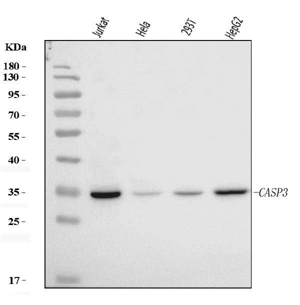
Western blot analysis of Caspase-3(P17)/CASP3 using anti-Caspase-3(P17)/CASP3 antibody (A00334-2).
Electrophoresis was performed on a 5-20% SDS-PAGE gel at 70V (Stacking gel) / 90V (Resolving gel) for 2-3 hours. The sample well of each lane was loaded with 30 ug of sample under reducing conditions.
Lane 1: human Jurkat whole cell lysates,
Lane 2: human Hela whole cell lysates,
Lane 3: human 293T whole cell lysates,
Lane 4: human HepG2 whole cell lysates.
After electrophoresis, proteins were transferred to a nitrocellulose membrane at 150 mA for 50-90 minutes. Blocked the membrane with 5% non-fat milk/TBS for 1.5 hour at RT. The membrane was incubated with rabbit anti-Caspase-3(P17)/CASP3 antigen affinity purified polyclonal antibody (Catalog # A00334-2) at 0.5 μg/mL overnight at 4°C, then washed with TBS-0.1%Tween 3 times with 5 minutes each and probed with a goat anti-rabbit IgG-HRP secondary antibody at a dilution of 1:5000 for 1.5 hour at RT. The signal is developed using an Enhanced Chemiluminescent detection (ECL) kit (Catalog # EK1002) with Tanon 5200 system. A specific band was detected for Caspase-3(P17)/CASP3 at approximately 32 kDa. The expected band size for Caspase-3(P17)/CASP3 is at 32 kDa.

Click image to see more details
IF analysis of Caspase-3(P17)/CASP3 using anti-Caspase-3(P17)/CASP3 antibody (A00334-2).
Caspase-3(P17)/CASP3 was detected in an immunocytochemical section of SiHa cells. Enzyme antigen retrieval was performed using IHC enzyme antigen retrieval reagent (AR0022) for 15 mins. The cells were blocked with 10% goat serum. And then incubated with 5 μg/mL rabbit anti-Caspase-3(P17)/CASP3 Antibody (A00334-2) overnight at 4°C. DyLight®488 Conjugated Goat Anti-Rabbit IgG (BA1127) was used as secondary antibody at 1:100 dilution and incubated for 30 minutes at 37°C. The section was counterstained with DAPI. Visualize using a fluorescence microscope and filter sets appropriate for the label used.

Click image to see more details
Flow Cytometry analysis of Caco-2 cells using anti-Caspase-3(P17)/CASP3 antibody (A00334-2).
Overlay histogram showing Caco-2 cells stained with A00334-2 (Blue line). To facilitate intracellular staining, cells were fixed with 4% paraformaldehyde and permeabilized with permeabilization buffer. The cells were blocked with 10% normal goat serum. And then incubated with rabbit anti-Caspase-3(P17)/CASP3 Antibody (A00334-2, 1 μg/1x106 cells) for 30 min at 20°C. DyLight®488 conjugated goat anti-rabbit IgG (BA1127, 5-10 μg/1x106 cells) was used as secondary antibody for 30 minutes at 20°C. Isotype control antibody (Green line) was rabbit IgG (1 μg/1x106) used under the same conditions. Unlabelled sample without incubation with primary antibody and secondary antibody (Red line) was used as a blank control.

Click image to see more details
Cv MC proliferation and apoptosis assays. The location of mantle tissue used for cell culture was indicated by dashed line (a) . Cv MCs after 48-h culture was shown (b) . Changes of medium pH in response to increased atmospheric CO 2 ( n = 4), (c) . Results of IFs with Ki-67 (d) and caspase 3 (e) were also quantified with the cells treated by ambient air and increased CO 2 concentration (2.5%). n = 8. Scale bar: 50 μm. Error bar: standard error of mean.
Index in Frontiersin under a CC BY license. DOI: 10.3389/fmars.2018.00203
Specific Publications For Anti-Caspase-3(p17)/CASP3 Antibody Picoband® (A00334-2)
Loading publications
Recommended Resources
Here are featured tools and databases that you might find useful.
- Boster's Pathways Library
- Protein Databases
- Bioscience Research Protocol Resources
- Data Processing & Analysis Software
- Photo Editing Software
- Scientific Literature Resources
- Research Paper Management Tools
- Molecular Biology Software
- Primer Design Tools
- Bioinformatics Tools
- Phylogenetic Tree Analysis
Customer Reviews
Have you used Anti-Caspase-3(p17)/CASP3 Antibody Picoband®?
Share your experimental results or join a short interview to earn up to $1,000 in product credits or other rewards.
0 Reviews For Anti-Caspase-3(p17)/CASP3 Antibody Picoband®
Customer Q&As
Have a question?
Find answers in Q&As, reviews.
Can't find your answer?
Submit your question
