Click image to see more details
-

-
-
-
-
+3
Product Info Summary
| SKU: | A00137-1 |
|---|---|
| Size: | 80 µl |
| Reactive Species: | Human |
| Host: | Rabbit |
| Application: | Flow Cytometry, IF, IHC-P, WB, IHC-P-Leica |
Customers Who Bought This Also Bought

Product info
Product Name
Anti-CD14 Antibody (N-term)
SKU/Catalog Number
A00137-1
Size
80 µl
Form
Liquid
Description
Boster Bio Anti-CD14 Antibody (N-term) (Catalog # A00137-1). Tested in IHC-P, Flow Cytometry, WB, IF, IHC-P-Leica application(s). This antibody reacts with Human.
Storage & Handling
Maintain refrigerated at 2-8°C for up to 2 weeks. For long-term storage, store at -20°C in small aliquots to prevent freeze-thaw cycles.
Cite This Product
Anti-CD14 Antibody (N-term) (Boster Biological Technology, Pleasanton CA, USA, Catalog # A00137-1)
Host
Rabbit
Contents
Purified polyclonal antibody supplied in PBS with 0.09% (W/V) sodium azide.
Clonality
Polyclonal
Isotype
Rabbit IgG
Immunogen
This CD14 antibody is generated from rabbits immunized with a KLH conjugated synthetic peptide between 54-83 amino acids from the N-terminal region of human CD14.
Cross-reactivity
No cross reactivity with other proteins.
Reactive Species
A00137-1 is reactive to CD14 in Human
Calculated molecular weight
40.1 kDa
Background of CD14
CD14 is a surface protein preferentially expressed on monocytes/macrophages. It binds lipopolysaccharide binding protein and recently has been shown to bind apoptotic cells.
Antibody Validation
Boster validates all antibodies on WB, IHC, ICC, Immunofluorescence, and ELISA with known positive control and negative samples to ensure specificity and high affinity, including thorough antibody incubations.
Application & Images
Applications
A00137-1 is guaranteed for Flow Cytometry, IF, IHC-P, WB, IHC-P-Leica Boster Guarantee
Assay Dilutions Recommendation
The recommendations below provide a starting point for assay optimization. The actual working concentration varies and should be decided by the user.
IF: 1:25
WB: 1:2000
IHC-P-Leica: 1:1000
FC: 1:10-1:50
Validation Images & Assay Conditions
Click image to see more details
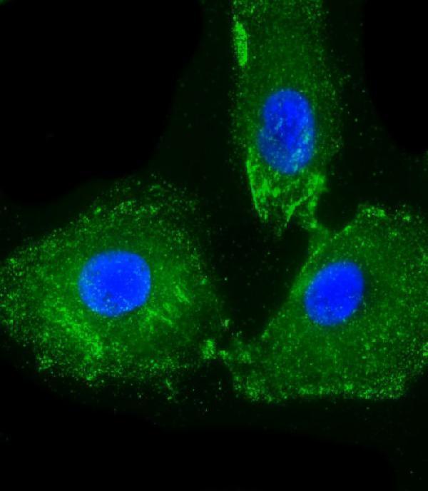
Immunofluorescent analysis of 4% paraformaldehyde-fixed, 0. 1% Triton X-100 permeabilized A549 cells labeling CD14 with A00137-1 at 1/25 dilution, followed by Dylight® 488-conjugated goat anti-Rabbit IgG secondary antibody at 1/200 dilution (green). Immunofluorescence image showing cytoplasm and membrance staining on A549 cell line. Cytoplasmic actin is detected with Dylight® 554 Phalloidin at 1/500 dilution (red). The nuclear counter stain is DAPI (blue).

Click image to see more details
All lanes : Anti-CD14 Antibody (N-term) at 1:2000 dilution
Lane 1: Human lung tissue lysate
Lane 2: Human liver tissue lysate
Lane 3: A549 whole cell lysate
Lysates/proteins at 20 µg per lane.
Secondary
Goat Anti-Rabbit IgG, (H+L), Peroxidase conjugated at 1/10000 dilution.
Predicted band size : 40 kDa
Blocking/Dilution buffer: 5% NFDM/TBST.

Click image to see more details
All lanes : Anti-CD14 Antibody (N-term) at 1:1000 dilution
Lane 1: Human lung tissue lysate
Lane 2: Human liver tissue lysate
Lysates/proteins at 20 µg per lane.
Secondary
Goat Anti-Rabbit IgG, (H+L), Peroxidase conjugated at 1/10000 dilution.
Predicted band size : 40 kDa
Blocking/Dilution buffer: 5% NFDM/TBST.

Click image to see more details
All lanes : Anti-CD14 Antibody (N-term) at 1:1000 dilution
Lane 1: THP-1 whole cell lysate
Lane 2: Human lung tissue lysate
Lane 3: Human liver tissue lysate
Lane 4: A549 whole cell lysate
Lysates/proteins at 20 µg per lane.
Secondary
Goat Anti-Rabbit IgG, (H+L), Peroxidase conjugated at 1/10000 dilution.
Predicted band size : 40 kDa
Blocking/Dilution buffer: 5% NFDM/TBST.

Click image to see more details
All lanes : Anti-CD14 Antibody (N-term) at 1:2000 dilution
Lane 1: Human liver lysate
Lane 2: Human lung lysate
Lysates/proteins at 20 µg per lane.
Secondary
Goat Anti-Rabbit IgG, (H+L), Peroxidase conjugated at 1/10000 dilution.
Predicted band size : 40 kDa
Blocking/Dilution buffer: 5% NFDM/TBST.

Click image to see more details
Immunohistochemical analysis of paraffin-embedded human liver tissue using A00137-1 performed on the Leica® BOND RXm. Tissue was fixed with formaldehyde at room temperature; antigen retrieval was by heat mediation with a EDTA buffer (pH9. 0). Samples were incubated with primary antibody (1:1000) for 1 hours at room temperature. A undiluted biotinylated CRF Anti-Polyvalent HRP Polymer antibody was used as the secondary antibody.

Click image to see more details
Overlay histogram showing Jurkat cells stained with A00137-1 (green line). The cells were fixed with 2% paraformaldehyde and then permeabilized with 90% methanol for 10 min. The cells were then icubated in 2% bovine serum albumin to block non-specific protein-protein interactions followed by the antibody (1:25 dilution) for 60 min at 37ºC. The secondary antibody used was Goat-Anti-Rabbit IgG, DyLight® 488 Conjugated Highly Cross-Adsorbed at 1/200 dilution for 40 min at Room temperature. Isotype control antibody (blue line) was rabbit IgG1 (1μg/1x10^6 cells) used under the same conditions. Acquisition of >10, 000 events was performed.
Specific Publications For Anti-CD14 Antibody (N-term) (A00137-1)
Loading publications
Recommended Resources
Here are featured tools and databases that you might find useful.
- Boster's Pathways Library
- Protein Databases
- Bioscience Research Protocol Resources
- Data Processing & Analysis Software
- Photo Editing Software
- Scientific Literature Resources
- Research Paper Management Tools
- Molecular Biology Software
- Primer Design Tools
- Bioinformatics Tools
- Phylogenetic Tree Analysis
Customer Reviews
Have you used Anti-CD14 Antibody (N-term)?
Share your experimental results or join a short interview to earn up to $1,000 in product credits or other rewards.
0 Reviews For Anti-CD14 Antibody (N-term)
Customer Q&As
Have a question?
Find answers in Q&As, reviews.
Can't find your answer?
Submit your question
