Click image to see more details
Product Info Summary
| SKU: | PA1443 |
|---|---|
| Size: | 100 μg/vial |
| Reactive Species: | Mouse |
| Host: | Rabbit |
| Application: | WB |
Customers Who Bought This Also Bought

Product info
Product Name
Anti-CD14 Antibody Picoband®
SKU/Catalog Number
PA1443
BA0719-2 is an alternative SKU for this antibody, used in previous lots.
Size
100 μg/vial
Form
Lyophilized
Description
Boster Bio Anti-CD14 Antibody catalog # PA1443. Tested in WB applications. This antibody reacts with Mouse. The brand Picoband indicates this is a premium antibody that guarantees superior quality, high affinity, and strong signals with minimal background in Western blot applications. Only our best-performing antibodies are designated as Picoband, ensuring unmatched performance.
Storage & Handling
Store at -20˚C for one year from date of receipt. After reconstitution, at 4˚C for one month. It can also be aliquotted and stored frozen at -20˚C for six months. Avoid repeated freeze-thaw cycles.
Cite This Product
Anti-CD14 Antibody Picoband® (Boster Biological Technology, Pleasanton CA, USA, Catalog # PA1443)
Host
Rabbit
Contents
Each vial contains 4 mg Trehalose, 0.9 mg NaCl and 0.2 mg Na2HPO4.
Clonality
Polyclonal
Isotype
Rabbit IgG
Immunogen
A synthetic peptide corresponding to a sequence in the middle region of mouse CD14.
Cross-reactivity
No cross-reactivity with other proteins
Reactive Species
PA1443 is reactive to Cd14 in Mouse
Observed Molecular Weight
50 kDa
Calculated molecular weight
39.2 kDa
Background of Cd14
CD14, Cluster of differentiation 14, single-copy gene encoding 2 protein forms: a 50- to 55-kD glycosylphosphatidylinositol-anchored membrane protein (mCD14) and a monocyte or liver-derived soluble serum protein (sCD14) that lacks the anchor. By in situ hybridization and study of somatic cell hybrid DNA that the gene is located at bands 5q23-q31. CD14 acts as a co-receptor (along with the Toll-like receptor TLR 4 and MD-2) for the detection of bacterial lipopolysaccharide (LPS). CD14 can bind LPS only in the presence of lipopolysaccharide-binding protein (LBP). Although LPS is considered its main ligand, CD14 also recognizes other pathogen-associated molecular patterns.
Antibody Validation
Boster validates all antibodies on WB, IHC, ICC, Immunofluorescence, and ELISA with known positive control and negative samples to ensure specificity and high affinity, including thorough antibody incubations.
Application & Images
Applications
PA1443 is guaranteed for WB Boster Guarantee
Recommend Dilution
| Application | Dilution | Species |
|---|---|---|
| Western blot | 0.1-0.5μg/ml | Mouse |
Tested application
Suggested blocking solution with 5% non-fat milk or BSA; (*)Recommended protein loading: 20-40 µg per lane
Validation Images & Assay Conditions
Click image to see more details
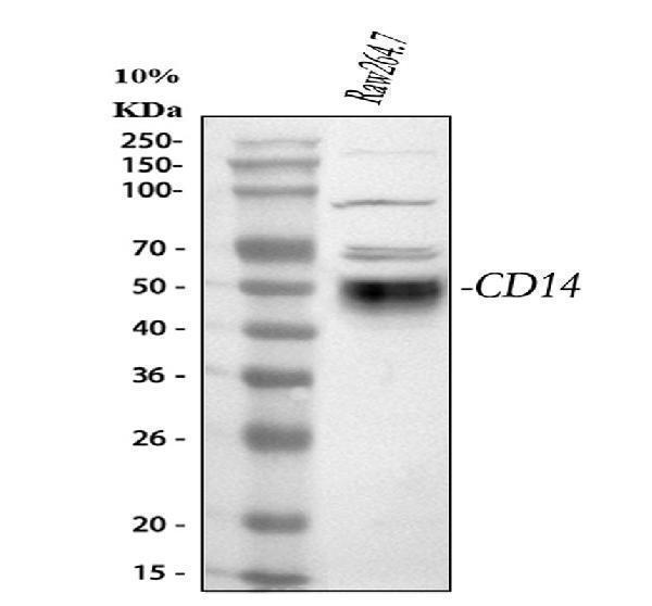
Western blot analysis of CD14 using anti-CD14 antibody (PA1443).
Electrophoresis was performed on a 5-20% SDS-PAGE gel at 70V (Stacking gel) / 90V (Resolving gel) for 2-3 hours. The sample well of each lane was loaded with 30 ug of sample under reducing conditions.
Lane 1: mouse RAW264.7 whole cell lysates.
After electrophoresis, proteins were transferred to a nitrocellulose membrane at 150 mA for 50-90 minutes. Blocked the membrane with 5% non-fat milk/TBS for 1.5 hour at RT. The membrane was incubated with rabbit anti-CD14 antigen affinity purified polyclonal antibody (Catalog # PA1443) at 0.5 μg/mL overnight at 4°C, then washed with TBS-0.1%Tween 3 times with 5 minutes each and probed with a goat anti-rabbit IgG-HRP secondary antibody at a dilution of 1:5000 for 1.5 hour at RT. The signal is developed using an Enhanced Chemiluminescent detection (ECL) kit (Catalog # EK1002) with Tanon 5200 system. A specific band was detected for CD14 at approximately 50 kDa. The expected band size for CD14 is at 39 kDa.
Specific Publications For Anti-CD14 Antibody Picoband® (PA1443)
Loading publications
Recommended Resources
Here are featured tools and databases that you might find useful.
- Boster's Pathways Library
- Protein Databases
- Bioscience Research Protocol Resources
- Data Processing & Analysis Software
- Photo Editing Software
- Scientific Literature Resources
- Research Paper Management Tools
- Molecular Biology Software
- Primer Design Tools
- Bioinformatics Tools
- Phylogenetic Tree Analysis
Customer Reviews
Have you used Anti-CD14 Antibody Picoband®?
Share your experimental results or join a short interview to earn up to $1,000 in product credits or other rewards.
0 Reviews For Anti-CD14 Antibody Picoband®
Customer Q&As
Have a question?
Find answers in Q&As, reviews.
Can't find your answer?
Submit your question
4 Customer Q&As for Anti-CD14 Antibody Picoband®
Question
We are currently using anti-CD14 antibody PA1443 for mouse tissue, and we are satisfied with the WB results. The species of reactivity given in the datasheet says mouse. Is it true that the antibody can work on bovine tissues as well?
S. Yang
Verified customer
Asked: 2019-11-21
Answer
The anti-CD14 antibody (PA1443) has not been tested for cross reactivity specifically with bovine tissues, but there is a good chance of cross reactivity. We have an innovator award program that if you test this antibody and show it works in bovine you can get your next antibody for free. Please contact me if I can help you with anything.
Boster Scientific Support
Answered: 2019-11-21
Question
Our lab were happy with the WB result of your anti-CD14 antibody. However we have observed positive staining in macrophage cell membrane using this antibody. Is that expected? Could you tell me where is CD14 supposed to be expressed?
Verified Customer
Verified customer
Asked: 2019-10-22
Answer
From literature, macrophage does express CD14. Generally CD14 expresses in cell membrane. Regarding which tissues have CD14 expression, here are a few articles citing expression in various tissues:
Brain, Pubmed ID: 15489334
Cerebrospinal fluid, Pubmed ID: 19838169
Glioblastoma, Pubmed ID: 2779588
Liver, Pubmed ID: 19159218, 24275569
Lymphocyte, Pubmed ID: 2453848
Macrophage, Pubmed ID: 2472171
Plasma, Pubmed ID: 16335952
Promyelocytic leukemia, Pubmed ID: 18810425
Boster Scientific Support
Answered: 2019-10-22
Question
We have observed staining in mouse glioblastoma. Any tips? Is anti-CD14 antibody supposed to stain glioblastoma positively?
Verified Customer
Verified customer
Asked: 2018-10-02
Answer
From literature glioblastoma does express CD14. From Uniprot.org, CD14 is expressed in liver, lymphocyte, macrophage, promyelocytic leukemia, brain, glioblastoma, plasma, cerebrospinal fluid, among other tissues. Regarding which tissues have CD14 expression, here are a few articles citing expression in various tissues:
Brain, Pubmed ID: 15489334
Cerebrospinal fluid, Pubmed ID: 19838169
Glioblastoma, Pubmed ID: 2779588
Liver, Pubmed ID: 19159218, 24275569
Lymphocyte, Pubmed ID: 2453848
Macrophage, Pubmed ID: 2472171
Plasma, Pubmed ID: 16335952
Promyelocytic leukemia, Pubmed ID: 18810425
Boster Scientific Support
Answered: 2018-10-02
Question
you antibody using your anti-CD14 antibody for toll-like receptor 4 signaling pathway studies. Has this antibody been tested with western blotting on spleen tissue? We would like to see some validation images before ordering.
P. Walker
Verified customer
Asked: 2017-07-26
Answer
We appreciate your inquiry. This PA1443 anti-CD14 antibody is validated on mouse thymus tissue, tissue lysate, spleen tissue. It is guaranteed to work for WB in mouse. Our Boster guarantee will cover your intended experiment even if the sample type has not been be directly tested.
Boster Scientific Support
Answered: 2017-07-26
