Click image to see more details
Product Info Summary
| SKU: | P03905 |
|---|---|
| Size: | 100ug |
| Reactive Species: | Human |
| Host: | Rabbit |
| Application: | ELISA, IP, WB |
Customers Who Bought This Also Bought

Product info
Product Name
Anti-CDC27 phospho T244 Antibody
View all CDC27, D0S1430E Antibodies
SKU/Catalog Number
P03905
Size
100ug
Form
Liquid (sterile filtered)
Description
Boster Bio Anti-CDC27 phospho T244 Antibody (Catalog # P03905). Tested in Dot blot, ELISA, WB applications. This antibody reacts with Human.
Storage & Handling
Store vial at -20°C prior to opening. Aliquot contents and freeze at -20°C or below for extended storage. Avoid cycles of freezing and thawing. Centrifuge product if not completely clear after standing at room temperature. This product is stable for several weeks at 4°C as an undiluted liquid. Dilute only prior to immediate use. Expiration date is one (1) year from date of opening. (Ship on dry ice.)
Cite This Product
Anti-CDC27 phospho T244 Antibody (Boster Biological Technology, Pleasanton CA, USA, Catalog # P03905)
Host
Rabbit
Contents
0.02 M Potassium Phosphate, 0.15 M Sodium Chloride, pH 7.2, 0.01% (w/v) Sodium Azide
Clonality
Polyclonal
Isotype
IgG
Immunogen
This affinity purified antibody was prepared from whole rabbit serum produced by repeated immunizations with a synthetic peptide corresponding to an internal portion surrounding T244 of Human CDC27.
Cross-reactivity
No cross reactivity with other proteins.
Reactive Species
P03905 is reactive to CDC27, D0S1430E in Human
Observed Molecular Weight
42 kDa
Calculated molecular weight
91.9 kDa
Background of CDC27, D0S1430E
Human CDC27 (also called Cell division cycle protein 27 homolog, CDC27Hs and H-NUC) shares strong similarity with Saccharomyces cerevisiae protein Cdc27. This protein is a component of anaphase-promoting complex (APC), which is composed of eight protein subunits and highly conserved in eukaryotic cells. APC catalyzes the formation of a cyclin B-ubiquitin conjugate that is responsible for the ubiquitin-mediated proteolysis of B-type cyclins. This protein and 3 other members of the APC complex contain the TPR (tetratricopeptide repeat), a protein domain important for protein-protein interaction. This protein was shown to interact with mitotic checkpoint proteins including Mad2, p55CDC and BUBR1, and thus may be involved in controlling the timing of mitosis.
Antibody Validation
Boster validates all antibodies on WB, IHC, ICC, Immunofluorescence, and ELISA with known positive control and negative samples to ensure specificity and high affinity, including thorough antibody incubations.
Application & Images
Applications
P03905 is guaranteed for ELISA, IP, WB Boster Guarantee
Recommend Dilution
| Application | Dilution | Species |
|---|---|---|
| ELISA: 1:8 | 000 - 1:30 | 000 |
| WB: 1:300 - 1:2 | 000 |
Validation Images & Assay Conditions
Click image to see more details
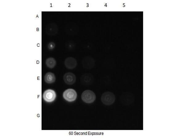
Dot Blot of Rabbit Anti-CDC27pT244 Antibody. Dilutions in Columns: (1) 100ng, (2) 33.33ng, (3) 11.11ng, (4) 3.7ng, (5) 1.23ng. Tested BSA Peptide Reactivity in Rows: (A) AKT1-BSA, (B) AKT1 pT308-BSA, (C) AKT1 S473-BSA, (D) AKT1 pS473-BSA, (E) CDC27 T244-BSA, (F) CDC27 pT244-BSA, (G) BSA control. Primary Antibody: Anti-CDC27pT244 at 1µg/mL overnight at 2-8°C. Secondary Antibody: Goat anti-Rabbit IgG HRP at 1:70,000 at RT for 30mins. Block: BlockOut Buffer .

Click image to see more details
Western blot using Boster's Affinity Purified anti-CDC27 pT244 antibody shows detection of a band ~92 kDa corresponding to phosphorylated human CDC27 (arrowhead). Lane 1 shows lysate from asynchronous cells. Lane 2 shows lysate from cells treated with nocodazole. Phosphorylated CDC27 is mostly present only in cell preparations arrested in mitosis. Each lane contains approximately 30 µg of HeLa whole cell lysates separated by 12.5% SDS-PAGE followed by transfer to nitrocellulose. After blocking with 5% non-fat dry milk in TTBS, the membrane was probed with the primary antibody diluted to 1:500 for 1 h at room temperature followed by washes and reaction with a 1:5,000 dilution of HRP Gt-a-Rabbit IgG [H&L] MX (611-103-122) for 45 min at room temperature. ECL reagent was used for detection. Other detection systems will yield similar results. Data contributed by Bing Li, UT Southwestern.
Specific Publications For Anti-CDC27 phospho T244 Antibody (P03905)
Loading publications
Recommended Resources
Here are featured tools and databases that you might find useful.
- Boster's Pathways Library
- Protein Databases
- Bioscience Research Protocol Resources
- Data Processing & Analysis Software
- Photo Editing Software
- Scientific Literature Resources
- Research Paper Management Tools
- Molecular Biology Software
- Primer Design Tools
- Bioinformatics Tools
- Phylogenetic Tree Analysis
Customer Reviews
Have you used Anti-CDC27 phospho T244 Antibody?
Share your experimental results or join a short interview to earn up to $1,000 in product credits or other rewards.
0 Reviews For Anti-CDC27 phospho T244 Antibody
Customer Q&As
Have a question?
Find answers in Q&As, reviews.
Can't find your answer?
Submit your question
1 Customer Q&As for Anti-CDC27 phospho T244 Antibody
Question
We are currently using anti-CDC27 phospho T244 antibody P03905 for human tissue, and we are content with the IP results. The species of reactivity given in the datasheet says human. Is it true that the antibody can work on feline tissues as well?
P. Huang
Verified customer
Asked: 2013-02-13
Answer
The anti-CDC27 phospho T244 antibody (P03905) has not been validated for cross reactivity specifically with feline tissues, though there is a good chance of cross reactivity. We have an innovator award program that if you test this antibody and show it works in feline you can get your next antibody for free. Please contact me if I can help you with anything.
Boster Scientific Support
Answered: 2013-02-13
