Click image to see more details
-

-
-
-
-
+9
Product Info Summary
| SKU: | PB9561 |
|---|---|
| Size: | 100 μg/vial |
| Reactive Species: | Human, Mouse, Rat |
| Host: | Rabbit |
| Application: | ELISA, IF, IHC, ICC, WB |
Customers Who Bought This Also Bought

Product info
Product Name
Anti-E Cadherin 1/CDH1 Antibody Picoband®
SKU/Catalog Number
PB9561
Size
100 μg/vial
Form
Lyophilized
Description
Boster Bio Anti-E Cadherin 1/CDH1 Antibody Picoband® catalog # PB9561. Tested in ELISA, ICC/IF, IHC, WB applications. This antibody reacts with Human, Mouse, Rat. The brand Picoband indicates this is a premium antibody that guarantees superior quality, high affinity, and strong signals with minimal background in Western blot applications. Only our best-performing antibodies are designated as Picoband, ensuring unmatched performance.
Storage & Handling
Store at -20˚C for one year from date of receipt. After reconstitution, at 4˚C for one month. It can also be aliquotted and stored frozen at -20˚C for six months. Avoid repeated freeze-thaw cycles.
Cite This Product
Anti-E Cadherin 1/CDH1 Antibody Picoband® (Boster Biological Technology, Pleasanton CA, USA, Catalog # PB9561)
Host
Rabbit
Contents
Each vial contains 4 mg Trehalose, 0.9 mg NaCl and 0.2 mg Na2HPO4.
Clonality
Polyclonal
Isotype
Rabbit IgG
Immunogen
E.coli-derived human E Cadherin recombinant protein (Position: A286-A703). Human E Cadherin shares 79.7% and 80.9% amino acid (aa) sequence identity with mouse and rat E Cadherin, respectively.
Cross-reactivity
No cross-reactivity with other proteins
Reactive Species
PB9561 is reactive to CDH1 in Human, Mouse, Rat
Observed Molecular Weight
130 kDa
Calculated molecular weight
97.5 kDa
Background of CDH1
CDH1 (Cadherin 1), also known as ECAD or UVO, is a protein that in humans is encoded by the CDH1 gene. Cadherin-1 is a classical member of the cadherin superfamily. By Southern analysis of DNA from a panel of mouse-human somatic cell hybrids, Mansouri et al. (1987, 1988) assigned the UVO gene to 16q (16p11-qter). Frebourg et al. (2006) found that in human embryos CDH1 is highly expressed at 4 and 5 weeks in the frontonasal prominence and at 6 weeks in the lateral and medial nasal prominences, and is therefore expressed during critical stages of lip and palate development. CDH1 is involved in mechanisms regulating cell-cell adhesions, mobility and proliferation of epithelial cells. Has a potent invasive suppressor role. It is a ligand for integrin alpha-E/beta-7.
Antibody Validation
Boster validates all antibodies on WB, IHC, ICC, Immunofluorescence, and ELISA with known positive control and negative samples to ensure specificity and high affinity, including thorough antibody incubations.
Application & Images
Applications
PB9561 is guaranteed for ELISA, IF, IHC, ICC, WB Boster Guarantee
Recommend Dilution
| Application | Dilution | Species |
|---|---|---|
| Western blot | 0.1-0.5μg/ml | Human |
| Immunohistochemistry (Paraffin-embedded Section) | 2-5μg/ml | Human, Mouse, Rat |
| Immunocytochemistry/Immunofluorescence | 5 μg/ml | Human |
| ELISA | 0.1-0.5μg/ml | - |
Tested application
Suggested blocking solution with 5% non-fat milk or BSA; (*)Recommended protein loading: 20-40 µg per lane
Use TE buffer pH 9.0 for antigen retrieval; (*) citrate buffer pH 6.0 is an alternative.
Validation Images & Assay Conditions
Click image to see more details
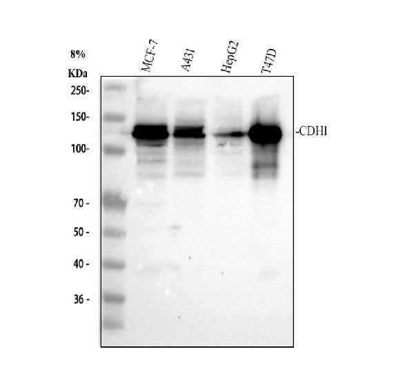
Western blot analysis of CDH1 using anti-CDH1 antibody (PB9561).
Electrophoresis was performed on a 8% SDS-PAGE gel at 80V (Stacking gel) / 120V (Resolving gel) for 2 hours. The sample well of each lane was loaded with 30 ug of sample under reducing conditions.
Lane 1: human MCF-7 whole cell lysates,
Lane 2: human A431 whole cell lysates,
Lane 3: human HepG2 whole cell lysates,
Lane 4: human T47D whole cell lysates.
After electrophoresis, proteins were transferred to a nitrocellulose membrane at 150 mA for 50-90 minutes. Blocked the membrane with 5% non-fat milk/TBS for 1.5 hour at RT. The membrane was incubated with rabbit anti-CDH1 antigen affinity purified polyclonal antibody (PB9561) at 0.5 μg/mL overnight at 4°C, then washed with TBS-0.1%Tween 3 times with 5 minutes each and probed with a goat anti-rabbit IgG-HRP secondary antibody (Catalog # BA1054) at a dilution of 1:5000 for 1.5 hour at RT. The signal is developed using an ECL Plus Western Blotting Substrate (Catalog # AR1196-200) with Tanon 5200 system. A specific band was detected for CDH1 at approximately 130 kDa. The expected band size for CDH1 is at 98 kDa.

Click image to see more details
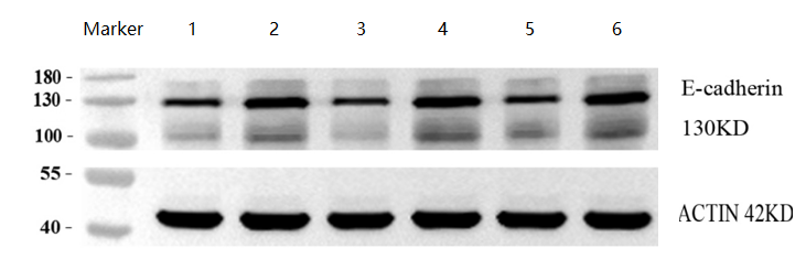
Western blot analysis of CDH1 using anti-CDH1 antibody (PB9561).
Electrophoresis was performed on a 8% SDS-PAGE gel at 80V (Stacking gel) / 120V (Resolving gel) for 2 hours. The sample well of each lane was loaded with 30 ug of sample under reducing conditions.
Lane 1: human cervical cancer tissue lysates,
Lane 2: human cervical cancer adjacent tissue lysates,
Lane 3: human cervical cancer tissue lysates,
Lane 4: human cervical cancer adjacent tissue lysates.
Lane 5: human cervical cancer tissue lysates,
Lane 6: human cervical cancer adjacent tissue lysates.
After electrophoresis, proteins were transferred to a nitrocellulose membrane at 150 mA for 50-90 minutes. Blocked the membrane with 5% non-fat milk/TBS for 1.5 hour at RT. The membrane was incubated with rabbit anti-CDH1 antigen affinity purified polyclonal antibody (PB9561) at 1:1000 overnight at 4°C, then washed with TBS-0.1%Tween 3 times with 5 minutes each and probed with a goat anti-rabbit IgG-HRP secondary antibody (Catalog # BA1054) at a dilution of 1:10000 for 1 hour at RT. The signal is developed using an ECL Plus Western Blotting Substrate (Catalog # AR1196-200) with ChemiDoc MP system. A specific band was detected for CDH1 at approximately 130 kDa. The expected band size for CDH1 is at 98 kDa.

Click image to see more details
IHC analysis of CDH1 using anti-CDH1 antibody (PB9561).
CDH1 was detected in a paraffin-embedded section of human placenta tissue. Heat mediated antigen retrieval was performed in EDTA buffer (pH 8.0, epitope retrieval solution). The tissue section was blocked with 10% goat serum. The tissue section was then incubated with 2 μg/ml rabbit anti-CDH1 Antibody (PB9561) overnight at 4°C. Peroxidase Conjugated Goat Anti-rabbit IgG was used as secondary antibody and incubated for 30 minutes at 37°C. The tissue section was developed using HRP Conjugated Rabbit IgG Super Vision Assay Kit (Catalog # SV0002) with DAB as the chromogen.

Click image to see more details
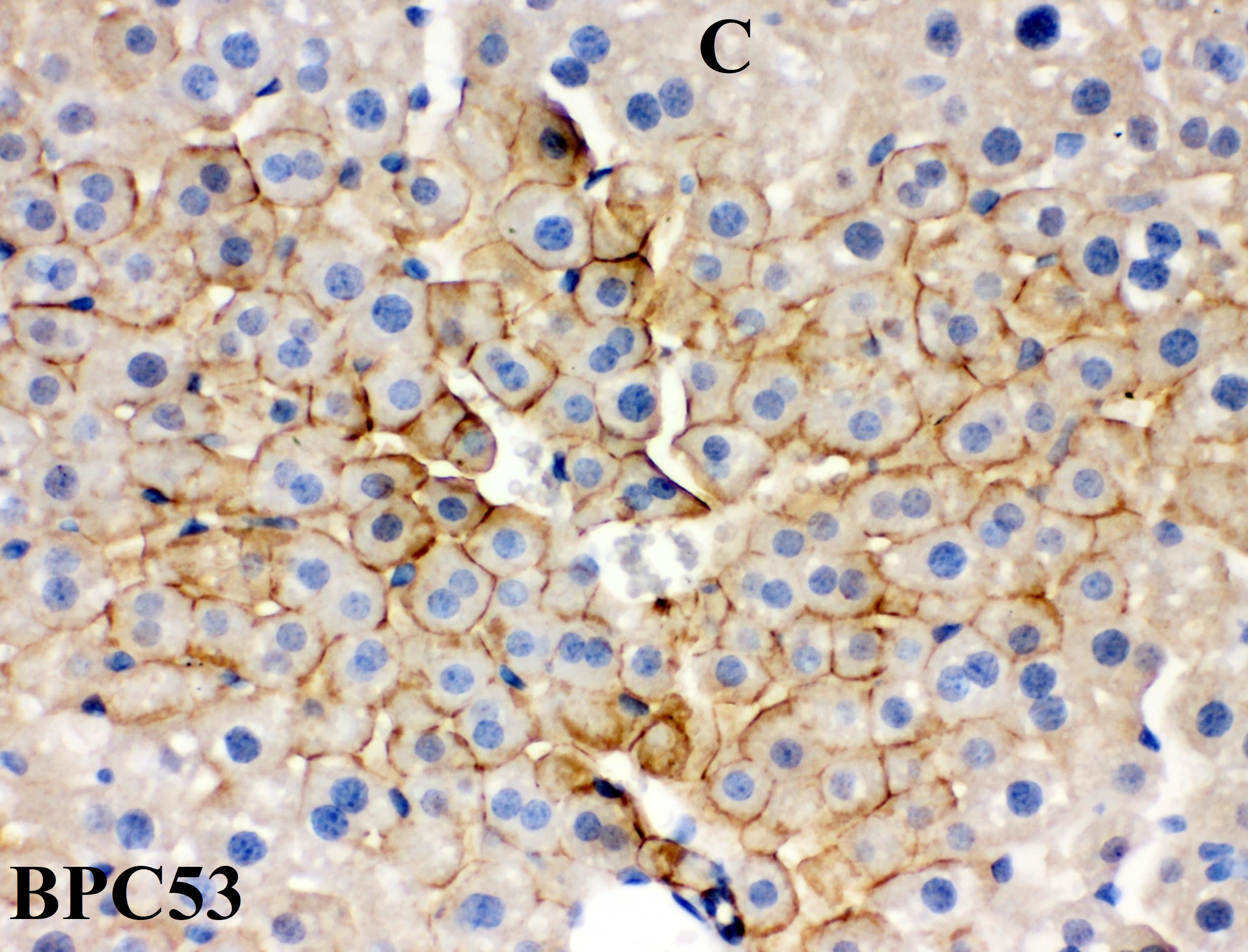
IHC analysis of CDH1 using anti-CDH1 antibody (PB9561).
CDH1 was detected in a paraffin-embedded section of human hepatocellular carcinoma tissue. Heat mediated antigen retrieval was performed in EDTA buffer (pH 8.0, epitope retrieval solution). The tissue section was blocked with 10% goat serum. The tissue section was then incubated with 2 μg/ml rabbit anti-CDH1 Antibody (PB9561) overnight at 4°C. Peroxidase Conjugated Goat Anti-rabbit IgG was used as secondary antibody and incubated for 30 minutes at 37°C. The tissue section was developed using HRP Conjugated Rabbit IgG Super Vision Assay Kit (Catalog # SV0002) with DAB as the chromogen.

Click image to see more details
IHC analysis of CDH1 using anti-CDH1 antibody (PB9561).
CDH1 was detected in a paraffin-embedded section of mouse colon tissue. Heat mediated antigen retrieval was performed in EDTA buffer (pH 8.0, epitope retrieval solution). The tissue section was blocked with 10% goat serum. The tissue section was then incubated with 2 μg/ml rabbit anti-CDH1 Antibody (PB9561) overnight at 4°C. Peroxidase Conjugated Goat Anti-rabbit IgG was used as secondary antibody and incubated for 30 minutes at 37°C. The tissue section was developed using HRP Conjugated Rabbit IgG Super Vision Assay Kit (Catalog # SV0002) with DAB as the chromogen.

Click image to see more details
IHC analysis of CDH1 using anti-CDH1 antibody (PB9561).
CDH1 was detected in a paraffin-embedded section of rat colon tissue. Heat mediated antigen retrieval was performed in EDTA buffer (pH 8.0, epitope retrieval solution). The tissue section was blocked with 10% goat serum. The tissue section was then incubated with 2 μg/ml rabbit anti-CDH1 Antibody (PB9561) overnight at 4°C. Peroxidase Conjugated Goat Anti-rabbit IgG was used as secondary antibody and incubated for 30 minutes at 37°C. The tissue section was developed using HRP Conjugated Rabbit IgG Super Vision Assay Kit (Catalog # SV0002) with DAB as the chromogen.

Click image to see more details
IF analysis of CDH1 using anti-CDH1 antibody (PB9561).
CDH1 was detected in an immunocytochemical section of MCF-7 cells. Enzyme antigen retrieval was performed using IHC enzyme antigen retrieval reagent (AR0022) for 15 mins. The cells were blocked with 10% goat serum. And then incubated with 5 μg/mL rabbit anti-CDH1 Antibody (PB9561) overnight at 4°C. Fluoro488488 Conjugated Goat Anti-Rabbit IgG (BA1127) was used as secondary antibody at 1:500 dilution and incubated for 30 minutes at 37°C. The section was counterstained with DAPI. Visualize using a fluorescence microscope and filter sets appropriate for the label used.

Click image to see more details
Immunohistochemical analysis of dorsolateral prostate E-cadherin, Vimtein, ERα and AR expression in aged rats. The expression of vimentin, ERα and AR increased, and the expression of E-cadherin decreased in BPA-treated groups. ( a – p ) Representative sections of comparable regions are shown for vehicle control rats ( a , e , i , m ), and animals exposed to BPA (10 μg/kg/day) ( b , f , j , n ), BPA (30 μg /kg/day) ( c , g , k , o ), and BPA (90 μg/kg/day) ( d , h , l , p ) (scale bar: 50 μm, ×400). BPA: bisphenol A; AR: androgen receptor; ERα:estrogen receptor-α.
Index in PubMed under a CC BY license. PMID: 29323181

Click image to see more details
LPC inhibits pulmonary metastasis of CT26 colon cancer. a Schematic view of the experimental procedures of CT26 pulmonary metastatic mouse model. b , c Image and corresponding H&E staining of lung tissue. d Percentage change of body weight. e – g Lung weight, the number of lung tumor nodule, and metastasis rate ( n = 6 mice). h Protein expression of PCNA, vimentin and E-cadherin in lungs ( n = 3 mice). Data were presented as mean ± SEM, * p < 0.05, ** p < 0.01.
Index in PubMed under a CC BY license. PMID: 36774343

Click image to see more details
Interfering with DLGAP4 inhibits the PPARβ/δ signalling pathway and the expression of proliferation- and metastasis-related proteins. Western blotting was performed to measure the protein expression of DLGAP4, PPARβ/δ, CyclinD1, E-cadherin and N-cadherin in shNC or shDLGAP4 HepG2 and HCCLM3 cells. The data were obtained from the average of three independent experiments. *P < 0.05.
Index in PubMed under a CC BY license. PMID: 36396671

Click image to see more details
PPI inhibits the metastasis of cervical cancer cells in vivo. ( A , B ) Bioluminescence imaging of various organs exhibiting metastatic or disseminated cancer cells ( A ) and a quantitative analysis of total bioluminescence in each group of organs ( B ). ( C ) H&E staining of various organs in each group. The scale is 100 μm. ( D , E ) The immunofluorescence staining of E-cadherin, VE-cadherin, and Vimentin was performed, followed by data quantification analysis, comparing the control group with the treated group. Six animal samples were used per group ( n = 6), and the results are presented as mean ± S.D. **: p < 0.01; ***: p < 0.001 vs. control. The scale is 100 μm.
Index in PubMed under a CC BY license. PMID: 40429774

Click image to see more details
PPI inhibits the growth, migration, and invasion of HO-8910PM cells by reversing the EMT progress. ( A ) Cell proliferation was evaluated at 48 h using the MTT assay, and the IC 50 values were determined from dose–response curves generated with GraphPad Prism 8. ( B ) The wound-healing assay was conducted in HO-8910PM cells to evaluate cell migration. Images were captured at 0 and 24 h. The scale is 500 μm. ( C ) The data quantification results of ( B ). ( D ) A transwell migration assay was performed to monitor the rate of cellular migration. The scale is 500 μm. ( E ) The quantification results of ( D ). ( F ) The protein expression levels of E-cadherin, VE-cadherin, and Vimentin in HO-8910PM cells. ( G ) The quantification results of ( F ). ( H ) An immunofluorescence assay was performed to detect E-cadherin, VE-cadherin, and Vimentin expression in HO-8910PM cells treated with PPI. The scale is 100 μm. ( I ) The quantification results of ( H ). Data from six independent experiments are presented as mean ± S.D. *: p < 0.05; **: p < 0.01; ***: p < 0.001 vs. control.
Index in PubMed under a CC BY license. PMID: 40429774
Specific Publications For Anti-E Cadherin 1/CDH1 Antibody Picoband® (PB9561)
Loading publications
Recommended Resources
Here are featured tools and databases that you might find useful.
- Boster's Pathways Library
- Protein Databases
- Bioscience Research Protocol Resources
- Data Processing & Analysis Software
- Photo Editing Software
- Scientific Literature Resources
- Research Paper Management Tools
- Molecular Biology Software
- Primer Design Tools
- Bioinformatics Tools
- Phylogenetic Tree Analysis
Customer Reviews
Have you used Anti-E Cadherin 1/CDH1 Antibody Picoband®?
Share your experimental results or join a short interview to earn up to $1,000 in product credits or other rewards.
2 Reviews For Anti-E Cadherin 1/CDH1 Antibody Picoband®
E-cadherin Antibody Picoband® (PB9561) shows clear, specific bands in human cervical cancer and adjacent tissues by WB, with higher expression in adjacent tissues than in cancer tissues, demonstrating excellent antibody performance.
Excellent
| SKU | PB9561 |
|---|---|
| Application | Western Blot |
| Sample | human cervical cancer and adjacent tissues |
| Sample Processing Description | Three samples each of human cervical cancer and adjacent tissues were collected, and total protein was extracted. |
| Other Reagents | RIPA lysis buffer, Protease inhibitor, Running buffer, Transfer buffer, Blocking buffer |
| Primary Antibody | E Cadherin 1/CDH1 Antibody Picoband® |
| Primary Incubation | 1:1000, overnight at 4 ℃ |
| Secondary Antibody | HRP Conjugated AffiniPure Goat Anti-Rabbit IgG (H+L) (BA1054) |
| Secondary Incubation | 1:10000, 1 h in RT |
| Detection | Substrate: ECL substrate, Image system: ChemiDoc MP |
| Results Summary | Based on this study, the expression of E-cadherin in human cervical cancer and adjacent tissues was compared, and results from three cases showed that E-cadherin levels were significantly higher in adjacent tissues than in cancer tissues. |
Xiaoyuan Qu, Shandong First Medical University
Verified customer
Submitted 2026-03-27
Immunocytochemistry for Anti-E Cadherin 1/CDH1 Antibody
Excellent
The antibody worked perfectly, Great sensitivity
| SKU | PB9561 |
|---|---|
| Application | Immunohistochemistry (paraffin-embedded) |
| Blocking step | 5% BSA as a blocking agent for 30 min at 37°C |
| Sample | Mouse Liver |
| Fixative | Fixed with 4% paraformaldehyde |
| Primary Ab Incubation | 4°C overnight |
| Primary Ab Incubation diluent | 5% BSA in TBS |
| Primary Ab Concentration | 2ug/ml |
| Secondary Antibody | SABC kit from Boster Bio, (SA1022) |
| Secondary Ab Dilution | The kit was ready to use, no dilution needed |
| Secondary Ab Incubation | 4°C overnight |
E.Hernandez
Verified customer
Submitted 2020-07-18
Customer Q&As
Have a question?
Find answers in Q&As, reviews.
Can't find your answer?
Submit your question
6 Customer Q&As for Anti-E Cadherin 1/CDH1 Antibody Picoband®
Question
Are there any optimizations that you suggest or Antigen retrieval for PB9561?
Verified customer
Asked: 2021-01-27
Answer
For the Anti-E Cadherin 1/CDH1 Antibody Picoband™ (PB9561), AR0022 is recommended to be used for Antigen retrieval. https://www.bosterbio.com/antigen-retrieval-reagent-ar0022-boster.html
Boster Scientific Support
Answered: 2021-01-28
Question
Our lab want to know about using your anti-E Cadherin/CDH1 antibody for regulation of gene expression studies. Has this antibody been tested with western blotting on placenta tissue? We would like to see some validation images before ordering.
Verified Customer
Verified customer
Asked: 2020-02-20
Answer
We appreciate your inquiry. This PB9561 anti-E Cadherin/CDH1 antibody is tested on human ileum tissue, placenta tissue, tissue lysate, hela whole cell lysate, mouse ileum tissue, ileum organoid tissue, colon organoid tissue. It is guaranteed to work for ELISA, IF, IHC-P, IHC-F, WB in human, mouse, rat. Our Boster guarantee will cover your intended experiment even if the sample type has not been be directly tested.
Boster Scientific Support
Answered: 2020-02-20
Question
Our lab used your anti-E Cadherin/CDH1 antibody for IHC-F on hippocampus tongue in a previous experiment. I am using mouse, and I plan to use the antibody for IHC-P next. We want examining hippocampus tongue as well as pancreas in our next experiment. Could you please give me some suggestion on which antibody would work the best for IHC-P?
Verified Customer
Verified customer
Asked: 2020-02-12
Answer
I viewed the website and datasheets of our anti-E Cadherin/CDH1 antibody and it appears that PB9561 has been tested on mouse in both IHC-F and IHC-P. Thus PB9561 should work for your application. Our Boster satisfaction guarantee will cover this product for IHC-P in mouse even if the specific tissue type has not been validated. We do have a comprehensive range of products for IHC-P detection and you can check out our website bosterbio.com to find out more information about them.
Boster Scientific Support
Answered: 2020-02-12
Question
We are currently using anti-E Cadherin/CDH1 antibody PB9561 for rat tissue, and we are satisfied with the WB results. The species of reactivity given in the datasheet says human, mouse, rat. Is it possible that the antibody can work on canine tissues as well?
Verified Customer
Verified customer
Asked: 2019-07-05
Answer
The anti-E Cadherin/CDH1 antibody (PB9561) has not been validated for cross reactivity specifically with canine tissues, though there is a good chance of cross reactivity. We have an innovator award program that if you test this antibody and show it works in canine you can get your next antibody for free. Please contact me if I can help you with anything.
Boster Scientific Support
Answered: 2019-07-05
Question
We have seen staining in rat liver. Do you have any suggestions? Is anti-E Cadherin/CDH1 antibody supposed to stain liver positively?
Verified Customer
Verified customer
Asked: 2019-06-17
Answer
Based on literature liver does express CDH1. Based on Uniprot.org, CDH1 is expressed in jejunal mucosa, pancreas, liver, epidermal carcinoma, hippocampus tongue, placenta, brain, among other tissues. Regarding which tissues have CDH1 expression, here are a few articles citing expression in various tissues:
Brain, Pubmed ID: 14595118
Epidermal carcinoma, Pubmed ID: 10597309
Hippocampus, and Tongue, Pubmed ID: 14702039
Liver, Pubmed ID: 3263290, 8185635, 24275569, 8033105, 11953314
Pancreas, Pubmed ID: 8459805
Placenta, Pubmed ID: 7543680, 7601454
Boster Scientific Support
Answered: 2019-06-17
Question
Our team were content with the WB result of your anti-E Cadherin/CDH1 antibody. However we have observed positive staining in epidermal carcinoma adherens junction using this antibody. Is that expected? Could you tell me where is CDH1 supposed to be expressed?
Verified Customer
Verified customer
Asked: 2018-12-17
Answer
From literature, epidermal carcinoma does express CDH1. Generally CDH1 expresses in cell junction, adherens junction. Regarding which tissues have CDH1 expression, here are a few articles citing expression in various tissues:
Brain, Pubmed ID: 14595118
Epidermal carcinoma, Pubmed ID: 10597309
Hippocampus, and Tongue, Pubmed ID: 14702039
Liver, Pubmed ID: 3263290, 8185635, 24275569, 8033105, 11953314
Pancreas, Pubmed ID: 8459805
Placenta, Pubmed ID: 7543680, 7601454
Boster Scientific Support
Answered: 2018-12-17
