Click image to see more details
-

-
-
-
-
+3
Product Info Summary
| SKU: | PB9491 |
|---|---|
| Size: | 100 μg/vial |
| Reactive Species: | Mouse, Rat |
| Host: | Rabbit |
| Application: | IF, IHC, IHC-F, WB |
Customers Who Bought This Also Bought

Product info
Product Name
Anti-CPT1B Antibody Picoband®
SKU/Catalog Number
PB9491
Size
100 μg/vial
Form
Lyophilized
Description
Boster Bio Anti-CPT1B Antibody Picoband® catalog # PB9491. Tested in IF, IHC, IHC-F, WB applications. This antibody reacts with Mouse, Rat. The brand Picoband indicates this is a premium antibody that guarantees superior quality, high affinity, and strong signals with minimal background in Western blot applications. Only our best-performing antibodies are designated as Picoband, ensuring unmatched performance.
Storage & Handling
Store at -20˚C for one year from date of receipt. After reconstitution, at 4˚C for one month. It can also be aliquotted and stored frozen at -20˚C for six months. Avoid repeated freeze-thaw cycles.
Cite This Product
Anti-CPT1B Antibody Picoband® (Boster Biological Technology, Pleasanton CA, USA, Catalog # PB9491)
Host
Rabbit
Contents
Each vial contains antibody formulated with stabilizing components, 0.9 mg NaCl, 0.2 mg Na2HPO4, and 0.05 mg NaN3.
*This antibody is supplied in a stabilized formulation.
Compatibility with conjugation reactions depends on the chemistry of the conjugation method used.
For conjugation methods that are not compatible with the stabilizing components present in this formulation, a carrier-free antibody format is required.
Clonality
Polyclonal
Isotype
Rabbit IgG
Immunogen
A synthetic peptide corresponding to a sequence at the N-terminus of human CPT1B, different from the related mouse sequence by two amino acids, and from the related rat sequence by four amino acids.
Cross-reactivity
No cross-reactivity with other proteins
Reactive Species
PB9491 is reactive to CPT1B in Mouse, Rat
Observed Molecular Weight
88 kDa
Calculated molecular weight
87.8 kDa
Background of CPT1B
CPT1B is located on 22q13.33. The protein encoded by this gene, a member of the carnitine/ choline acetyltransferase family, is the rate-controlling enzyme of the long-chain fatty acid beta-oxidation pathway in muscle mitochondria. This enzyme is required for the net transport of long-chain fatty acyl-CoAs from the cytoplasm into the mitochondria. Multiple transcript variants encoding different isoforms have been found for this gene, and read-through transcripts are expressed from the upstream locus that include exons from this gene.
Antibody Validation
Boster validates all antibodies on WB, IHC, ICC, Immunofluorescence, and ELISA with known positive control and negative samples to ensure specificity and high affinity, including thorough antibody incubations.
Application & Images
Applications
PB9491 is guaranteed for IF, IHC, IHC-F, WB Boster Guarantee
Recommend Dilution
| Application | Dilution | Species |
|---|---|---|
| Western blot | 0.1-0.5μg/ml | |
| Immunohistochemistry (Paraffin-embedded Section) | 0.5-1μg/ml | |
| Immunohistochemistry (Frozen Section) | 0.5-1μg/ml | |
| Immunofluorescence | 5 μg/ml |
Tested application
Suggested blocking solution with 5% non-fat milk or BSA; (*)Recommended protein loading: 20-40 µg per lane
Use TE buffer pH 9.0 for antigen retrieval; (*) citrate buffer pH 6.0 is an alternative.
Validation Images & Assay Conditions
Click image to see more details
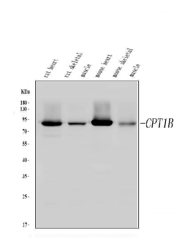
Western blot analysis of CPT1B using anti-CPT1B antibody (PB9491).
Electrophoresis was performed on a 5-20% SDS-PAGE gel at 70V (Stacking gel) / 90V (Resolving gel) for 2-3 hours. The sample well of each lane was loaded with 30 ug of sample under reducing conditions.
Lane 1: rat heart tissue lysates,
Lane 2: rat skeletal muscle tissue lysates,
Lane 3: mouse heart tissue lysates,
Lane 4: mouse skeletal muscle tissue lysates.
After electrophoresis, proteins were transferred to a nitrocellulose membrane at 150 mA for 50-90 minutes. Blocked the membrane with 5% non-fat milk/TBS for 1.5 hour at RT. The membrane was incubated with rabbit anti-CPT1B antigen affinity purified polyclonal antibody (Catalog # PB9491) at 0.5 μg/mL overnight at 4°C, then washed with TBS-0.1%Tween 3 times with 5 minutes each and probed with a goat anti-rabbit IgG-HRP secondary antibody at a dilution of 1:5000 for 1.5 hour at RT. The signal is developed using an Enhanced Chemiluminescent detection (ECL) kit (Catalog # EK1002) with Tanon 5200 system. A specific band was detected for CPT1B at approximately 88 kDa. The expected band size for CPT1B is at 88 kDa.

Click image to see more details
IHC analysis of CPT1B using anti-CPT1B antibody (PB9491).
CPT1B was detected in paraffin-embedded section of Mouse Skeletal Muscle Tissue. Heat mediated antigen retrieval was performed in citrate buffer (pH6, epitope retrieval solution) for 20 mins. The tissue section was blocked with 10% goat serum. The tissue section was then incubated with 1μg/ml rabbit anti-CPT1B Antibody (PB9491) overnight at 4°C. Biotinylated goat anti-rabbit IgG was used as secondary antibody and incubated for 30 minutes at 37°C. The tissue section was developed using Strepavidin-Biotin-Complex (SABC)(Catalog # SA1022) with DAB as the chromogen.

Click image to see more details
IHC analysis of CPT1B using anti-CPT1B antibody (PB9491).
CPT1B was detected in paraffin-embedded section of Rat Cardiac Muscle Tissue. Heat mediated antigen retrieval was performed in citrate buffer (pH6, epitope retrieval solution) for 20 mins. The tissue section was blocked with 10% goat serum. The tissue section was then incubated with 1μg/ml rabbit anti-CPT1B Antibody (PB9491) overnight at 4°C. Biotinylated goat anti-rabbit IgG was used as secondary antibody and incubated for 30 minutes at 37°C. The tissue section was developed using Strepavidin-Biotin-Complex (SABC)(Catalog # SA1022) with DAB as the chromogen.

Click image to see more details
IHC analysis of CPT1B using anti-CPT1B antibody (PB9491).
CPT1B was detected in frozen section of mouse cardiac muscle tissue. The tissue section was blocked with 10% goat serum. The tissue section was then incubated with 1μg/ml rabbit anti-CPT1B Antibody (PB9491) overnight at 4°C. Biotinylated goat anti-rabbit IgG was used as secondary antibody and incubated for 30 minutes at 37°C. The tissue section was developed using Strepavidin-Biotin-Complex (SABC)(Catalog # SA1022) with DAB as the chromogen.

Click image to see more details
IHC analysis of CPT1B using anti-CPT1B antibody (PB9491).
CPT1B was detected in frozen section of rat cardiac muscle tissue. The tissue section was blocked with 10% goat serum. The tissue section was then incubated with 1μg/ml rabbit anti-CPT1B Antibody (PB9491) overnight at 4°C. Biotinylated goat anti-rabbit IgG was used as secondary antibody and incubated for 30 minutes at 37°C. The tissue section was developed using Strepavidin-Biotin-Complex (SABC)(Catalog # SA1022) with DAB as the chromogen.

Click image to see more details
IF analysis of CPT1B using anti-CPT1B antibody (PB9491).
CPT1B was detected in a paraffin-embedded section of mouse cardiac tissue. Heat mediated antigen retrieval was performed in EDTA buffer (pH 8.0, epitope retrieval solution). The tissue section was blocked with 10% goat serum. The tissue section was then incubated with 5 μg/mL rabbit anti-CPT1B Antibody (PB9491) overnight at 4°C. Biotin conjugated goat anti-rabbit IgG (BA1003) was used as secondary antibody and incubated for 30 minutes at 37°C. The tissue section was developed using DyLight®488 Conjugated Avidin (BA1128). The section was counterstained with DAPI. Visualize using a fluorescence microscope and filter sets appropriate for the label used.

Click image to see more details
IF analysis of CPT1B using anti-CPT1B antibody (PB9491).
CPT1B was detected in a paraffin-embedded section of rat cardiac tissue. Heat mediated antigen retrieval was performed in EDTA buffer (pH 8.0, epitope retrieval solution). The tissue section was blocked with 10% goat serum. The tissue section was then incubated with 5 μg/mL rabbit anti-CPT1B Antibody (PB9491) overnight at 4°C. Biotin conjugated goat anti-rabbit IgG (BA1003) was used as secondary antibody and incubated for 30 minutes at 37°C. The tissue section was developed using DyLight®488 Conjugated Avidin (BA1128). The section was counterstained with DAPI. Visualize using a fluorescence microscope and filter sets appropriate for the label used.
Specific Publications For Anti-CPT1B Antibody Picoband® (PB9491)
Loading publications
Recommended Resources
Here are featured tools and databases that you might find useful.
- Boster's Pathways Library
- Protein Databases
- Bioscience Research Protocol Resources
- Data Processing & Analysis Software
- Photo Editing Software
- Scientific Literature Resources
- Research Paper Management Tools
- Molecular Biology Software
- Primer Design Tools
- Bioinformatics Tools
- Phylogenetic Tree Analysis
Customer Reviews
Have you used Anti-CPT1B Antibody Picoband®?
Share your experimental results or join a short interview to earn up to $1,000 in product credits or other rewards.
0 Reviews For Anti-CPT1B Antibody Picoband®
Customer Q&As
Have a question?
Find answers in Q&As, reviews.
Can't find your answer?
Submit your question
2 Customer Q&As for Anti-CPT1B Antibody Picoband®
Question
Does it work well for rat tissue in the WB method (LI-COR system)? Can you confirm the specificity of this antibody for the isoform CPT1C?
Verified Customer
Verified customer
Asked: 2019-12-18
Answer
Yes. This antibody works well for rat tissues in WB (LI-COR system) and has no cross reactivity with CPT1C.
Boster Scientific Support
Answered: 2019-12-18
Question
We are currently using anti-CPT1B antibody PB9491 for rat tissue, and we are content with the WB results. The species of reactivity given in the datasheet says human, mouse, rat. Is it possible that the antibody can work on primate tissues as well?
Verified Customer
Verified customer
Asked: 2018-06-11
Answer
The anti-CPT1B antibody (PB9491) has not been validated for cross reactivity specifically with primate tissues, but there is a good chance of cross reactivity. We have an innovator award program that if you test this antibody and show it works in primate you can get your next antibody for free. Please contact me if I can help you with anything.
Boster Scientific Support
Answered: 2018-06-11
