Click image to see more details
-

-
-
-
-
+1
Product Info Summary
| SKU: | M00252 |
|---|---|
| Size: | 100 μl |
| Reactive Species: | Human, Mouse, Rat |
| Host: | Rabbit |
| Application: | IF, ICC, WB |
Customers Who Bought This Also Bought

Product info
Product Name
Anti-FoxO3a Rabbit Monoclonal Antibody
SKU/Catalog Number
M00252
BM4734 is an alternative SKU for this antibody, used in previous lots.
Size
100 μl
Form
Liquid
Description
Boster Bio Anti-FoxO3a Rabbit Monoclonal Antibody catalog # M00252. Tested in WB, ICC/IF applications. This antibody reacts with Human, Mouse, Rat.
Storage & Handling
Store at -20°C for one year. For short term storage and frequent use, store at 4°C for up to one month. Avoid repeated freeze-thaw cycles.
Cite This Product
Anti-FoxO3a Rabbit Monoclonal Antibody (Boster Biological Technology, Pleasanton CA, USA, Catalog # M00252)
Host
Rabbit
Contents
Rabbit IgG in stabilizing components, phosphate buffered saline, pH 7.4, 150mM NaCl, 0.02% sodium azide and 50% glycerol.
*This antibody is supplied in a stabilized formulation.
Compatibility with conjugation reactions depends on the chemistry of the conjugation method used.
For conjugation methods that are not compatible with the stabilizing components present in this formulation, a carrier-free antibody format is required.
Clonality
Monoclonal
Clone Number
HFD-6
Isotype
Rabbit IgG
Immunogen
A synthesized peptide derived from human FoxO3a
Reactive Species
M00252 is reactive to FOXO3 in Human, Mouse, Rat
Observed Molecular Weight
90 kDa
Calculated molecular weight
71.3 kDa
Antibody Validation
Boster validates all antibodies on WB, IHC, ICC, Immunofluorescence, and ELISA with known positive control and negative samples to ensure specificity and high affinity, including thorough antibody incubations.
Application & Images
Applications
M00252 is guaranteed for IF, ICC, WB Boster Guarantee
Assay Dilutions Recommendation
The recommendations below provide a starting point for assay optimization. The actual working concentration varies and should be decided by the user.
WB 1:500-2000
ICC/IF 1:50-200
Positive Control
ICC/IF: Hela cell
Validation Images & Assay Conditions
Click image to see more details
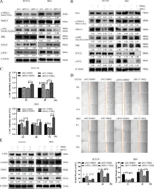
RPL5 regulates colon cancer cell proliferation and migration through MAPK/ERK signaling pathway. A HCT116 and RKO cells were treated with RPL5-targeting siRNAs (siRPL5–1 and siRPL5–2) or negative control siRNA (siNC). After 48 h, cell lysates were harvested, and the protein samples were separated by SDS-PAGE. The levels of MAPK/ERK signaling pathway related proteins P-MEK1/2, MEK1/2, P-ERK, ERK, FOXO3 and c-Myc were detected by western blotting. The band intensities of P-MEK1/2, MEK1/2, P-ERK, ERK, FOXO3 and c-Myc proteins were quantified relative to β-Tubulin and normalized to the siNC sample. The blots were cut prior to hybridization with antibodies during blotting. B TBHQ (50 μM) was added to HCT116 and RKO cells after 6 h transfection of siRPL5, and the proteins of each group were extracted 48 h later. Changes of proteins related to MAPK/ERK signaling pathway were detected by western blotting. The blots were cut prior to hybridization with antibodies during blotting. C HCT116 and RKO cells were transfected with siRPL5 and then added TBHQ (20 μM) to detect the effect of TBHQ on the proliferation inhibition of colon cancer cells caused by siRPL5 at 24 h and 48 h. D HCT116 and RKO cells were transfected with siRPL5 and then added TBHQ (20uM) to detect the effect of TBHQ on the inhibition of colon cancer cell migration caused by siRPL5 at 24 h and 48 h. (E) TBHQ (50 μM) was added to HCT116 and RKO cells after transfection of siRPL5 for 6 h, and cell cycle-related proteins (CDK4 and CyclinD1) and migration-related proteins (MMP2 and MMP9) were detected by western blotting. The band intensities of CDK4, CyclinD1, MMP2 and MMP9 proteins were quantified relative to β-Tubulin and normalized to the siNC sample. The blots were cut prior to hybridization with antibodies during blotting. The data are representative of three independent experiments. (* P < 0.05, ** P < 0.01, *** P < 0.001)
Index in PubMed under a CC BY license. PMID: 36384455

Click image to see more details
All lanes use the Antibody at 1:1K dilution for 1 hour at room temperature.

Click image to see more details
Western blot analysis of FOXO3A expression in MCF-7 cell lysate.

Click image to see more details
Immunofluorescent analysis of Hela cells, using FoxO3a Antibody.

Click image to see more details
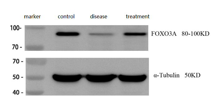
Western blot analysis of FoxO3a using anti-FoxO3a antibody (M00252).
Electrophoresis was performed on a 5-20% SDS-PAGE gel at 70V (Stacking gel) / 90V (Resolving gel) for 2-3 hours. The sample well of each lane was loaded with 30 ug of sample under reducing conditions.
Lane 1: control group-normal mouse hippocampal tissue lysates,
Lane 2: hippocampal tissue from Alzheimer’s disease model mouse,
Lane 3: hippocampal tissue from Alzheimer’s disease model mouse treated with a self-developed drug.
After electrophoresis, proteins were transferred to a nitrocellulose membrane at 150 mA for 50-90 minutes. Blocked the membrane with 5% non-fat milk/TBS for 1.5 hour at RT. The membrane was incubated with rabbit anti-FoxO3a antigen affinity purified polyclonal antibody (Catalog # PA1079) at 1:2000 overnight at 4°C, then washed with TBS-0.1%Tween 3 times with 5 minutes each and probed with a goat anti-rabbit IgG-HRP secondary antibody at a dilution of 1:10000 for 1 hour at RT. The signal is developed using an Enhanced Chemiluminescent detection (ECL) kit (Catalog # EK1002) with ChemiDoc MP system. A specific band was detected for FoxO3a at approximately 80-100 kDa. The expected band size for FoxO3a is at 71.3 kDa.
Specific Publications For Anti-FoxO3a Rabbit Monoclonal Antibody (M00252)
Loading publications
Recommended Resources
Here are featured tools and databases that you might find useful.
- Boster's Pathways Library
- Protein Databases
- Bioscience Research Protocol Resources
- Data Processing & Analysis Software
- Photo Editing Software
- Scientific Literature Resources
- Research Paper Management Tools
- Molecular Biology Software
- Primer Design Tools
- Bioinformatics Tools
- Phylogenetic Tree Analysis
Customer Reviews
Have you used Anti-FoxO3a Rabbit Monoclonal Antibody?
Share your experimental results or join a short interview to earn up to $1,000 in product credits or other rewards.
1 Reviews For Anti-FoxO3a Rabbit Monoclonal Antibody
The Anti-FOXO3A antibody (M00252) produced clear, specific bands with low background, and reliably detected expected changes in FOXO3A levels in normal, Alzheimer’s model, and treated mouse hippocampal tissues, demonstrating excellent performance.
Excellent
| SKU | M00252 |
|---|---|
| Application | Western Blot |
| Sample | mouse brain tissue |
| Sample Processing Description | ① Normal mouse hippocampal tissue, ② Hippocampal tissue from Alzheimer’s disease model mouse, ③ Hippocampal tissue from Alzheimer’s disease model mouse treated with a self-developed drug. Total protein was extracted from all samples. |
| Other Reagents | RIPA lysis buffer, Protease inhibitor, Running buffer, Transfer buffer, Blocking buffer |
| Primary Antibody | TNF alpha Antibody Picoband® |
| Primary Incubation | 1:2000, overnight at 4 ℃ |
| Secondary Antibody | HRP Conjugated AffiniPure Goat Anti-Rabbit IgG (H+L) (BA1054) |
| Secondary Incubation | 1:10000, 1 h in RT |
| Detection | Substrate: ECL substrate, Image system: ChemiDoc MP |
| Results Summary | FOXO3a is a key member of the FOXO transcription factor family, playing a central regulatory role in cellular processes such as stress response, apoptosis, autophagy, metabolism, and antioxidation. In Alzheimer’s disease, FOXO3a is known to exert neuroprotective effects. Experimental results show that FOXO3a protein levels are significantly decreased in the brains of Alzheimer’s model mice, while treatment leads to a noticeable increase. |
Huili Yin, Shandong First Medical University
Verified customer
Submitted 2026-03-30
Customer Q&As
Have a question?
Find answers in Q&As, reviews.
Can't find your answer?
Submit your question
15 Customer Q&As for Anti-FoxO3a Rabbit Monoclonal Antibody
Question
We have been able to see staining in mouse stomach. Do you have any suggestions? Is anti-FoxO3a Rabbit Monoclonal antibody supposed to stain stomach positively?
Verified Customer
Verified customer
Asked: 2020-01-15
Answer
From literature stomach does express FOXO3. From Uniprot.org, FOXO3 is expressed in trabecular bone tissue, rhabdomyosarcoma, stomach, muscle placenta, cervix carcinoma, leukemic t-cell, cervix carcinoma erythroleukemia, among other tissues. Regarding which tissues have FOXO3 expression, here are a few articles citing expression in various tissues:
Cervix carcinoma, Pubmed ID: 18220336, 18669648
Cervix carcinoma, and Erythroleukemia, Pubmed ID: 23186163
Leukemic T-cell, Pubmed ID: 19690332
Muscle, and Placenta, Pubmed ID: 15489334
Rhabdomyosarcoma, Pubmed ID: 9479491
Stomach, Pubmed ID: 14702039
Boster Scientific Support
Answered: 2020-01-15
Question
Is there a BSA free version of anti-FoxO3a Rabbit Monoclonal antibody M00252 available?
Verified Customer
Verified customer
Asked: 2019-09-23
Answer
I appreciate your recent telephone inquiry. I can confirm that some lots of this anti-FoxO3a Rabbit Monoclonal antibody M00252 are BSA free. For now, these lots are available and we can make a BSA free formula for you free of charge. It will take 3 extra days to prepare. If you require this antibody BSA free again in future, please do not hesitate to contact me and I will be pleased to check which lots we have in stock that are BSA free.
Boster Scientific Support
Answered: 2019-09-23
Question
We appreciate helping with my inquiry over the phone. Here are the WB image, lot number and protocol we used for leukemic t-cell using anti-FoxO3a Rabbit Monoclonal antibody M00252. Let me know if you need anything else.
R. Kulkarni
Verified customer
Asked: 2019-08-30
Answer
Thanks for the data. You have provided everything we needed. Our lab team are working to resolve your inquiry as quickly as possible, and we appreciate your patience and understanding! Please let me know if there is anything you need in the meantime.
Boster Scientific Support
Answered: 2019-08-30
Question
I was wanting to use your anti-FoxO3a Rabbit Monoclonal antibody for IF for rat leukemic t-cell on frozen tissues, but I want to know if it has been validated for this particular application. Has this antibody been validated and is this antibody a good choice for rat leukemic t-cell identification?
Verified Customer
Verified customer
Asked: 2019-08-09
Answer
As indicated on the product datasheet, M00252 anti-FoxO3a Rabbit Monoclonal antibody has been tested for IF, WB on human, mouse, rat tissues. We have an innovator award program that if you test this antibody and show it works in rat leukemic t-cell in IHC-frozen, you can get your next antibody for free.
Boster Scientific Support
Answered: 2019-08-09
Question
My team were happy with the WB result of your anti-FoxO3a Rabbit Monoclonal antibody. However we have been able to see positive staining in cervix carcinoma cytosol using this antibody. Is that expected? Could you tell me where is FOXO3 supposed to be expressed?
Verified Customer
Verified customer
Asked: 2019-07-09
Answer
From literature, cervix carcinoma does express FOXO3. Generally FOXO3 expresses in cytoplasm, cytosol. Regarding which tissues have FOXO3 expression, here are a few articles citing expression in various tissues:
Cervix carcinoma, Pubmed ID: 18220336, 18669648
Cervix carcinoma, and Erythroleukemia, Pubmed ID: 23186163
Leukemic T-cell, Pubmed ID: 19690332
Muscle, and Placenta, Pubmed ID: 15489334
Rhabdomyosarcoma, Pubmed ID: 9479491
Stomach, Pubmed ID: 14702039
Boster Scientific Support
Answered: 2019-07-09
Question
See attached the WB image, lot number and protocol we used for leukemic t-cell using anti-FoxO3a Rabbit Monoclonal antibody M00252. Please let me know if you require anything else.
Verified Customer
Verified customer
Asked: 2019-06-17
Answer
Thank you very much for the data. Our lab team are working to resolve this as quickly as possible, and we appreciate your patience and understanding! You have provided everything we needed. Please let me know if there is anything you need in the meantime.
Boster Scientific Support
Answered: 2019-06-17
Question
We are currently using anti-FoxO3a Rabbit Monoclonal antibody M00252 for human tissue, and we are happy with the IF results. The species of reactivity given in the datasheet says human, mouse, rat. Is it likely that the antibody can work on canine tissues as well?
Verified Customer
Verified customer
Asked: 2019-01-18
Answer
The anti-FoxO3a Rabbit Monoclonal antibody (M00252) has not been tested for cross reactivity specifically with canine tissues, but there is a good chance of cross reactivity. We have an innovator award program that if you test this antibody and show it works in canine you can get your next antibody for free. Please contact me if I can help you with anything.
Boster Scientific Support
Answered: 2019-01-18
Question
Would anti-FoxO3a Rabbit Monoclonal antibody M00252 work for IF with leukemic t-cell?
K. Evans
Verified customer
Asked: 2018-07-23
Answer
According to the expression profile of leukemic t-cell, FOXO3 is highly expressed in leukemic t-cell. So, it is likely that anti-FoxO3a Rabbit Monoclonal antibody M00252 will work for IF with leukemic t-cell.
Boster Scientific Support
Answered: 2018-07-23
Question
Our lab used your anti-FoxO3a Rabbit Monoclonal antibody for IF on stomach in the past. I am using rat, and We intend to use the antibody for WB next. I am interested in examining stomach as well as cervix carcinoma in our next experiment. Could you please give me some suggestion on which antibody would work the best for WB?
Verified Customer
Verified customer
Asked: 2018-06-21
Answer
I looked at the website and datasheets of our anti-FoxO3a Rabbit Monoclonal antibody and it appears that M00252 has been validated on rat in both IF and WB. Thus M00252 should work for your application. Our Boster satisfaction guarantee will cover this product for WB in rat even if the specific tissue type has not been validated. We do have a comprehensive range of products for WB detection and you can check out our website bosterbio.com to find out more information about them.
Boster Scientific Support
Answered: 2018-06-21
Question
Does M00252 anti-FoxO3a Rabbit Monoclonal antibody work on parafin embedded sections? If so, which fixation method do you recommend we use (PFA, paraformaldehyde, other)?
Verified Customer
Verified customer
Asked: 2018-05-03
Answer
As indicated on the product datasheet, M00252 anti-FoxO3a Rabbit Monoclonal antibody as been tested on IF. It is best to use PFA for fixation because it has better tissue penetration ability. PFA needs to be prepared fresh before use. Long term stored PFA turns into formalin, as the PFA molecules congregate and become formalin.
Boster Scientific Support
Answered: 2018-05-03
Question
I see that the anti-FoxO3a Rabbit Monoclonal antibody M00252 works with IF, what is the protocol used to produce the result images on the product page?
O. Zhao
Verified customer
Asked: 2017-12-28
Answer
You can find protocols for IF on the "support/technical resources" section of our navigation menu. If you have any further questions, please send an email to support@bosterbio.com
Boster Scientific Support
Answered: 2017-12-28
Question
My question regards to test anti-FoxO3a Rabbit Monoclonal antibody M00252 on rat leukemic t-cell for research purposes, then I may be interested in using anti-FoxO3a Rabbit Monoclonal antibody M00252 for diagnostic purposes as well. Is the antibody suitable for diagnostic purposes?
Verified Customer
Verified customer
Asked: 2017-08-14
Answer
The products we sell, including anti-FoxO3a Rabbit Monoclonal antibody M00252, are only intended for research use. They would not be suitable for use in diagnostic work. If you have the means to develop a product into diagnostic use, and are interested in collaborating with us and develop our product into an IVD product, please contact us for more discussions.
Boster Scientific Support
Answered: 2017-08-14
Question
Is a blocking peptide available for product anti-FoxO3a Rabbit Monoclonal antibody (M00252)?
R. Anderson
Verified customer
Asked: 2016-07-22
Answer
We do provide the blocking peptide for product anti-FoxO3a Rabbit Monoclonal antibody (M00252). If you would like to place an order for it please contact support@bosterbio.com and make a special request.
Boster Scientific Support
Answered: 2016-07-22
Question
Is this M00252 anti-FoxO3a Rabbit Monoclonal antibody reactive to the isotypes of FOXO3?
V. Huang
Verified customer
Asked: 2015-12-17
Answer
The immunogen of M00252 anti-FoxO3a Rabbit Monoclonal antibody is A synthesized peptide derived from human FoxO3a . Could you tell me which isotype you are interested in so I can help see if the immunogen is part of this isotype?
Boster Scientific Support
Answered: 2015-12-17
Question
I have a question about product M00252, anti-FoxO3a Rabbit Monoclonal antibody. I was wondering if it would be possible to conjugate this antibody with biotin. I would need it to be without BSA or sodium azide. I am planning on using a buffer exchange of sodium azide with PBS only. Would there be problems for me to conjugate the antibody and store it in -20 degrees in small aliquots?
A. Mitchell
Verified customer
Asked: 2013-02-01
Answer
We suggest not storing this antibody with PBS buffer only in -20 degrees. If you want to store it in -20 degrees it is best to add some cryoprotectant like glycerol. If you want carrier free M00252 anti-FoxO3a Rabbit Monoclonal antibody, we can provide it to you in a special formula with trehalose and/or glycerol. These molecules will not interfere with conjugation chemistry and provide a good level of protection for the antibody from degradation. Please be sure to specify this in your purchase order.
Boster Scientific Support
Answered: 2013-02-01
