Click image to see more details
-

-
-
-
-
+2
Product Info Summary
| SKU: | A00256-1 |
|---|---|
| Size: | 100ug |
| Reactive Species: | Human |
| Host: | Rabbit |
| Application: | ELISA, IF, IHC, WB |
Customers Who Bought This Also Bought

Product info
Product Name
Anti-HDAC-1 (C-terminus) Antibody
SKU/Catalog Number
A00256-1
Size
100ug
Form
Liquid (sterile filtered)
Description
Boster Bio Anti-HDAC-1 (C-terminus) Antibody (Catalog # A00256-1). Tested in ELISA, IF, IHC, WB applications. This antibody reacts with Human.
Storage & Handling
Store vial at -20°C prior to opening. Aliquot contents and freeze at -20°C or below for extended storage. Avoid cycles of freezing and thawing. Centrifuge product if not completely clear after standing at room temperature. This product is stable for several weeks at 4°C as an undiluted liquid. Dilute only prior to immediate use. Expiration date is one (1) year from date of opening. (Ship on dry ice.)
Cite This Product
Anti-HDAC-1 (C-terminus) Antibody (Boster Biological Technology, Pleasanton CA, USA, Catalog # A00256-1)
Host
Rabbit
Contents
0.02 M Potassium Phosphate, 0.15 M Sodium Chloride, pH 7.2, 0.01% (w/v) Sodium Azide
Clonality
Polyclonal
Isotype
IgG
Immunogen
Anti-HDAC-1 antibody was prepared from whole rabbit serum produced by repeated immunizations with a synthetic peptide corresponding to a C-Terminal region near amino acids 450-482 of Human HDAC-1.
Reactive Species
A00256-1 is reactive to HDAC1 in Human
Calculated molecular weight
55.1 kDa
Background of HDAC1
HDAC-1 antibody recognizes HDAC1 (also known as HD1, histone deacetylase 1, RPD3, RPD3L1) which belongs to the histone deacetylase/acuc/apha family and is a component of the histone deacetylase complex. Histone acetylation and deacetylation, catalyzed by multisubunit complexes, play a key role in the regulation of eukaryotic gene expression. It also interacts with retinoblastoma tumor-suppressor protein and this complex is a key element in the control of cell proliferation and differentiation. Together with metastasis-associated protein-2, it deacetylates p53 and modulates its effect on cell growth and apoptosis.
Antibody Validation
Boster validates all antibodies on WB, IHC, ICC, Immunofluorescence, and ELISA with known positive control and negative samples to ensure specificity and high affinity, including thorough antibody incubations.
Application & Images
Applications
A00256-1 is guaranteed for ELISA, IF, IHC, WB Boster Guarantee
Recommend Dilution
| Application | Dilution | Species |
|---|---|---|
| ELISA: 1:10 | 000 - 1:50 | 000 |
| IHC: 1:200 - 1:1 | 000 | |
| WB: 1:1 | 000 - 1:5 | 000 |
| Anti-HDAC-1 Antibody has been tested for use in ELISA | immunohistochemistry | immunofluorescence, and western blot. Specific conditions for reactivity should be optimized by the end user. Specific nuclear staining is observed by IHC. Expect bands at 65 kDa in size corresponding to HDAC-1 by western blotting in the appropriate cell lysate or extract. |
Validation Images & Assay Conditions
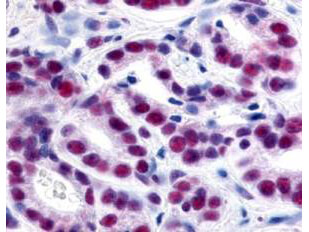
Click image to see more details
Immunohistochemistry of Rabbit Anti-HDAC-1 Antibody. Tissue: human prostate cancer tissue. Fixation: formalin fixed paraffin embedded. Antigen retrieval: not required. Primary antibody: HDAC-1 antibody at 1:500 for 1 h at RT. Secondary antibody: Peroxidase rabbit secondary antibody at 1:10,000 for 45 min at RT. Localization: HDAC-1 is nuclear. Staining: HDAC-1 precipitated purple with blue counterstain. Personal Communication, Alan Yen, LifeSpanBiosciences, Seattle, WA.

Click image to see more details
Western Blot of Rabbit Anti-HDAC-1 Antibody. Lane 1: 293 whole cell lysate . Load: 35 µg per lane. Primary antibody: HDAC-1 antibody at 1:3,500 for overnight at 4°C. Secondary antibody: IRDye800™ rabbit secondary antibody at 1:10,000 for 45 min at RT. Block: 5% BLOTTO overnight at 4°C. Predicted/Observed size: ~65 kDa corresponding to human HDAC1. Other band(s): none.

Click image to see more details
Immunohistochemistry of Rabbit Anti-HDAC-1 Antibody. Tissue: human lung tissue. Fixation: formalin fixed paraffin embedded. Antigen retrieval: not required. Primary antibody: HDAC-1 antibody at 10 µg/mL for 1 h at RT. Secondary antibody: Peroxidase rabbit secondary antibody at 1:10,000 for 45 min at RT. Localization: HDAC-1 is nuclear. Staining: HDAC-1 as brown color indicates presence of protein, blue color shows cell nuclei.

Click image to see more details
Immunofluorescence Microscopy of Rabbit Anti-HDAC-1 antibody. Fixation: 0.5% PFA. Antigen retrieval: not required. Primary antibody: HDAC-1 antibody at 10 µg/mL for 1 h at RT. Secondary antibody: rabbit secondary antibody at 1:10,000 for 45 min at RT. Localization: HDAC-1 is nuclear. Staining: HDAC-1 was used with Atto 425 (shown in red). Anti-Keratin monoclonal antibody was used with Dylight 488 (shown in green) to detect Keratin. Data was collected on a STED-CW TCS-SP5 Confocal system (Leica Microsystems).

Click image to see more details
Immunofluorescence Microscopy of Rabbit anti-HDAC1 Antibody. Tissue: A431 cells. Fixation: methanol. Antigen retrieval: blocked with normal goat serum. Primary antibody: HDAC1 antibody at 4 µg/mL for 1 h at RT. Secondary antibody: rabbit secondary antibody at 0.2 µg/mL for 45 min at RT. Localization: HDAC1 is nuclear. Staining: HDAC1 as green fluorescent signal. A-tubulin monoclonal antibody detected with ATTO 425 (colored RED). 2-color STED image, Leica Microsystems.

Click image to see more details
Western Blot of Rabbit Anti-HDAC-1 Antibody. Lane 1: LNCaP prostate cancer cells. Load: 50 µg per lane. Primary antibody: HDAC-1 antibody at 1:1000 for overnight at 4°C. Secondary antibody: IRDye800™ rabbit secondary antibody at 1:10,000 for 45 min at RT. Block: 5% BLOTTO overnight at 4°C. Predicted/Observed size: 55kDa for HDAC-1.
Specific Publications For Anti-HDAC-1 (C-terminus) Antibody (A00256-1)
Loading publications
Recommended Resources
Here are featured tools and databases that you might find useful.
- Boster's Pathways Library
- Protein Databases
- Bioscience Research Protocol Resources
- Data Processing & Analysis Software
- Photo Editing Software
- Scientific Literature Resources
- Research Paper Management Tools
- Molecular Biology Software
- Primer Design Tools
- Bioinformatics Tools
- Phylogenetic Tree Analysis
Customer Reviews
Have you used Anti-HDAC-1 (C-terminus) Antibody?
Share your experimental results or join a short interview to earn up to $1,000 in product credits or other rewards.
0 Reviews For Anti-HDAC-1 (C-terminus) Antibody
Customer Q&As
Have a question?
Find answers in Q&As, reviews.
Can't find your answer?
Submit your question
