Click image to see more details
-

-
-
-
-
+1
Product Info Summary
| SKU: | RP1062 |
|---|---|
| Size: | 100 μg/vial |
| Reactive Species: | Human, Mouse, Rat |
| Host: | Rabbit |
| Application: | WB |
Customers Who Bought This Also Bought

Product info
Product Name
Anti-Hepatocyte growth factor HGF Antibody Picoband®
SKU/Catalog Number
RP1062
PB0298 is an alternative SKU for this antibody, used in previous lots.
Size
100 μg/vial
Form
Lyophilized
Description
Boster Bio Anti-Hepatocyte growth factor HGF Antibody catalog # RP1062. Tested in WB applications. This antibody reacts with Human, Mouse, Rat. The brand Picoband indicates this is a premium antibody that guarantees superior quality, high affinity, and strong signals with minimal background in Western blot applications. Only our best-performing antibodies are designated as Picoband, ensuring unmatched performance. HGF (hepatocyte growth factor/scatter factor) is described as a paracrine cellular growth, motility, and morphogenic factor secreted by mesenchymal cells, acting primarily on epithelial and endothelial cells; states HGF activates a tyrosine kinase signaling cascade after binding the proto-oncogenic c-Met receptor, and notes roles in embryonic organ development, adult organ regeneration, and wound healing. Assay context: Human/Mouse/Rat-reactive, Western blot-validated antibody (observed MW 70 kDa; calculated 82.9 kDa; immunogen described as N-terminus peptide of mouse HGF with limited differences vs human/rat); regenerative/remodeling studies may contextualize HGF with ECM output readouts such as FN1 (fibronectin) and mitogenic ligands like EGF (parallel growth-factor axis framing), and tissue localization can be handled in an IHC context when morphology is part of the question (method-context).
Storage & Handling
Store at -20˚C for one year from date of receipt. After reconstitution, at 4˚C for one month. It can also be aliquotted and stored frozen at -20˚C for six months. Avoid repeated freeze-thaw cycles.
Cite This Product
Anti-Hepatocyte growth factor HGF Antibody Picoband® (Boster Biological Technology, Pleasanton CA, USA, Catalog # RP1062)
Host
Rabbit
Contents
Each vial contains 4 mg Trehalose, 0.9 mg NaCl and 0.2 mg Na2HPO4.
Clonality
Polyclonal
Isotype
Rabbit IgG
Immunogen
A synthetic peptide corresponding to a sequence at the N-terminus of mouse HGF, different from the related human sequence by five amino acids, and from the related rat sequence by one amino acid.
Cross-reactivity
No cross-reactivity with other proteins
Reactive Species
RP1062 is reactive to Hgf in Human, Mouse, Rat
Observed Molecular Weight
70 kDa
Calculated molecular weight
82.9 kDa
Background of Hgf
Hepatocyte growth factor/scatter factor (HGF/SF) is a paracrine cellular growth, motility and morphogenic factor. This gene is mapped to 7q21.11. It is secreted by mesenchymal cells and targets and acts primarily uponepithelial cells and endothelial cells, but also acts on haemopoietic progenitor cells. It has been shown to have a major role in embryonic organ development, specifically in myogenesis, in adult organ regeneration and in wound healing. HGF can regulate cell growth, cell motility, and morphogenesis by activating a tyrosine kinase signaling cascade after binding to the proto-oncogenic c-Met receptor. HGF also serves as a paracrine mediator to control placental development and growth. HGF was identified as one of the liver sinusoidal endothelial cell-derived paracrine mediators promoting hepatocyte growth.
Antibody Validation
Boster validates all antibodies on WB, IHC, ICC, Immunofluorescence, and ELISA with known positive control and negative samples to ensure specificity and high affinity, including thorough antibody incubations.
Application & Images
Applications
RP1062 is guaranteed for WB Boster Guarantee
Recommend Dilution
| Application | Dilution | Species |
|---|---|---|
| Western blot | 0.1-0.5μg/ml | Human, Mouse, Rat |
Tested application
Suggested blocking solution with 5% non-fat milk or BSA; (*)Recommended protein loading: 20-40 µg per lane
Validation Images & Assay Conditions
Click image to see more details
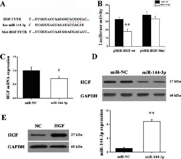
HGF is a direct target of miR-144-3p. a HGF 3′-UTR was a direct target of miR-144-3p. b Luciferase reporter assay detected the luciferase activity of cells co-transfected with the wild-type (WT) or mutant (Mut) HGF 3′-UTR reporter genes or negative control miRNA mimics (miR-NC). ** P < 0.01 versus miR-NC. c RT-qPCR was used to quantify HGF expression at mRNA levels. ** P < 0.01 versus negative control. d Western blot was used to measure HGF protein expression in A549 cells co-transfected with miR-144-3p or controls. e Western blot was used to detect the expression of HGF protein, and RT-qPCR was used to detect expression of miR-144-3p. ** P < 0.01 versus miR-NC
Index in PubMed under a CC BY license. PMID: 35568918

Click image to see more details
HGF expression levels in lung cancer tissues and various lung cancer lines. a Immunohistochemical staining was used to measure HGF protein expression. b RT-qPCR was used to quantify HGF expression at the mRNA level in tumor tissues and paired normal tissues. c RT-qPCR was used to analyze HGF expression in lung cancer cell lines and a normal lung cell line. ** P < 0.01 versus controls. Scale bar = 50 μm
Index in PubMed under a CC BY license. PMID: 35568918

Click image to see more details
Function of miR-144-3p in A549 cells. a Proliferation of A549 cells transfected with control or miR-144-3p mimics together with HGF was detected by CCK-8 assay. * P < 0.05 versus negative control miRNA mimics (miR-NC). ## P < 0.01 versus miR-144-3p + Vector. b Flow cytometry was used to analyze the cell cycle. * P < 0.05 versus miR-NC. ## P < 0.01 versus miR-144-3p + Vector. Representative images and counts of cell migration following transfection with control or miR-144-3p together with HGF, c Transwell assay and d Wound-healing assay. * P < 0.05 versus miR-NC. ## P < 0.01 versus miR-144-3p + Vector. Scale bar = 25 μm. miR-NC, miR-144, miR-144 + vector and miR-144 + HGF need to be explained in a figure
Index in PubMed under a CC BY license. PMID: 35568918

Click image to see more details
Function of miR-144-3p in the phenotype of NCI-H1299 cells. a Proliferation of NCI-H1299 cells transfected with control or miR-144-3p mimics together with HGF, as determined by CCK-8 assay. ** P < 0.01 versus miR-NC. ## P < 0.01 versus miR-144-3p + Vector. b Flow cytometry was used to analyze the cell cycle. ** P < 0.01 versus miR-NC. ## P < 0.01 versus miR-144-3p + Vector. Represented images and counts of cell migration following transfection with control or miR-144-3p mimics together with HGF, as detected by c Transwell assay and d Wound-healing assay. ** P < 0.01 versus miR-NC. ## P < 0.01 versus miR-144-3p + Vector. Scale bar = 25 μm. miR-NC, miR-144, miR-144 + vector and miR-144 + HGF need to be explained in a figure
Index in PubMed under a CC BY license. PMID: 35568918

Click image to see more details
Western blot analysis of HGF using anti-HGF antibody (RP1062).
Electrophoresis was performed on a 10% SDS-PAGE gel at 80V (Stacking gel) / 120V (Resolving gel) for 2 hours. The sample well of each lane was loaded with 30 ug of sample under reducing conditions.
Lane 1: human hepatocellular carcinoma tumor tissue (HCCT) lysates,
Lane 2: human hepatocellular carcinoma paracancerous tissue (HCCP) lysates,
Lane 3: human HepG2 whole cell lysates,
Lane 4: human HUH7 whole cell lysates,
Lane 5: rat liver tissue lysates,
Lane 6: rat RH35 whole cell lysates,
Lane 7: mouse liver tissue lysates.
After electrophoresis, proteins were transferred to a nitrocellulose membrane at 150 mA for 50-90 minutes. Blocked the membrane with 5% non-fat milk/TBS for 1.5 hour at RT. The membrane was incubated with rabbit anti-HGF antigen affinity purified polyclonal antibody (Catalog # RP1062) at 0.5 μg/mL overnight at 4°C, then washed with TBS-0.1%Tween 3 times with 5 minutes each and probed with a goat anti-rabbit IgG-HRP secondary antibody at a dilution of 1:5000 for 1.5 hour at RT. The signal is developed using an ECL Plus Western Blotting Substrate (Catalog # AR1196-200) with Tanon 5200 system. A specific band was detected for HGF at approximately 70 kDa. The expected band size for HGF is at 83 kDa.
Specific Publications For Anti-Hepatocyte growth factor HGF Antibody Picoband® (RP1062)
Loading publications
Recommended Resources
Here are featured tools and databases that you might find useful.
- Boster's Pathways Library
- Protein Databases
- Bioscience Research Protocol Resources
- Data Processing & Analysis Software
- Photo Editing Software
- Scientific Literature Resources
- Research Paper Management Tools
- Molecular Biology Software
- Primer Design Tools
- Bioinformatics Tools
- Phylogenetic Tree Analysis
Customer Reviews
Have you used Anti-Hepatocyte growth factor HGF Antibody Picoband®?
Share your experimental results or join a short interview to earn up to $1,000 in product credits or other rewards.
0 Reviews For Anti-Hepatocyte growth factor HGF Antibody Picoband®
Customer Q&As
Have a question?
Find answers in Q&As, reviews.
Can't find your answer?
Submit your question
14 Customer Q&As for Anti-Hepatocyte growth factor HGF Antibody Picoband®
Question
My question regarding product RP1062, anti-HGF antibody. I was wondering if it would be possible to conjugate this antibody with biotin. I would need it to be without BSA or sodium azide. I am planning on using a buffer exchange of sodium azide with PBS only. Would there be problems for me to conjugate the antibody and store it in -20 degrees in small aliquots?
Verified Customer
Verified customer
Asked: 2020-03-25
Answer
We do not recommend storing this antibody with PBS buffer only in -20 degrees. If you want to store it in -20 degrees it is best to add some cryoprotectant like glycerol. If you want carrier free RP1062 anti-HGF antibody, we can provide it to you in a special formula with trehalose and/or glycerol. These molecules will not interfere with conjugation chemistry and provide a good level of protection for the antibody from degradation. Please be sure to specify this in your purchase order.
Boster Scientific Support
Answered: 2020-03-25
Question
Would anti-HGF antibody RP1062 work for WB with leukocyte?
Verified Customer
Verified customer
Asked: 2020-03-19
Answer
According to the expression profile of leukocyte, HGF is highly expressed in leukocyte. So, it is likely that anti-HGF antibody RP1062 will work for WB with leukocyte.
Boster Scientific Support
Answered: 2020-03-19
Question
Is this RP1062 anti-HGF antibody reactive to the isotypes of HGF?
Verified Customer
Verified customer
Asked: 2020-03-04
Answer
The immunogen of RP1062 anti-HGF antibody is A synthetic peptide corresponding to a sequence at the N-terminus of mouse HGF (33-64aa QKKRRNTLHEFKKSAKTTLTKEDPLLKIKTKK), different from the related human sequence by five amino acids, and from the related rat sequence by one amino acid. Could you tell me which isotype you are interested in so I can help see if the immunogen is part of this isotype?
Boster Scientific Support
Answered: 2020-03-04
Question
Will RP1062 anti-HGF antibody work on parafin embedded sections? If so, which fixation method do you recommend we use (PFA, paraformaldehyde, other)?
Verified Customer
Verified customer
Asked: 2020-02-13
Answer
You can see on the product datasheet, RP1062 anti-HGF antibody as been validated on WB. It is best to use PFA for fixation because it has better tissue penetration ability. PFA needs to be prepared fresh before use. Long term stored PFA turns into formalin, as the PFA molecules congregate and become formalin.
Boster Scientific Support
Answered: 2020-02-13
Question
I was wanting to use your anti-HGF antibody for WB for mouse leukocyte on frozen tissues, but I want to know if it has been tested for this particular application. Has this antibody been tested and is this antibody a good choice for mouse leukocyte identification?
P. Edwards
Verified customer
Asked: 2019-12-27
Answer
It shows on the product datasheet, RP1062 anti-HGF antibody has been validated for WB on mouse tissues. We have an innovator award program that if you test this antibody and show it works in mouse leukocyte in IHC-frozen, you can get your next antibody for free.
Boster Scientific Support
Answered: 2019-12-27
Question
We are currently using anti-HGF antibody RP1062 for mouse tissue, and we are happy with the WB results. The species of reactivity given in the datasheet says mouse. Is it possible that the antibody can work on zebrafish tissues as well?
Verified Customer
Verified customer
Asked: 2019-06-26
Answer
The anti-HGF antibody (RP1062) has not been validated for cross reactivity specifically with zebrafish tissues, though there is a good chance of cross reactivity. We have an innovator award program that if you test this antibody and show it works in zebrafish you can get your next antibody for free. Please contact me if I can help you with anything.
Boster Scientific Support
Answered: 2019-06-26
Question
I see that the anti-HGF antibody RP1062 works with WB, what is the protocol used to produce the result images on the product page?
Verified Customer
Verified customer
Asked: 2019-02-13
Answer
You can find protocols for WB on the "support/technical resources" section of our navigation menu. If you have any further questions, please send an email to support@bosterbio.com
Boster Scientific Support
Answered: 2019-02-13
Question
We have been able to see staining in mouse embryonic fibroblast. Are there any suggestions? Is anti-HGF antibody supposed to stain embryonic fibroblast positively?
Verified Customer
Verified customer
Asked: 2018-07-09
Answer
From literature embryonic fibroblast does express HGF. From Uniprot.org, HGF is expressed in placenta, liver, leukocyte, lung fibroblast, embryonic fibroblast, brain, among other tissues. Regarding which tissues have HGF expression, here are a few articles citing expression in various tissues:
Brain, Pubmed ID: 15489334
Embryonic fibroblast, Pubmed ID: 1831266
Leukocyte, Pubmed ID: 2145836
Liver, Pubmed ID: 2531289
Lung fibroblast, Pubmed ID: 1824873
Placenta, Pubmed ID: 1826653, 2528952
Boster Scientific Support
Answered: 2018-07-09
Question
Is a blocking peptide available for product anti-HGF antibody (RP1062)?
Verified Customer
Verified customer
Asked: 2018-06-13
Answer
We do provide the blocking peptide for product anti-HGF antibody (RP1062). If you would like to place an order for it please contact support@bosterbio.com and make a special request.
Boster Scientific Support
Answered: 2018-06-13
Question
Our lab want to know about using your anti-HGF antibody for r-mmu-5673001; raf/map kinase cascade studies. Has this antibody been tested with western blotting on nih whole cell lysate? We would like to see some validation images before ordering.
Verified Customer
Verified customer
Asked: 2018-02-27
Answer
Thanks for your inquiry. This RP1062 anti-HGF antibody is validated on nih whole cell lysate. It is guaranteed to work for WB in mouse. Our Boster guarantee will cover your intended experiment even if the sample type has not been be directly tested.
Boster Scientific Support
Answered: 2018-02-27
Question
Do you have a BSA free version of anti-HGF antibody RP1062 available?
F. Zhang
Verified customer
Asked: 2017-09-13
Answer
I appreciate your recent telephone inquiry. I can confirm that some lots of this anti-HGF antibody RP1062 are BSA free. For now, these lots are available and we can make a BSA free formula for you free of charge. It will take 3 extra days to prepare. If you require this antibody BSA free again in future, please do not hesitate to contact me and I will be pleased to check which lots we have in stock that are BSA free.
Boster Scientific Support
Answered: 2017-09-13
Question
you antibody to test anti-HGF antibody RP1062 on mouse leukocyte for research purposes, then I may be interested in using anti-HGF antibody RP1062 for diagnostic purposes as well. Is the antibody suitable for diagnostic purposes?
Verified Customer
Verified customer
Asked: 2017-07-07
Answer
The products we sell, including anti-HGF antibody RP1062, are only intended for research use. They would not be suitable for use in diagnostic work. If you have the means to develop a product into diagnostic use, and are interested in collaborating with us and develop our product into an IVD product, please contact us for more discussions.
Boster Scientific Support
Answered: 2017-07-07
Question
We appreciate helping with my inquiry over the phone. Here are the WB image, lot number and protocol we used for leukocyte using anti-HGF antibody RP1062. Let me know if you need anything else.
Z. Li
Verified customer
Asked: 2017-04-10
Answer
Thanks for the data. You have provided everything we needed. Our lab team are working to resolve your inquiry as quickly as possible, and we appreciate your patience and understanding! Please let me know if there is anything you need in the meantime.
Boster Scientific Support
Answered: 2017-04-10
Question
Here is the WB image, lot number and protocol we used for leukocyte using anti-HGF antibody RP1062. Please let me know if you require anything else.
A. Roberts
Verified customer
Asked: 2013-06-07
Answer
Thank you very much for the data. Our lab team are working to resolve this as quickly as possible, and we appreciate your patience and understanding! You have provided everything we needed. Please let me know if there is anything you need in the meantime.
Boster Scientific Support
Answered: 2013-06-07
