Click image to see more details
Product Info Summary
| SKU: | PA1533 |
|---|---|
| Size: | 100 μg/vial |
| Reactive Species: | Rat |
| Host: | Rabbit |
| Application: | ELISA, WB |
Customers Who Bought This Also Bought

Product info
Product Name
Anti-IL1 beta/IL1B Antibody Picoband®
SKU/Catalog Number
PA1533
BA14789 is an alternative SKU for this antibody, used in previous lots.
Size
100 μg/vial
Form
Lyophilized
Description
Boster Bio Anti-IL1 beta/IL1B Antibody catalog # PA1533. Tested in ELISA, WB applications. This antibody reacts with Rat. The brand Picoband indicates this is a premium antibody that guarantees superior quality, high affinity, and strong signals with minimal background in Western blot applications. Only our best-performing antibodies are designated as Picoband, ensuring unmatched performance.
Storage & Handling
Store at -20˚C for one year from date of receipt. After reconstitution, at 4˚C for one month. It can also be aliquotted and stored frozen at -20˚C for six months. Avoid repeated freeze-thaw cycles.
Cite This Product
Anti-IL1 beta/IL1B Antibody Picoband® (Boster Biological Technology, Pleasanton CA, USA, Catalog # PA1533)
Host
Rabbit
Contents
Each vial contains 4 mg Trehalose, 0.9 mg NaCl and 0.2 mg Na2HPO4.
Clonality
Polyclonal
Isotype
Rabbit IgG
Immunogen
A synthetic peptide corresponding to a sequence at the C-terminus of rat IL-1 beta.
Cross-reactivity
No cross-reactivity with other proteins
Reactive Species
PA1533 is reactive to Il1b in Rat
Observed Molecular Weight
17 kDa
Calculated molecular weight
30.6 kDa
Background of Il1b
Interleukin-1 beta (IL-1beta) also know as catabolin, is a cytokine protein that in humans is encoded by the IL1B gene, which localizes to the long arm of chromosome 2 at position 2q13-2q21 between two fragile sites. Interleukin 1 (IL-1) is a protein with several biological activities regulating host defense and immune responses. The human IL-1 family plays an important role in the pathogenesis of many diseases and functions as a key mediator of the host response to various infectious, inflammatory, and immunologic challenges.
Antibody Validation
Boster validates all antibodies on WB, IHC, ICC, Immunofluorescence, and ELISA with known positive control and negative samples to ensure specificity and high affinity, including thorough antibody incubations.
Application & Images
Applications
PA1533 is guaranteed for ELISA, WB Boster Guarantee
Recommend Dilution
| Application | Dilution | Species |
|---|---|---|
| ELISA | 0.5-1μg/ml | Rat, - |
| Western blot | 0.1-0.5μg/ml | Rat |
Tested application
Suggested blocking solution with 5% non-fat milk or BSA; (*)Recommended protein loading: 20-40 µg per lane
Validation Images & Assay Conditions
Click image to see more details
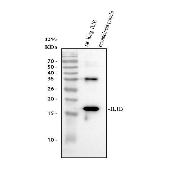
Western blot analysis of IL1B using anti-IL1B antibody (PA1533).
Electrophoresis was performed on a 13% SDS-PAGE gel at 80V (Stacking gel) / 120V (Resolving gel) for 2 hours.
Lane 1: recombinant rat IL1B protein 10 ng.
After electrophoresis, proteins were transferred to a nitrocellulose membrane at 150 mA for 50-90 minutes. Blocked the membrane with 5% non-fat milk/TBS for 1.5 hour at RT. The membrane was incubated with rabbit anti-IL1B antigen affinity purified polyclonal antibody (PA1533) at 0.5 μg/mL overnight at 4°C, then washed with TBS-0.1%Tween 3 times with 5 minutes each and probed with a goat anti-rabbit IgG-HRP secondary antibody (Catalog # BA1054) at a dilution of 1:5000 for 1.5 hour at RT. The signal is developed using an ECL Plus Western Blotting Substrate (Catalog # AR1196-200) with Tanon 5200 system. A specific band was detected for IL1B at approximately 17 kDa.
Specific Publications For Anti-IL1 beta/IL1B Antibody Picoband® (PA1533)
Loading publications
Recommended Resources
Here are featured tools and databases that you might find useful.
- Boster's Pathways Library
- Protein Databases
- Bioscience Research Protocol Resources
- Data Processing & Analysis Software
- Photo Editing Software
- Scientific Literature Resources
- Research Paper Management Tools
- Molecular Biology Software
- Primer Design Tools
- Bioinformatics Tools
- Phylogenetic Tree Analysis
Customer Reviews
Have you used Anti-IL1 beta/IL1B Antibody Picoband®?
Share your experimental results or join a short interview to earn up to $1,000 in product credits or other rewards.
0 Reviews For Anti-IL1 beta/IL1B Antibody Picoband®
Customer Q&As
Have a question?
Find answers in Q&As, reviews.
Can't find your answer?
Submit your question
5 Customer Q&As for Anti-IL1 beta/IL1B Antibody Picoband®
Question
We are currently using anti-IL1 beta/IL1B antibody PA1533 for rat tissue, and we are content with the WB results. The species of reactivity given in the datasheet says rat. Is it likely that the antibody can work on goat tissues as well?
Verified Customer
Verified customer
Asked: 2020-03-11
Answer
The anti-IL1 beta/IL1B antibody (PA1533) has not been tested for cross reactivity specifically with goat tissues, but there is a good chance of cross reactivity. We have an innovator award program that if you test this antibody and show it works in goat you can get your next antibody for free. Please contact me if I can help you with anything.
Boster Scientific Support
Answered: 2020-03-11
Question
My question regards using your anti-IL1 beta/IL1B antibody for positive regulation of cell division studies. Has this antibody been tested with western blotting on rat thymus tissue? We would like to see some validation images before ordering.
Verified Customer
Verified customer
Asked: 2020-03-05
Answer
We appreciate your inquiry. This PA1533 anti-IL1 beta/IL1B antibody is validated on rat thymus tissue, spleen tissue, tissue lysate. It is guaranteed to work for ELISA, WB in rat. Our Boster guarantee will cover your intended experiment even if the sample type has not been be directly tested.
Boster Scientific Support
Answered: 2020-03-05
Question
We ordered your anti-IL1 beta/IL1B antibody for WB on leukocyte in the past. I am using rat, and I plan to use the antibody for ELISA next. We want examining leukocyte as well as histiocytic lymphoma in our next experiment. Could give a recommendation on which antibody would work the best for ELISA?
Verified Customer
Verified customer
Asked: 2020-02-03
Answer
I looked at the website and datasheets of our anti-IL1 beta/IL1B antibody and it appears that PA1533 has been validated on rat in both WB and ELISA. Thus PA1533 should work for your application. Our Boster satisfaction guarantee will cover this product for ELISA in rat even if the specific tissue type has not been validated. We do have a comprehensive range of products for ELISA detection and you can check out our website bosterbio.com to find out more information about them.
Boster Scientific Support
Answered: 2020-02-03
Question
My boss were well pleased with the WB result of your anti-IL1 beta/IL1B antibody. However we have observed positive staining in macrophage cytoplasm using this antibody. Is that expected? Could you tell me where is IL1B supposed to be expressed?
Verified Customer
Verified customer
Asked: 2019-06-05
Answer
According to literature, macrophage does express IL1B. Generally IL1B expresses in cytoplasm, cytosol. Regarding which tissues have IL1B expression, here are a few articles citing expression in various tissues:
Histiocytic lymphoma, Pubmed ID: 3493774
Leukocyte, Pubmed ID: 3490654
Lung, Pubmed ID: 15489334
Macrophage, Pubmed ID: 20148899
Monocyte, Pubmed ID: 2635664, 11991722
Skin, Pubmed ID: 1919436
Boster Scientific Support
Answered: 2019-06-05
Question
We have observed staining in rat macrophage. Any tips? Is anti-IL1 beta/IL1B antibody supposed to stain macrophage positively?
Verified Customer
Verified customer
Asked: 2019-01-22
Answer
From what I have seen in literature macrophage does express IL1B. From what I have seen in Uniprot.org, IL1B is expressed in smooth muscle tissue, leukocyte, histiocytic lymphoma, monocyte, lung, skin, macrophage, among other tissues. Regarding which tissues have IL1B expression, here are a few articles citing expression in various tissues:
Histiocytic lymphoma, Pubmed ID: 3493774
Leukocyte, Pubmed ID: 3490654
Lung, Pubmed ID: 15489334
Macrophage, Pubmed ID: 20148899
Monocyte, Pubmed ID: 2635664, 11991722
Skin, Pubmed ID: 1919436
Boster Scientific Support
Answered: 2019-01-22
