Click image to see more details
-

-
-
-
-
+10
Product Info Summary
| SKU: | PA1352 |
|---|---|
| Size: | 100 μg/vial |
| Reactive Species: | Mouse, Rat |
| Host: | Rabbit |
| Application: | IHC, WB |
Customers Who Bought This Also Bought

Product info
Product Name
Anti-Interleukin-6 IL6 Antibody Picoband®
SKU/Catalog Number
PA1352
BA4339 is an alternative SKU for this antibody, used in previous lots.
Size
100 μg/vial
Form
Lyophilized
Description
Boster Bio Anti-Interleukin-6 IL6 Antibody catalog # PA1352. Tested in IHC, WB applications. This antibody reacts with Mouse, Rat. The brand Picoband indicates this is a premium antibody that guarantees superior quality, high affinity, and strong signals with minimal background in Western blot applications. Only our best-performing antibodies are designated as Picoband, ensuring unmatched performance.
Storage & Handling
Store at -20˚C for one year from date of receipt. After reconstitution, at 4˚C for one month. It can also be aliquotted and stored frozen at -20˚C for six months. Avoid repeated freeze-thaw cycles.
Cite This Product
Anti-Interleukin-6 IL6 Antibody Picoband® (Boster Biological Technology, Pleasanton CA, USA, Catalog # PA1352)
Host
Rabbit
Contents
Each vial contains 4 mg Trehalose, 0.9 mg NaCl and 0.2 mg Na2HPO4.
Clonality
Polyclonal
Isotype
Rabbit IgG
Immunogen
A synthetic peptide corresponding to a sequence at the C-terminus of rat IL6, different from the related mouse sequence by two amino acids.
Cross-reactivity
No cross-reactivity with other proteins
Reactive Species
PA1352 is reactive to Il6 in Mouse, Rat
Observed Molecular Weight
28-30 kDa
Calculated molecular weight
24.4 kDa
Background of Il6
Interleukin-6 (IL-6) is a protein that in humans is encoded by the IL6 gene. IL-6 is an interleukin that acts as both a pro-inflammatory and anti-inflammatory cytokine. It is secreted by T cells and macrophages to stimulate immune response to trauma, especially burns or other tissue damage leading to inflammation. IL-6 is one of the most important mediators of fever and of the acute phase response. IL-6 is also essential for hybridoma growth and is found in many supplemental cloning media such as briclone. Bowcock et al. (1988) assigned the IL6 gene to chromosome 7p21. By in situ hybridization and Southern blot analysis of mouse-human hybrid cell lines, Sutherland et al. (1988) mapped the IL6 gene to chromosome 7p15.
Antibody Validation
Boster validates all antibodies on WB, IHC, ICC, Immunofluorescence, and ELISA with known positive control and negative samples to ensure specificity and high affinity, including thorough antibody incubations.
Application & Images
Applications
PA1352 is guaranteed for IHC, WB Boster Guarantee
Assay Dilutions Recommendation
The recommendations below provide a starting point for assay optimization. The actual working concentration varies and should be decided by the user.
Western blot, 0.1-0.5μg/ml, Mouse, Rat
Immunohistochemistry (Paraffin-embedded Section), 2-5μg/ml, Human
Positive Control
WB: recombinant rat IL6 protein, mouse spleen tissue, rat thymus tissue, rat spleen tissue
IHC: rat spleen tissue
Validation Images & Assay Conditions
Click image to see more details
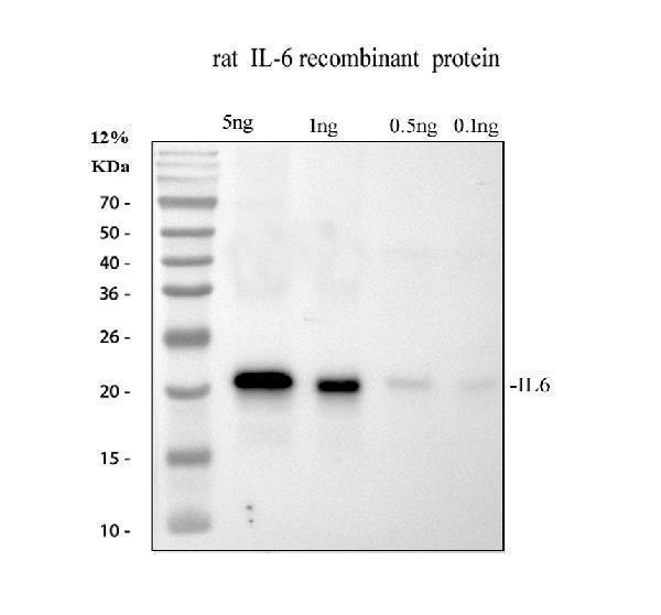
Western blot analysis of IL6 using anti-IL6 antibody (PA1352).
Electrophoresis was performed on a 12% SDS-PAGE gel at 80V (Stacking gel) / 120V (Resolving gel) for 2 hours.
Lane 1: recombinant rat IL6 protein 5 ng.
Lane 2: recombinant rat IL6 protein 10 ng.
Lane 3: recombinant rat IL6 protein 0.5 ng.
Lane 4: recombinant rat IL6 protein 0.1 ng.
After electrophoresis, proteins were transferred to a nitrocellulose membrane at 150 mA for 50-90 minutes. Blocked the membrane with 5% non-fat milk/TBS for 1.5 hour at RT. The membrane was incubated with rabbit anti-IL6 antigen affinity purified polyclonal antibody (PA1352) at 0.5 μg/mL overnight at 4°C, then washed with TBS-0.1%Tween 3 times with 5 minutes each and probed with a goat anti-rabbit IgG-HRP secondary antibody (Catalog # BA1054) at a dilution of 1:5000 for 1.5 hour at RT. The signal is developed using an ECL Plus Western Blotting Substrate (Catalog # AR1196-200) with Tanon 5200 system. A specific band was detected for IL6 at approximately 22 kDa.

Click image to see more details
Western blot analysis of IL6 using anti-IL6 antibody (PA1352).
Electrophoresis was performed on a 12% SDS-PAGE gel at 80V (Stacking gel) / 120V (Resolving gel) for 2 hours. The sample well of each lane was loaded with 30 ug of sample under reducing conditions.
Lane 1: mouse spleen tissue lysates.
After electrophoresis, proteins were transferred to a nitrocellulose membrane at 150 mA for 50-90 minutes. Blocked the membrane with 5% non-fat milk/TBS for 1.5 hour at RT. The membrane was incubated with rabbit anti-IL6 antigen affinity purified polyclonal antibody (PA1352) at 0.5 μg/mL overnight at 4°C, then washed with TBS-0.1%Tween 3 times with 5 minutes each and probed with a goat anti-rabbit IgG-HRP secondary antibody (Catalog # BA1054) at a dilution of 1:5000 for 1.5 hour at RT. The signal is developed using an ECL Plus Western Blotting Substrate (Catalog # AR1196-200) with Tanon 5200 system. A specific band was detected for IL6 at approximately 28-30 kDa. The expected band size for IL6 is at 24 kDa.

Click image to see more details
Western blot analysis of IL6 using anti-IL6 antibody (PA1352).
Electrophoresis was performed on a 12% SDS-PAGE gel at 80V (Stacking gel) / 120V (Resolving gel) for 2 hours. The sample well of each lane was loaded with 30 ug of sample under reducing conditions.
Lane 1: rat thymus tissue lysates,
Lane 2: rat spleen tissue lysates.
After electrophoresis, proteins were transferred to a nitrocellulose membrane at 150 mA for 50-90 minutes. Blocked the membrane with 5% non-fat milk/TBS for 1.5 hour at RT. The membrane was incubated with rabbit anti-IL6 antigen affinity purified polyclonal antibody (PA1352) at 0.5 μg/mL overnight at 4°C, then washed with TBS-0.1%Tween 3 times with 5 minutes each and probed with a goat anti-rabbit IgG-HRP secondary antibody (Catalog # BA1054) at a dilution of 1:5000 for 1.5 hour at RT. The signal is developed using an ECL Plus Western Blotting Substrate (Catalog # AR1196-200) with Tanon 5200 system. A specific band was detected for IL6 at approximately 28-30 kDa. The expected band size for IL6 is at 24 kDa.

Click image to see more details
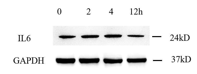
Western blot analysis of IL6 using anti-IL6 antibody (PA1352).
Electrophoresis was performed on a 12% SDS-PAGE gel at 80V (Stacking gel) / 120V (Resolving gel) for 2 hours. The sample well of each lane was loaded with 30 ug of sample under reducing conditions.
Lane 1: human MCF-7 whole cell lysates,
Lane 2: 2h drug treated-human MCF-7 whole cell lysates,
Lane 3: 4h drug treated-human MCF-7 whole cell lysates,
Lane 4: 12h drug treated-human MCF-7 whole cell lysates.
After electrophoresis, proteins were transferred to a nitrocellulose membrane at 150 mA for 50-90 minutes. Blocked the membrane with 5% non-fat milk/TBS for 1.5 hour at RT. The membrane was incubated with rabbit anti-IL6 antigen affinity purified polyclonal antibody (PA1352) at 1:500 overnight at 4°C, then washed with TBS-0.1%Tween 3 times with 5 minutes each and probed with a goat anti-rabbit IgG-HRP secondary antibody (Catalog # BA1054) at a dilution of 1:2000 for 1 hour at RT. The signal is developed using an ECL Plus Western Blotting Substrate (Catalog # AR1196-200) with ChemiDoc MP system system. A specific band was detected for IL6 at 24 kDa. The expected band size for IL6 is at 24 kDa.

Click image to see more details
IHC analysis of IL6 using anti-IL6 antibody (PA1352).
IL6 was detected in a paraffin-embedded section of rat spleen tissue. Heat mediated antigen retrieval was performed in EDTA buffer (pH 8.0, epitope retrieval solution). The tissue section was blocked with 10% goat serum. The tissue section was then incubated with 2 μg/ml rabbit anti-IL6 Antibody (PA1352) overnight at 4°C. Peroxidase Conjugated Goat Anti-rabbit IgG was used as secondary antibody and incubated for 30 minutes at 37°C. The tissue section was developed using HRP Conjugated Rabbit IgG Super Vision Assay Kit (Catalog # SV0002) with DAB as the chromogen.

Click image to see more details
Histological evaluation and statistical analysis of diabetic wound after Mn3O4 treatment by immunohistochemical staining. Immunohistochemical staining of (A) IL-6, (B) CD86 and (C) CD206 in the wound bed at day 7. (D–F) Statistical results of immunohistochemical staining in each group. Data are presented as mean ± SD. Statistical significance was determined using t-test (n = 3, ns = no significance, ***P < 0.001, ****P < 0.0001).
Index in Regenerative Biomaterials under a CC BY license. DOI: 10.1093/rb/rbaf089

Click image to see more details
Collagen deposition, inflammation, and angiogenesis analyses in acute large-area wounds. (A) Masson staining and immunofluorescence of IL-6 (red) and CD31 (red) and a-SMA (green) in wound samples. Scale bars: 100 μm (top) and 50 μm (middle and bottom). (B to D) Quantification assessment of (B) collagen deposition, (C) IL-6 expression, and (D) CD31 expression. **P < 0.01, ***P < 0.001.
Index in PubMed under a CC BY license. PMID: 39109247

Click image to see more details
Relative expression of IL-6, caspase-3, and cleaved-caspase-3. (A) Immunoblot results of IL-6, caspase-3, and cleaved-caspase-3. (B) Analysis of IL-6 relative expression. One-way ANOVA showed differences among the five groups, F = 10.86, p < 0.0001. (C) Analysis of caspase-3 relative expression. One-way ANOVA showed differences among the five groups, F = 8.50, p = 0.0004. (D) Analysis of cleaved-caspase-3 relative expression. One-way ANOVA showed differences among the five groups, F = 17.36, p < 0.0001. ANOVA, analysis of variance; caspase-3, cysteinyl aspartate specific proteinase 3; IL-6, interleukin 6.
Index in PubMed under a CC BY license. PMID: 34975719

Click image to see more details
Experimental workflow. One hundred four rats were randomly divided into five groups: group S (sham, n = 20), group M (middle cerebral artery occlusion [MCAO], n = 28), group H2M (intermittent hypobaric hypoxia preconditioned MCAO group, 2 h/day, n = 20), group H6M (intermittent hypobaric hypoxia preconditioned MCAO group, 6 h/day, n = 28), and group HpM (persistent hypobaric hypoxia preconditioned MCAO group, n = 28). Behavioral tests and morphological staining (TTC staining) were used to analyze the severity of infarction. Total protein expression of NeuN (a specific marker of mature neurons), caspase-3, cleaved-caspase-3, and IL-6 was estimated using western blotting, which explained the severity of injury from different perspectives. Ultrastructural changes were observed under a transmission electron microscope. The most effective pretreatment group was selected for further label-free proteomic study and provided a reliable direction for mechanism exploration. Western blotting was used to verify the expression of the target protein, and key markers for the biological process were detected using immunofluorescence. caspase-3, cysteinyl aspartate specific proteinase 3; IL-6, interleukin 6; NeuN, neuron-specific nuclear protein; TTC, 2,3,5-triphenyl tetrazolium chloride.
Index in PubMed under a CC BY license. PMID: 34975719

Click image to see more details
Inhibitive effect of nanofiber membranes on anti-inflammation in Il-1β induced chondrocytes. a Relative mRNA expression levels of Il6, Tnf-a, iNos, Mmp1, Mmp3, and Mmp13 in IL-1β induced chondrocytes cultured on P, PGF, PF, PPF, or PPGF nanofiber membranes. b Protein expression of MMP13, TNF-a, and IL6 in IL-1β induced chondrocytes cultured on P, PGF, PF, PPF, or PPGF nanofiber membranes (Scale bars, 100 µm). The values were presented as mean ± SD ( n = 3; statistics: one-way ANOVA; # means p < 0.01, ** , ## means p < 0.01, *** , ### means p < 0.001, * is the statistical difference compare with P group and # is the statistical difference between the pairwise comparison).
Index in PubMed under a CC BY license. PMID: 36323709

Click image to see more details
MT inhibited the production of pro-inflammatory proteins in sleep-deprived rats. (A) Western blot bands showing the protein expression levels of IL-1β, IL-6, TNF-α, iNOS, and COX2 in the HP, respectively. (B–F) Relative protein expression level of IL-1β, IL-6, TNF-α, iNOS, and COX2 in the HP, respectively. The data are expressed as the means ± SEM. # p < 0.05, ## p < 0.01, ### p < 0.001 vs Control group; * p < 0.05, ** p < 0.01 vs. Model group.
Index in PubMed under a CC BY license. PMID: 39101143

Click image to see more details
Therapeutic effectiveness evaluated after administering AE@SiO 2 -MTX. (A) Knee joints were stained with HE and safranin O. Scale bar: 100 μm. (B) TNF-α, IL-6, MMP3 and MMP13 and DAPI staining applied to knee sections. (C) Line profile analysis verified the co-expression and co-localization of TNF-α and IL-6, MMP3 and MMP13. Scale bar: 50 μm.
Index in PubMed under a CC BY license. PMID: 40521182

Click image to see more details
Intravenous administration of AE@SiO 2 -MTX alleviated arthritis symptoms in CIA mice. (A) Visual outline of the in vivo treatment procedure. (B-D). Quantification of paw thickness, paw volume and arthritis score at multiple intervals after treatments. (E) Illustrative images of hindlimbs from each group before and after treatments. (F) Micro-CT images in 3D of fore paws, hind paws, and knee joints for different treatment groups. (G-H) The evaluation of BMD levels in six groups post-treatments. (I-J) Plasma levels of IL-6 and IL-10 after treatments. Data were expressed as mean±SD. Data were analyzed for statistical significance via One-way ANOVA. * P < 0.05, ** P < 0.01, *** P < 0.001, **** P < 0.0001.
Index in PubMed under a CC BY license. PMID: 40521182

Click image to see more details
PF-127/hADSCs-Exos complex treatment inhibits inflammatory reaction. a Representative images of TNF-α immunostaining at 4, 7, and 10 days after treatment. Scale bar = 20 µm. b Quantification of TNF-α + IHC stained tissues. c Representative images illustrating IHC results of IL-6 at 4, 7, and 10 days after surgery. Scale bar = 20 µm. d Quantification of IL-6 + IHC stained tissues. e IHC images of wound sections stained with CD68 on days 4, 7, and 10 post-wounding. Scale bar = 20 µm. f Quantification of the number of CD68 positive cells in the wound area on days 4, 7, and 10. g IHC images of wound sections stained with CD206 at days 4, 7, and 10 post-wounding. Scale bar = 20 µm. h Quantification of the number of CD206 positive cells in the wound area on days 4, 7, and 10. In b, d, and f , data are shown as mean ± SEM; n = 6 for each group. * p < 0.05, ** p < 0.01, *** p < 0.001, and **** p < 0.0001 versus vehicle control group
Index in PubMed under a CC BY license. PMID: 35941707
Specific Publications For Anti-Interleukin-6 IL6 Antibody Picoband® (PA1352)
Loading publications
Recommended Resources
Here are featured tools and databases that you might find useful.
- Boster's Pathways Library
- Protein Databases
- Bioscience Research Protocol Resources
- Data Processing & Analysis Software
- Photo Editing Software
- Scientific Literature Resources
- Research Paper Management Tools
- Molecular Biology Software
- Primer Design Tools
- Bioinformatics Tools
- Phylogenetic Tree Analysis
Customer Reviews
Have you used Anti-Interleukin-6 IL6 Antibody Picoband®?
Share your experimental results or join a short interview to earn up to $1,000 in product credits or other rewards.
1 Reviews For Anti-Interleukin-6 IL6 Antibody Picoband®
This antibody exhibits high specificity and efficiency and is suitable for Western blot detection of IL6 protein in MCF-7 cells, yielding clean and well-defined target bands.
Excellent
| SKU | PA1352 |
|---|---|
| Application | Western Blot |
| Sample | Human mcf-7 cells |
| Sample Processing Description | RIPA lysis buffer supplemented with a protease inhibitor cocktail was added to the cell culture dish, and the cells were lysed on ice for 30 min. The lysates were then centrifuged, and the supernatants were collected. Protein concentration was determined, and samples were adjusted to equal concentrations. Subsequently, 5× protein loading buffer was added, and the samples were denatured at 100 °C for 10 min. A total of 15 μL of protein sample was loaded per lane for SDS-PAGE electrophoresis. |
| Other Reagents | Blocking buffer |
| Primary Antibody | CRCP Rabbit Monoclonal Antibody |
| Primary Incubation | 1:2000, overnight at 4 ℃ |
| Secondary Antibody | HRP Conjugated AffiniPure Goat Anti-Rabbit IgG (H+L) |
| Secondary Incubation | 1 hour in room temperature |
| Detection | Substrate: ECL, Imaging system:ChemiDoc MP |
| Results Summary | As shown in the figure, the expression levels of CRCP protein in MCF-7 cells at different drug treatment time points are presented. The Western blot results obtained with this antibody are clear; although there are a few faint nonspecific bands above, they do not affect result interpretation. It can be observed that the expression of the target protein in MCF-7 cells increases with longer drug treatment times. |
Siyuan Bu, LUTCM
Verified customer
Submitted 2025-12-26
Customer Q&As
Have a question?
Find answers in Q&As, reviews.
Can't find your answer?
Submit your question
15 Customer Q&As for Anti-Interleukin-6 IL6 Antibody Picoband®
Question
We appreciate helping with my inquiry over the phone. Here are the WB image, lot number and protocol we used for lung using anti-IL6 antibody PA1352. Let me know if you need anything else.
A. Moore
Verified customer
Asked: 2020-04-16
Answer
We appreciate the data. You have provided everything we needed. Our lab team are working to resolve your inquiry as quickly as possible, and we appreciate your patience and understanding! Please let me know if there is anything you need in the meantime.
Boster Scientific Support
Answered: 2020-04-16
Question
Is this PA1352 anti-IL6 antibody reactive to the isotypes of IL6?
Verified Customer
Verified customer
Asked: 2020-03-27
Answer
The immunogen of PA1352 anti-IL6 antibody is A synthetic peptide corresponding to a sequence at the C-terminus of rat IL6(195-211aa KALEEFLKVTMRSTRQT), different from the related mouse sequence by two amino acids. Could you tell me which isotype you are interested in so I can help see if the immunogen is part of this isotype?
Boster Scientific Support
Answered: 2020-03-27
Question
Do you have a BSA free version of anti-IL6 antibody PA1352 available?
Verified Customer
Verified customer
Asked: 2020-03-24
Answer
I appreciate your recent telephone inquiry. I can confirm that some lots of this anti-IL6 antibody PA1352 are BSA free. For now, these lots are available and we can make a BSA free formula for you free of charge. It will take 3 extra days to prepare. If you require this antibody BSA free again in future, please do not hesitate to contact me and I will be pleased to check which lots we have in stock that are BSA free.
Boster Scientific Support
Answered: 2020-03-24
Question
Will anti-IL6 antibody PA1352 work on zebrafish WB with left coronary artery?
Verified Customer
Verified customer
Asked: 2020-02-14
Answer
Our lab technicians have not validated anti-IL6 antibody PA1352 on zebrafish. You can run a BLAST between zebrafish and the immunogen sequence of anti-IL6 antibody PA1352 to see if they may cross-react. If the sequence homology is close, then you can perform a pilot test. Keep in mind that since we have not validated zebrafish samples, this use of the antibody is not covered by our guarantee. However we have an innovator award program that if you test this antibody and show it works in zebrafish left coronary artery in WB, you can get your next antibody for free.
Boster Scientific Support
Answered: 2020-02-14
Question
I was wanting to use your anti-IL6 antibody for WB for mouse lung on frozen tissues, but I want to know if it has been validated for this particular application. Has this antibody been validated and is this antibody a good choice for mouse lung identification?
Verified Customer
Verified customer
Asked: 2020-01-15
Answer
It shows on the product datasheet, PA1352 anti-IL6 antibody has been validated for WB on mouse, rat tissues. We have an innovator award program that if you test this antibody and show it works in mouse lung in IHC-frozen, you can get your next antibody for free.
Boster Scientific Support
Answered: 2020-01-15
Question
Is a blocking peptide available for product anti-IL6 antibody (PA1352)?
Verified Customer
Verified customer
Asked: 2019-09-16
Answer
We do provide the blocking peptide for product anti-IL6 antibody (PA1352). If you would like to place an order for it please contact support@bosterbio.com and make a special request.
Boster Scientific Support
Answered: 2019-09-16
Question
We are currently using anti-IL6 antibody PA1352 for rat tissue, and we are satisfied with the WB results. The species of reactivity given in the datasheet says mouse, rat. Is it likely that the antibody can work on goat tissues as well?
L. Gonzalez
Verified customer
Asked: 2019-09-03
Answer
The anti-IL6 antibody (PA1352) has not been tested for cross reactivity specifically with goat tissues, though there is a good chance of cross reactivity. We have an innovator award program that if you test this antibody and show it works in goat you can get your next antibody for free. Please contact me if I can help you with anything.
Boster Scientific Support
Answered: 2019-09-03
Question
We need to test anti-IL6 antibody PA1352 on mouse lung for research purposes, then I may be interested in using anti-IL6 antibody PA1352 for diagnostic purposes as well. Is the antibody suitable for diagnostic purposes?
Verified Customer
Verified customer
Asked: 2019-08-06
Answer
The products we sell, including anti-IL6 antibody PA1352, are only intended for research use. They would not be suitable for use in diagnostic work. If you have the means to develop a product into diagnostic use, and are interested in collaborating with us and develop our product into an IVD product, please contact us for more discussions.
Boster Scientific Support
Answered: 2019-08-06
Question
See below the WB image, lot number and protocol we used for lung using anti-IL6 antibody PA1352. Please let me know if you require anything else.
Verified Customer
Verified customer
Asked: 2019-06-07
Answer
Thank you very much for the data. Our lab team are working to resolve this as quickly as possible, and we appreciate your patience and understanding! You have provided everything we needed. Please let me know if there is anything you need in the meantime.
Boster Scientific Support
Answered: 2019-06-07
Question
I see that the anti-IL6 antibody PA1352 works with WB, what is the protocol used to produce the result images on the product page?
Verified Customer
Verified customer
Asked: 2019-03-08
Answer
You can find protocols for WB on the "support/technical resources" section of our navigation menu. If you have any further questions, please send an email to support@bosterbio.com
Boster Scientific Support
Answered: 2019-03-08
Question
Does anti-IL6 antibody PA1352 work for WB with lung?
Verified Customer
Verified customer
Asked: 2018-12-03
Answer
According to the expression profile of lung, IL6 is highly expressed in lung. So, it is likely that anti-IL6 antibody PA1352 will work for WB with lung.
Boster Scientific Support
Answered: 2018-12-03
Question
I have a question about product PA1352, anti-IL6 antibody. I was wondering if it would be possible to conjugate this antibody with biotin. I would need it to be without BSA or sodium azide. I am planning on using a buffer exchange of sodium azide with PBS only. Would there be problems for me to conjugate the antibody and store it in -20 degrees in small aliquots?
Verified Customer
Verified customer
Asked: 2018-10-03
Answer
We do not recommend storing this antibody with PBS buffer only in -20 degrees. If you want to store it in -20 degrees it is best to add some cryoprotectant like glycerol. If you want carrier free PA1352 anti-IL6 antibody, we can provide it to you in a special formula with trehalose and/or glycerol. These molecules will not interfere with conjugation chemistry and provide a good level of protection for the antibody from degradation. Please be sure to specify this in your purchase order.
Boster Scientific Support
Answered: 2018-10-03
Question
We have seen staining in mouse left coronary artery. Are there any suggestions? Is anti-IL6 antibody supposed to stain left coronary artery positively?
G. Mangal
Verified customer
Asked: 2015-07-30
Answer
From literature left coronary artery does express IL6. From Uniprot.org, IL6 is expressed in left coronary artery, fibroblast, lung, among other tissues. Regarding which tissues have IL6 expression, here are a few articles citing expression in various tissues:
Fibroblast, Pubmed ID: 3758081
Lung, Pubmed ID: 15489334
Boster Scientific Support
Answered: 2015-07-30
Question
Our team were satisfied with the WB result of your anti-IL6 antibody. However we have been able to see positive staining in left coronary artery secreted. using this antibody. Is that expected? Could you tell me where is IL6 supposed to be expressed?
L. Jones
Verified customer
Asked: 2014-07-09
Answer
From what I have seen in literature, left coronary artery does express IL6. Generally IL6 expresses in secreted. Regarding which tissues have IL6 expression, here are a few articles citing expression in various tissues:
Fibroblast, Pubmed ID: 3758081
Lung, Pubmed ID: 15489334
Boster Scientific Support
Answered: 2014-07-09
Question
Would PA1352 anti-IL6 antibody work on parafin embedded sections? If so, which fixation method do you recommend we use (PFA, paraformaldehyde, other)?
B. Carter
Verified customer
Asked: 2013-01-31
Answer
It shows on the product datasheet, PA1352 anti-IL6 antibody as been validated on WB. It is best to use PFA for fixation because it has better tissue penetration ability. PFA needs to be prepared fresh before use. Long term stored PFA turns into formalin, as the PFA molecules congregate and become formalin.
Boster Scientific Support
Answered: 2013-01-31
