Click image to see more details
-

-
-
-
-
+7
Product Info Summary
| SKU: | A00683 |
|---|---|
| Size: | 0.1 mg |
| Reactive Species: | Human, Mouse, Rat |
| Host: | Rabbit |
| Application: | ELISA, IF, IHC-P, ICC, WB |
Customers Who Bought This Also Bought

Product info
Product Name
Anti-IRE1p ERN1 Antibody
SKU/Catalog Number
A00683
Size
0.1 mg
Form
Liquid
Description
Boster Bio Anti-IRE1p ERN1 Antibody (Catalog # A00683). Tested in ELISA, WB, ICC, IF, IHC-P applications. This antibody reacts with Human, Mouse, Rat.
Storage & Handling
IRE1p antibody can be stored at 4°C for three months and -20°C, stable for up to one year. Avoid repeated freeze-thaw cycles. Antibodies should not be exposed to prolonged high temperatures.
Cite This Product
Anti-IRE1p ERN1 Antibody (Boster Biological Technology, Pleasanton CA, USA, Catalog # A00683)
Host
Rabbit
Contents
IRE1p Antibody is supplied in PBS containing 0.02% sodium azide.
Clonality
Polyclonal
Isotype
IgG
Immunogen
Anti-IRE1p antibody was raised against a peptide corresponding to 16 amino acids near the carboxy terminus of human IRE1P. The immunogen is located within the last 50 amino acids of IRE1p.
Reactive Species
A00683 is reactive to ERN1 in Human, Mouse, Rat
Observed Molecular Weight
68 kDa
Calculated molecular weight
109.7 kDa
Background of ERN1
Accumulation of malfolded proteins in the endoplasmic reticulum (ER) activates the unfolded protein response (UPR) and the upregulation of the ER molecular chaperones GRP78 and GRP 94. These proteins are normally bound to ER transmembrane proteins such as IRE1p and ATF6 but ER stress causes their dissociation. This allows IRE1p, a serine-threonine protein kinase to transduce the unfolded protein signal from the ER to the nucleus. IRE1p also has an endoribonuclease activity that is required to splice X-box binding protein (XBP1) mRNA converting it to a potent UPR transcriptional activation. Depletion of IRE1p through the expression of a dominant negative form of IRE1p has no effect on transfected cells, but cell death via apoptosis occurs under stress conditions that cause unfolded proteins to accumulate in the ER. Two alternatively spliced transcript variants encoding different isoforms have been found for this gene.
Antibody Validation
Boster validates all antibodies on WB, IHC, ICC, Immunofluorescence, and ELISA with known positive control and negative samples to ensure specificity and high affinity, including thorough antibody incubations.
Application & Images
Applications
A00683 is guaranteed for ELISA, IF, IHC-P, ICC, WB Boster Guarantee
Recommend Dilution
| Application | Dilution | Species |
|---|---|---|
| Antibody validated: Western Blot in human | mouse and rat samples; Immunocytochemistry in mouse samples; Immunofluorescence in human | mouse and rat samples; Immunohistochemistry in human and rat samples. All other applications and species not yet tested. Optimal dilutions for each application should be determined by the researcher. |
Validation Images & Assay Conditions
Click image to see more details
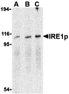
Western Blot Validation in Mouse A20 Cell Lysate
Loading: 15 μg of lysates per lane.
Antibodies: IRE1p A00683 (A: 0.5 μg/mL, B: 1 μg/mL, C: 2 μg/mL), 1h incubation at RT in 5% NFDM/TBST.
Secondary: Goat anti-rabbit IgG HRP conjugate at 1:10000 dilution.

Click image to see more details
KO Validation in HeLa Cells
Loading: 10 μg of WT cell lysates (lane 1) or IRE1P KO cell lysates (lane 2).
Antibodies: IRE1P A00683 (0.5 μg/mL) and beta-actin (1 μg/mL), 1h incubation at RT in 5% NFDM/TBST.
Secondary: Goat anti-rabbit IgG HRP conjugate at 1:10000 dilution.

Click image to see more details
Western Blot Validation in Human Cell Lines
Loading: 15 μg of lysates per lane.
Antibodies: IRE1p A00683 (0.4 μg/mL), 1h incubation at RT in 5% NFDM/TBST.
Secondary: Goat anti-rabbit IgG HRP conjugate at 1:10000 dilution.
Lane 1: Caco-2,
Lane2: SK-N-SH

Click image to see more details
Western Blot Validation in Rat Brain Tissue Lysate
Loading: 15 μg of lysates per lane.
Antibodies: IRE1p A00683 (A: 0.5 μg/mL, B: 1 μg/mL), 1h incubation at RT in 5% NFDM/TBST.
Secondary: Goat anti-rabbit IgG HRP conjugate at 1:10000 dilution.

Click image to see more details
Immunofluorescence Validation of IRE1p in Mouse A20 Cells
Immunofluorescent analysis of 4% paraformaldehyde-fixed A20 Cells labeling IRE1P with A00683 at 2 μg/mL, followed by goat anti-rabbit IgG secondary antibody at 1/500 dilution (red).

Click image to see more details
Immunocytochemistry Validation of IRE1p in Mouse A20 Cells
Immunocytochemical analysis of A20 cells using anti-IRE1p antibody (A00683) at 1 μg/ml. Cells was fixed with formaldehyde and blocked with 10% serum for 1 h at RT; antigen retrieval was by heat mediation with a citrate buffer (pH6). Samples were incubated with primary antibody overnight at 4˚C. A goat anti-rabbit IgG H&L (HRP) at 1/250 was used as secondary. Counter stained with Hematoxylin.

Click image to see more details
Immunofluorescence Validation of IRE1p in Human Small Intestine Tissue
Immunofluorescent analysis of 4% paraformaldehyde-fixed Human Small Intestine Tissue labeling IRE1p with A00683 at 20 μg/mL, followed by goat anti-rabbit IgG secondary antibody at 1/500 dilution (green) and DAPI staining (blue).

Click image to see more details
Immunofluorescence Validation of IRE1p in Rat Small Intestine Tissue
Immunofluorescent analysis of 4% paraformaldehyde-fixed Rat Small Intestine Tissue labeling IRE1p with A00683 at 20 μg/mL, followed by goat anti-rabbit IgG secondary antibody at 1/500 dilution (green) and DAPI staining (blue).

Click image to see more details
Induced Expression Validation of IRE1p in human umbilical vein endothelial cells (HUVECs) (Wang et al., 2019)
IRE1p expression was examined by Western blot analysis with anti-IRE1p antibodies (A00683). IRE1p was increased in HUVEC cells treated with 10 μM 20(S)‐PPD for 6 to 8 hours compared with control cells.

Click image to see more details
Immunohistochemistry Validation of IRE1p in Human Small Intestine Tissue
Immunohistochemical analysis of paraffin-embedded Human Small Intestine Tissue using anti-IRE1P antibody (A00683) at 2 μg/ml. Tissue was fixed with formaldehyde and blocked with 10% serum for 1 h at RT; antigen retrieval was by heat mediation with a citrate buffer (pH6). Samples were incubated with primary antibody overnight at 4˚C. A goat anti-rabbit IgG H&L (HRP) at 1/250 was used as secondary. Counter stained with Hematoxylin.

Click image to see more details
Immunohistochemistry Validation of IRE1p in Rat Small Intestine Tissue
Immunohistochemical analysis of paraffin-embedded Rat Small Intestine Tissue using anti-IRE1P antibody (A00683) at 2 μg/ml. Tissue was fixed with formaldehyde and blocked with 10% serum for 1 h at RT; antigen retrieval was by heat mediation with a citrate buffer (pH6). Samples were incubated with primary antibody overnight at 4˚C. A goat anti-rabbit IgG H&L (HRP) at 1/250 was used as secondary. Counter stained with Hematoxylin.
Specific Publications For Anti-IRE1p ERN1 Antibody (A00683)
Loading publications
Recommended Resources
Here are featured tools and databases that you might find useful.
- Boster's Pathways Library
- Protein Databases
- Bioscience Research Protocol Resources
- Data Processing & Analysis Software
- Photo Editing Software
- Scientific Literature Resources
- Research Paper Management Tools
- Molecular Biology Software
- Primer Design Tools
- Bioinformatics Tools
- Phylogenetic Tree Analysis
Customer Reviews
Have you used Anti-IRE1p ERN1 Antibody?
Share your experimental results or join a short interview to earn up to $1,000 in product credits or other rewards.
0 Reviews For Anti-IRE1p ERN1 Antibody
Customer Q&As
Have a question?
Find answers in Q&As, reviews.
Can't find your answer?
Submit your question
