Click image to see more details
-

-
-
-
-
+8
Product Info Summary
| SKU: | M00254-9 |
|---|---|
| Size: | 100 μg/vial |
| Reactive Species: | Human |
| Host: | Mouse |
| Application: | Flow Cytometry, IF, IHC, ICC, WB |
Customers Who Bought This Also Bought

Product info
Product Name
Anti-Ki67 Antibody Picoband® (monoclonal, 5C7)
SKU/Catalog Number
M00254-9
Size
100 μg/vial
Form
Lyophilized
Description
Boster Bio Anti-Ki67 Antibody Picoband® (monoclonal, 5C7) catalog # M00254-9. Tested in Flow Cytometry, IF, IHC, ICC, WB applications. This antibody reacts with Human. The brand Picoband indicates this is a premium antibody that guarantees superior quality, high affinity, and strong signals with minimal background in Western blot applications. Only our best-performing antibodies are designated as Picoband, ensuring unmatched performance.
Storage & Handling
At -20°C for one year from date of receipt. After reconstitution, at 4°C for one month. It can also be aliquotted and stored frozen at -20°C for six months. Avoid repeated freezing and thawing.
Cite This Product
Anti-Ki67 Antibody Picoband® (monoclonal, 5C7) (Boster Biological Technology, Pleasanton CA, USA, Catalog # M00254-9)
Host
Mouse
Contents
Each vial contains 4 mg Trehalose, 0.9 mg NaCl and 0.2 mg Na2HPO4.
Clonality
Monoclonal
Clone Number
5C7
Isotype
Mouse IgG2b
Immunogen
E. coli-derived human Ki67 recombinant protein (Position: K2860-I3256).
Cross-reactivity
No cross-reactivity with other proteins.
Reactive Species
M00254-9 is reactive to MKI67 in Human
Observed Molecular Weight
358 kDa
Calculated molecular weight
358.7 kDa
Background of MKI67
Ki-67(Proliferation-related Ki-67 antigen), also known as MKI67 or KIA, is a protein that in humans is encoded by the MKI67 gene. From study of a panel of human-rodent somatic cell hybrids, it has been demonstrated that a gene involved in the expression of the MKI67 antigen is located on chromosome 10. By in situ hybridization, Fonatsch et al. (1991) regionalized the MKI67 gene to chromosome 10q25-qter. By FISH, Traut et al. (1998) mapped the mouse Mki67 gene to chromosome 7F3-F5. Antigen KI-67 is a nuclear protein that is associated with and may be necessary for cellular proliferation. Furthermore it is associated with ribosomal RNA transcription. Inactivation of antigen KI-67 leads to inhibition of ribosomal RNA synthesis.
Antibody Validation
Boster validates all antibodies on WB, IHC, ICC, Immunofluorescence, and ELISA with known positive control and negative samples to ensure specificity and high affinity, including thorough antibody incubations.
Application & Images
Applications
M00254-9 is guaranteed for Flow Cytometry, IF, IHC, ICC, WB Boster Guarantee
Recommend Dilution
| Application | Dilution | Species |
|---|---|---|
| Western blot | 0.25-0.5 μg/ml | Human |
| Immunohistochemistry(Paraffin-embedded Section) | 2-5 μg/ml | Human |
| Immunocytochemistry/Immunofluorescence | 5 μg/ml | Human |
| Flow Cytometry (Fixed) | 1-3 μg/1x106 cells | Human |
Tested application
Suggested blocking solution with 5% non-fat milk or BSA; (*)Recommended protein loading: 20-40 µg per lane
Use TE buffer pH 9.0 for antigen retrieval; (*) citrate buffer pH 6.0 is an alternative.
Validation Images & Assay Conditions
Click image to see more details
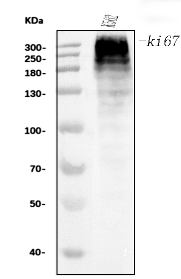
Western blot analysis of Ki67 using anti-Ki67 antibody (M00254-9).
Electrophoresis was performed on a 5-20% SDS-PAGE gel at 70V (Stacking gel) / 90V (Resolving gel) for 2-3 hours. The sample well of each lane was loaded with 30 ug of sample under reducing conditions.
Lane 1: human K562 whole cell lysates.
After electrophoresis, proteins were transferred to a nitrocellulose membrane at 150 mA for 50-90 minutes. Blocked the membrane with 5% non-fat milk/TBS for 1.5 hour at RT. The membrane was incubated with mouse anti-Ki67 antigen affinity purified monoclonal antibody (Catalog # M00254-9) at 0.5 μg/mL overnight at 4°C, then washed with TBS-0.1%Tween 3 times with 5 minutes each and probed with a goat anti-mouse IgG-HRP secondary antibody at a dilution of 1:10000 for 1.5 hour at RT. The signal is developed using an Enhanced Chemiluminescent detection (ECL) kit (Catalog # EK1001) with Tanon 5200 system. A specific band was detected for Ki67 at approximately 358 kDa. The expected band size for Ki67 is at 358 kDa.

Click image to see more details
IHC analysis of Ki67 using anti-Ki67 antibody (M00254-9).
Ki67 was detected in a paraffin-embedded section of human lung cancer tissue. Heat mediated antigen retrieval was performed in EDTA buffer (pH 8.0, epitope retrieval solution). The tissue section was blocked with 10% goat serum. The tissue section was then incubated with 2 μg/ml mouse anti-Ki67 Antibody (M00254-9) overnight at 4°C. Biotinylated goat anti-mouse IgG was used as secondary antibody and incubated for 30 minutes at 37°C. The tissue section was developed using Strepavidin-Biotin-Complex (SABC) (Catalog # SA1021) with DAB as the chromogen.

Click image to see more details
Effects of upregulation and downregulation of MALAT1 on U87 and U251 cell proliferation. ( a and b ) Cellular proliferation of untransfected or transfected U87 and U251 cells was measured using a CCK-8 assay daily for 3 days. ( c ) Cellular proliferation of untransfected or transfected U87 and U251 cells was measured by testing the expression of Ki-67. ( d and e ) The percentage of Ki-67-positive cells was calculated. Results are expressed as mean±S.D. from three independent experiments ( P <0.01). ( f ) Untransfected or transfected U87 and U251 cells were stained by propidium iodide and analyzed using flow cytometry. ( g and h ) The percentage of cells in the G0/G1, S and G2/M phases of the cell cycle was calculated. Results are expressed as mean±S.D. from three independent experiments ( P <0.01). Abbreviations: CCK-8, Cell Counting Kit-8; MALAT1, metastasis-associated lung adenocarcinoma transcript 1; S.D., standard deviation
Index in PubMed under a CC BY license. PMID: 26938295

Click image to see more details
Effects of overexpression of MALAT1 on proliferation in vivo . ( a and b ) Overexpression of MALAT1 reduced the growth of glioma in a subcutaneous glioma nude model. ( c and d ) MALAT1 expression in subcutaneous and incratranial tumors ( P <0.01). ( e ) Immunohistochemistry showed that overexpression of MALAT1 reduced the expression of Ki-67 and MMP2 in intracranial xenograft of U87. ( f and g ) Western blotting showed that overexpression of MALAT1 reduced the expression of P-ERK ( P <0.01). ( h and i ) The percentage of Ki-67- and MMP2-positive cells, respectively, calculated from densitometry immunohistochemistry signaling. Results are expressed as mean±S.D. from three independent experiments ( P <0.01). ( j ) Kaplan–Meier survival curves for nude mice implanted intracranially with U87 and its MALAT1 transfectants. MALAT1, metastasis-associated lung adenocarcinoma transcript 1
Index in PubMed under a CC BY license. PMID: 26938295

Click image to see more details
Cripto-1 gene silencing suppresses cell proliferation in vivo . (a) representative mouse bearing tumors (up was in CNE-2 control group, down was in CNE-2/GFP + /Cripto-1 - group). ( b) external whole-body fluorescence images of the same mouse. ( c) the external images of xenotransplant tumors (top was in CNE-2/GFP + /Cripto-1 - group, bottom was in CNE-2 control group). ( d) H&E stains of xenotransplant tumors, (×400). (e) expression of Ki67 in xenotransplant tumors of CNE-2 control group (×400). (f) expression of Ki67 in xenotransplant tumors of CNE-2/GFP + /Cripto-1 - group (×400).
Index in PubMed under a CC BY license. PMID: 19732464

Click image to see more details
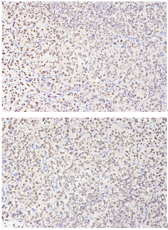
IHC analysis of Ki67 using anti-Ki67 antibody (M00254-9).
Ki67 was detected in a paraffin-embedded section of human lymphomas tissue. Heat mediated antigen retrieval was performed in EDTA buffer (pH 8.0, epitope retrieval solution). The tissue section was blocked with 10% goat serum. The tissue section was then incubated with 2 μg/ml mouse anti-Ki67 Antibody (M00254-9) overnight at 4°C. Biotinylated goat anti-mouse IgG was used as secondary antibody and incubated for 30 minutes at 37°C. The tissue section was developed using Strepavidin-Biotin-Complex (SABC) (Catalog # SA1021) with DAB as the chromogen.

Click image to see more details
IHC analysis of Ki67 using anti-Ki67 antibody (M00254-9).
Ki67 was detected in a paraffin-embedded section of human liver cancer tissue. Heat mediated antigen retrieval was performed in EDTA buffer (pH 8.0, epitope retrieval solution). The tissue section was blocked with 10% goat serum. The tissue section was then incubated with 2 μg/ml mouse anti-Ki67 Antibody (M00254-9) overnight at 4°C. Biotinylated goat anti-mouse IgG was used as secondary antibody and incubated for 30 minutes at 37°C. The tissue section was developed using Strepavidin-Biotin-Complex (SABC) (Catalog # SA1021) with DAB as the chromogen.

Click image to see more details
IHC analysis of Ki67 using anti-Ki67 antibody (M00254-9).
Ki67 was detected in a paraffin-embedded section of human esophageal squamous carcinoma tissue. Heat mediated antigen retrieval was performed in EDTA buffer (pH 8.0, epitope retrieval solution). The tissue section was blocked with 10% goat serum. The tissue section was then incubated with 2 μg/ml mouse anti-Ki67 Antibody (M00254-9) overnight at 4°C. Biotinylated goat anti-mouse IgG was used as secondary antibody and incubated for 30 minutes at 37°C. The tissue section was developed using Strepavidin-Biotin-Complex (SABC) (Catalog # SA1021) with DAB as the chromogen.

Click image to see more details
IHC analysis of Ki67 using anti-Ki67 antibody (M00254-9).
Ki67 was detected in a paraffin-embedded section of human cervical cancer tissue. Heat mediated antigen retrieval was performed in EDTA buffer (pH 8.0, epitope retrieval solution). The tissue section was blocked with 10% goat serum. The tissue section was then incubated with 2 μg/ml mouse anti-Ki67 Antibody (M00254-9) overnight at 4°C. Biotinylated goat anti-mouse IgG was used as secondary antibody and incubated for 30 minutes at 37°C. The tissue section was developed using Strepavidin-Biotin-Complex (SABC) (Catalog # SA1021) with DAB as the chromogen.

Click image to see more details
IF analysis of Ki67 using anti-Ki67 antibody (M00254-9).
Ki67 was detected in an immunocytochemical section of A431 cells. Enzyme antigen retrieval was performed using IHC enzyme antigen retrieval reagent (AR0022) for 15 mins. The cells were blocked with 10% goat serum. And then incubated with 5 μg/mL mouse anti-Ki67 Antibody (M00254-9) overnight at 4°C. DyLight®488 Conjugated Goat Anti-Mouse IgG (BA1126) was used as secondary antibody at 1:100 dilution and incubated for 30 minutes at 37°C. The section was counterstained with DAPI. Visualize using a fluorescence microscope and filter sets appropriate for the label used.

Click image to see more details
Flow Cytometry analysis of Jurkat cells using anti-Ki67 antibody (M00254-9).
Overlay histogram showing Jurkat cells stained with M00254-9 (Blue line). To facilitate intracellular staining, cells were fixed with 4% paraformaldehyde and permeabilized with permeabilization buffer. The cells were blocked with 10% normal goat serum. And then incubated with mouse anti-Ki67 Antibody (M00254-9, 1 μg/1x106 cells) for 30 min at 20°C. DyLight®488 conjugated goat anti-mouse IgG (BA1126, 5-10 μg/1x106 cells) was used as secondary antibody for 30 minutes at 20°C. Isotype control antibody (Green line) was mouse IgG (1 μg/1x106) used under the same conditions. Unlabelled sample without incubation with primary antibody and secondary antibody (Red line) was used as a blank control.

Click image to see more details
IHC analysis of Ki67 using anti-Ki67 antibody (M00254-9).
Ki67 was detected in a paraffin-embedded section of HepG2 subcutaneous xenograft in nude mouse tissue. Heat mediated antigen retrieval was performed in EDTA buffer (pH 8.0, epitope retrieval solution). The tissue section was blocked with 10% goat serum. The tissue section was then incubated with 1:200 mouse anti-Ki67 Antibody (M00254-9) overnight at 4°C. Two step IHC kit was used as secondary antibody and incubated for 30 minutes at 37°C. The tissue section was developed using Strepavidin-Biotin-Complex (SABC) (Catalog # SA1021) with DAB as the chromogen.
Specific Publications For Anti-Ki67 Antibody Picoband® (monoclonal, 5C7) (M00254-9)
Loading publications
Recommended Resources
Here are featured tools and databases that you might find useful.
- Boster's Pathways Library
- Protein Databases
- Bioscience Research Protocol Resources
- Data Processing & Analysis Software
- Photo Editing Software
- Scientific Literature Resources
- Research Paper Management Tools
- Molecular Biology Software
- Primer Design Tools
- Bioinformatics Tools
- Phylogenetic Tree Analysis
Customer Reviews
Have you used Anti-Ki67 Antibody Picoband® (monoclonal, 5C7)?
Share your experimental results or join a short interview to earn up to $1,000 in product credits or other rewards.
1 Reviews For Anti-Ki67 Antibody Picoband® (monoclonal, 5C7)
IHC with Anti-Ki67 antibody (M00254-9) in HepG2 xenograft tumors showed clear nuclear staining in proliferating cells, accurately reflecting the proliferation index with low background.
Excellent
| SKU | M00254-9 |
|---|---|
| Application | Immunohistochemistry |
| Sample | HepG2 subcutaneous xenograft in nude mice |
| Sample Processing Description | HepG2 cells were expanded in culture and subcutaneously implanted into nude mice. After 2 weeks of tumor formation, tumor tissues were harvested, fixed in 4% formaldehyde for 48 hours, and then processed for paraffin embedding and sectioning. |
| Other Reagents | Goat serum, DAB chromogen solution |
| Primary Antibody | Ki67 Antibody Picoband® (monoclonal, 5C7) |
| Primary Incubation | 1:200, overnight at 4 ℃ |
| Secondary Antibody | Two-step IHC detection kit |
| Secondary Incubation | 30 min in 37℃ |
| Detection | Image system: Leica DM2500 |
| Results Summary | Ki67 is a key marker of cell proliferation with high specificity, as it is expressed only in actively dividing cells and has a very short half-life, allowing accurate assessment of the proliferation index at the time of sampling. It is widely used in tumor prognosis evaluation (e.g., high Ki67 index in breast cancer is often associated with poor prognosis) and assessment of tissue regenerative activity. In this experiment, clear and distinct nuclear positivity was observed with well-defined staining. |
Fengtong Wang, The First Affiliated Hospital of Xinjiang Medical University
Verified customer
Submitted 2026-04-21
Customer Q&As
Have a question?
Find answers in Q&As, reviews.
Can't find your answer?
Submit your question
