Click image to see more details
-

-
-
-
-
+5
Product Info Summary
| SKU: | A00633-1 |
|---|---|
| Size: | 100 μg/vial |
| Reactive Species: | Human, Mouse, Rat |
| Host: | Rabbit |
| Application: | ELISA, IF, IHC, WB |
Customers Who Bought This Also Bought

Product info
Product Name
Anti-Lactoferrin/LTF Antibody Picoband®
SKU/Catalog Number
A00633-1
Size
100 μg/vial
Form
Lyophilized
Description
Boster Bio Anti-Lactoferrin/LTF Antibody Picoband® catalog # A00633-1. Tested in ELISA, IF, IHC, WB applications. This antibody reacts with Human, Mouse, Rat. The brand Picoband indicates this is a premium antibody that guarantees superior quality, high affinity, and strong signals with minimal background in Western blot applications. Only our best-performing antibodies are designated as Picoband, ensuring unmatched performance.
Storage & Handling
Store at -20˚C for one year from date of receipt. After reconstitution, at 4˚C for one month. It can also be aliquotted and stored frozen at -20˚C for six months. Avoid repeated freeze-thaw cycles.
Cite This Product
Anti-Lactoferrin/LTF Antibody Picoband® (Boster Biological Technology, Pleasanton CA, USA, Catalog # A00633-1)
Host
Rabbit
Contents
Each vial contains 4 mg Trehalose, 0.9 mg NaCl and 0.2 mg Na2HPO4.
Clonality
Polyclonal
Isotype
Rabbit IgG
Immunogen
E. coli-derived human Lactoferrin recombinant protein (Position: D529-K710).
Cross-reactivity
No cross-reactivity with other proteins.
Reactive Species
A00633-1 is reactive to LTF in Human, Mouse, Rat
Observed Molecular Weight
85 kDa
Calculated molecular weight
78.2 kDa
Background of LTF
Lactoferrin (LF), also known as lactotransferrin (LTF), is a multifunctional protein of the transferrin family. The protein is a major iron-binding protein in milk and body secretions with an antimicrobial activity, making it an important component of the non-specific immune system. The protein demonstrates a broad spectrum of properties, including regulation of iron homeostasis, host defense against a broad range of microbial infections, anti-inflammatory activity, regulation of cellular growth and differentiation and protection against cancer development and metastasis. Antimicrobial, antiviral, antifungal and antiparasitic activity has been found for this protein and its peptides.
Antibody Validation
Boster validates all antibodies on WB, IHC, ICC, Immunofluorescence, and ELISA with known positive control and negative samples to ensure specificity and high affinity, including thorough antibody incubations.
Application & Images
Applications
A00633-1 is guaranteed for ELISA, IF, IHC, WB Boster Guarantee
Recommend Dilution
| Application | Dilution | Species |
|---|---|---|
| Western blot | 0.1-0.5μg/ml | Human |
| Immunohistochemistry (Paraffin-embedded Section) | 2-5μg/ml | Human |
| Immunofluorescence | 5μg/ml | Human |
| ELISA | 0.1-0.5μg/ml | - |
Tested application
Suggested blocking solution with 5% non-fat milk or BSA; (*)Recommended protein loading: 20-40 µg per lane
Use TE buffer pH 9.0 for antigen retrieval; (*) citrate buffer pH 6.0 is an alternative.
Validation Images & Assay Conditions
Click image to see more details
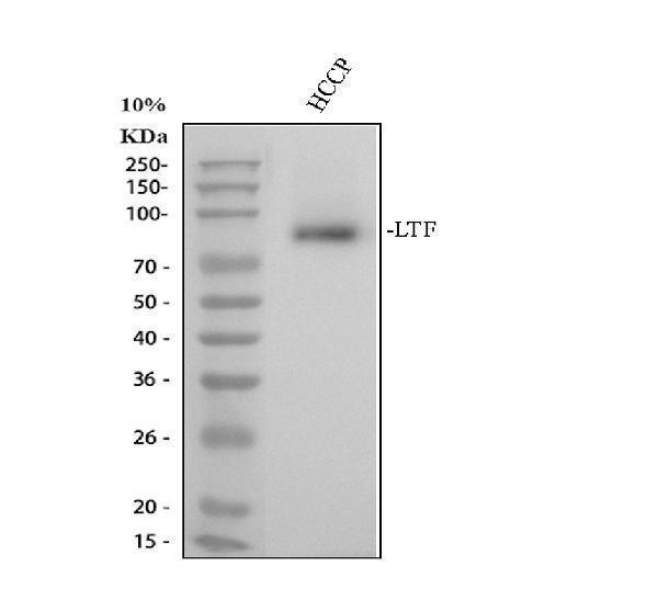
Western blot analysis of LTF using anti-LTF antibody (A00633-1).
Electrophoresis was performed on a 10% SDS-PAGE gel at 80V (Stacking gel) / 120V (Resolving gel) for 2 hours. The sample well of each lane was loaded with 30 ug of sample under reducing conditions.
Lane 1: human hepatocellular carcinoma paracancerous tissue (HCCP) lysates.
After electrophoresis, proteins were transferred to a nitrocellulose membrane at 150 mA for 50-90 minutes. Blocked the membrane with 5% non-fat milk/TBS for 1.5 hour at RT. The membrane was incubated with rabbit anti-LTF antigen affinity purified polyclonal antibody (A00633-1) at 0.5 μg/mL overnight at 4°C, then washed with TBS-0.1%Tween 3 times with 5 minutes each and probed with a goat anti-rabbit IgG-HRP secondary antibody (Catalog # BA1054) at a dilution of 1:5000 for 1.5 hour at RT. The signal is developed using an ECL Plus Western Blotting Substrate (Catalog # AR1196-200) with Tanon 5200 system. A specific band was detected for LTF at approximately 85 kDa. The expected band size for LTF is at 78 kDa.

Click image to see more details
IHC analysis of LTF using anti-LTF antibody (A00633-1).
LTF was detected in a paraffin-embedded section of human prostatic cancer tissue. Heat mediated antigen retrieval was performed in EDTA buffer (pH 8.0, epitope retrieval solution). The tissue section was blocked with 10% goat serum. The tissue section was then incubated with 2 μg/ml rabbit anti-LTF Antibody (A00633-1) overnight at 4°C. Peroxidase Conjugated Goat Anti-rabbit IgG was used as secondary antibody and incubated for 30 minutes at 37°C. The tissue section was developed using HRP Conjugated Rabbit IgG Super Vision Assay Kit (Catalog # SV0002) with DAB as the chromogen.

Click image to see more details
LTF, a ferroptosis-related gene, is identified in aging cochleae. (A) The volcano diagram of shows 43385 DEGs between the old group ( ~864316) and young group ( ~864308). (B) The heatmap shows 28 statistically significant DEGs. (C) Functional analysis of the statistically significant DEGs. (D) LTF, the hub gene, is obtained from the intersection of Up-Gens, Down-Gens (based on the statistically significant DEGs), and FRGs. (E) The PPI network shows that 12 proteins are interacting with LTF.
Index in PubMed under a CC BY license. PMID: 38282692

Click image to see more details
TF-miRNA-mRNA network in cochlear ferroptosis. (A) The volcano diagram of shows 7512 DEMs between the old group ( ~1095954) and young group ( ~1095948). (B) The heatmap shows 12 statistically significant DEMs. (C–F) TFs predicted by mmu-mir-130b (C) , mmu-mir-205 (D) , hsa-mir-130b (E) , and hsa-mir-205 (F) . (G) CEBPA and CEBPB were predicted by mir-130b. (H) STAT3 was predicted by mir-205. (I,J) The top 20 TFs predicted by LTF in mice (I) and humans (J) , CEBPA (Red box) was identified through the intersection of (I,J) , and TFs predicted by miRNAs in (G,H) . (K) The regulatory network was constructed.
Index in PubMed under a CC BY license. PMID: 38282692

Click image to see more details
Ferroptosis in aging HEI-OC1 cells. (A) SA-β-gal staining in HEI-OC1 cells treated with different concentrations of D-gal for 48 h. (B) Quantification of SA-β-positive cells in (A) . Compared with the control group, the percentage of positive cells is observed to have a statistical difference at 20 mg/mL D-gal ( p < 0.001; N = 3). (C–E) Compared with the control group, the expression of Fe 2+ (C) and MDA (D) is increased in aging HEI-OC1 cells with 20 mg/mL D-gal ( p = 0.006 and p < 0.001; N = 3). Oppositely, the expression of LTF (E) is decreased ( p < 0.001; N = 3). More importantly, Liproxstatin-1 reversed the above phenomenon of ferroptosis in aging HEI-OC1 cells with 20 mg/mL D-gal ( p = 0.038, p = 0.012, and p = 0.046; N = 3). (F) Immunofluorescence staining also shows decreased expression of LTF in aging HEI-OC1 cells with 20 mg/mL D-gal (LTF-green, DAPI-blue). Ctrl, control; Lip, Liproxstatin-1.
Index in PubMed under a CC BY license. PMID: 38282692

Click image to see more details
Ferroptosis in aging cochlear explants. (A) SA-β-gal staining in the basement membrane treated with different concentrations of D-gal for 48 h. (B) Quantification of SA-β-positive cells in (A) . Compared with the control group, the percentage of positive cells is observed to have a statistical difference at 30 mg/mL D-gal ( p < 0.001; N = 3). (C–E) Compared with the control group, the expression of Fe 2+ (C) and MDA (D) is increased in aging cochlear explants with 30 mg/mL D-gal ( p < 0.001; N = 5). Oppositely, the expression of LTF (E) is decreased in aging cochlear explants with 30 mg/mL D-gal ( p < 0.001; N = 5). OHC, outer hair cell; IHC, inner hair cell; Ctrl, control.
Index in PubMed under a CC BY license. PMID: 38282692

Click image to see more details
Ferroptosis in aging cochleae. (A) Compared with young mice (2mo), the distinctly increased ABR thresholds are shown in old mice (10mo) at all frequencies ( p < 0.001; N = 6 mice). (B–D) Compared with young mice, the expression of Fe 2+ (B) and MDA (C) is increased in aging cochleae ( p < 0.001; N = 5). Oppositely, the expression of LTF (D) is decreased ( p < 0.005; N = 3).
Index in PubMed under a CC BY license. PMID: 38282692

Click image to see more details
IF analysis of LTF using anti-LTF antibody (A00633-1).
LTF was detected in a paraffin-embedded section of human prostatic cancer tissue. Heat mediated antigen retrieval was performed in EDTA buffer (pH 8.0, epitope retrieval solution). The tissue section was blocked with 10% goat serum. The tissue section was then incubated with 5 μg/mL rabbit anti-LTF Antibody (A00633-1) overnight at 4°C. DyLight®488 Conjugated Goat Anti-Rabbit IgG (BA1127) was used as secondary antibody at 1:500 dilution and incubated for 30 minutes at 37°C. The section was counterstained with DAPI. Visualize using a fluorescence microscope and filter sets appropriate for the label used.

Click image to see more details
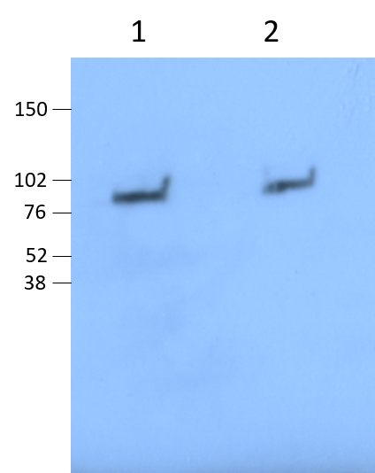
Western blot analysis of Lactoferrin/LTF using anti-Lactoferrin/LTF Antibody (A00633-1).
Electrophoresis was performed on a 5-20% SDS-PAGE gel at 70V (Stacking gel) / 90V (Resolving gel) for 2-3 hours. The sample well of each lane was loaded with 30 ug of sample under reducing conditions.
Lane 1: Total LTF from mouse neutrophil cell lysates.
Lane 2: degranulated LTF. Ratio 1:20.
After electrophoresis, proteins were transferred to a nitrocellulose membrane at 150 mA for 50-90 minutes. Blocked the membrane with 5% BSA for 1.5 hour at RT. The membrane was incubated with rabbit anti-Lactoferrin/LTF antigen affinity purified polyclonal antibody (A00633-1) at 1:2000 1h overnight at 4°C, then washed with TBS-0.1%Tween 3 times with 5 minutes each and probed with a goat anti-rabbit IgG-HRP secondary antibody at a dilution of 1:3000 dilution at RT for 1 hour. The signal is developed using HRP. A specific band was detected for Lactoferrin/LTF at approximately 77 kDa. The expected band size for Lactoferrin/LTF is at 77 kDa.
Specific Publications For Anti-Lactoferrin/LTF Antibody Picoband® (A00633-1)
Loading publications
Recommended Resources
Here are featured tools and databases that you might find useful.
- Boster's Pathways Library
- Protein Databases
- Bioscience Research Protocol Resources
- Data Processing & Analysis Software
- Photo Editing Software
- Scientific Literature Resources
- Research Paper Management Tools
- Molecular Biology Software
- Primer Design Tools
- Bioinformatics Tools
- Phylogenetic Tree Analysis
Customer Reviews
Have you used Anti-Lactoferrin/LTF Antibody Picoband®?
Share your experimental results or join a short interview to earn up to $1,000 in product credits or other rewards.
1 Reviews For Anti-Lactoferrin/LTF Antibody Picoband®
Very Clean Anti-Lactoferrin/LTF Antibody Picoband
Excellent
Source: Biocompare.com
| SKU | A00633-1 |
|---|---|
| Application | Western Blot |
| Sample | Mouse Neutrophils |
| Primary Incubation | 1:2000 1 hr |
| Blocking Agent | 5% BSA |
| Secondary Incubation | 1:3000 |
| Detection | HRP |
| Results Summary | Nicely compared total LTF in neutrophils to degranulated LTF. In the image below, Lane 1: Total LTF from mouse neutrophil cell lysates. Lane 2: degranulated LTF. Ratio 1:20. |
"Nicely detects degranulated LTF in supernatant mouse neutrophils stimulated with bacteria. Used at 1:2000 in 5% BSA."
Stephen Chetwynd
Verified customer
Submitted 2024-07-15
Customer Q&As
Have a question?
Find answers in Q&As, reviews.
Can't find your answer?
Submit your question
16 Customer Q&As for Anti-Lactoferrin/LTF Antibody Picoband®
Question
Is a blocking peptide available for product anti-Lactoferrin/LTF antibody (A00633-1)?
Verified Customer
Verified customer
Asked: 2020-04-29
Answer
We do provide the blocking peptide for product anti-Lactoferrin/LTF antibody (A00633-1). If you would like to place an order for it please contact support@bosterbio.com and make a special request.
Boster Scientific Support
Answered: 2020-04-29
Question
Thanks for helping with my inquiry over the phone. Here are the WB image, lot number and protocol we used for bone marrow using anti-Lactoferrin/LTF antibody A00633-1. Let me know if you need anything else.
Verified Customer
Verified customer
Asked: 2020-03-31
Answer
I appreciate the data. You have provided everything we needed. Our lab team are working to resolve your inquiry as quickly as possible, and we appreciate your patience and understanding! Please let me know if there is anything you need in the meantime.
Boster Scientific Support
Answered: 2020-03-31
Question
Does anti-Lactoferrin/LTF antibody A00633-1 work for IHC-P with bone marrow?
Verified Customer
Verified customer
Asked: 2020-03-03
Answer
According to the expression profile of bone marrow, LTF is highly expressed in bone marrow. So, it is likely that anti-Lactoferrin/LTF antibody A00633-1 will work for IHC-P with bone marrow.
Boster Scientific Support
Answered: 2020-03-03
Question
Can you help my question with product A00633-1, anti-Lactoferrin/LTF antibody. I was wondering if it would be possible to conjugate this antibody with biotin. I would need it to be without BSA or sodium azide. I am planning on using a buffer exchange of sodium azide with PBS only. Would there be problems for me to conjugate the antibody and store it in -20 degrees in small aliquots?
Verified Customer
Verified customer
Asked: 2020-02-06
Answer
We do not advise storing this antibody with PBS buffer only in -20 degrees. If you want to store it in -20 degrees it is best to add some cryoprotectant like glycerol. If you want carrier free A00633-1 anti-Lactoferrin/LTF antibody, we can provide it to you in a special formula with trehalose and/or glycerol. These molecules will not interfere with conjugation chemistry and provide a good level of protection for the antibody from degradation. Please be sure to specify this in your purchase order.
Boster Scientific Support
Answered: 2020-02-06
Question
We are currently using anti-Lactoferrin/LTF antibody A00633-1 for human tissue, and we are well pleased with the IHC-F results. The species of reactivity given in the datasheet says human, mouse, rat. Is it true that the antibody can work on goat tissues as well?
Verified Customer
Verified customer
Asked: 2020-01-16
Answer
The anti-Lactoferrin/LTF antibody (A00633-1) has not been tested for cross reactivity specifically with goat tissues, but there is a good chance of cross reactivity. We have an innovator award program that if you test this antibody and show it works in goat you can get your next antibody for free. Please contact me if I can help you with anything.
Boster Scientific Support
Answered: 2020-01-16
Question
See attached the WB image, lot number and protocol we used for bone marrow using anti-Lactoferrin/LTF antibody A00633-1. Please let me know if you require anything else.
Verified Customer
Verified customer
Asked: 2019-12-16
Answer
Thank you very much for the data. Our lab team are working to resolve this as quickly as possible, and we appreciate your patience and understanding! You have provided everything we needed. Please let me know if there is anything you need in the meantime.
Boster Scientific Support
Answered: 2019-12-16
Question
Is there a BSA free version of anti-Lactoferrin/LTF antibody A00633-1 available?
Verified Customer
Verified customer
Asked: 2019-11-01
Answer
I appreciate your recent telephone inquiry. I can confirm that some lots of this anti-Lactoferrin/LTF antibody A00633-1 are BSA free. For now, these lots are available and we can make a BSA free formula for you free of charge. It will take 3 extra days to prepare. If you require this antibody BSA free again in future, please do not hesitate to contact me and I will be pleased to check which lots we have in stock that are BSA free.
Boster Scientific Support
Answered: 2019-11-01
Question
I see that the anti-Lactoferrin/LTF antibody A00633-1 works with IHC-P, what is the protocol used to produce the result images on the product page?
Verified Customer
Verified customer
Asked: 2019-08-28
Answer
You can find protocols for IHC-P on the "support/technical resources" section of our navigation menu. If you have any further questions, please send an email to support@bosterbio.com
Boster Scientific Support
Answered: 2019-08-28
Question
I was wanting to use your anti-Lactoferrin/LTF antibody for IHC-P for mouse bone marrow on frozen tissues, but I want to know if it has been tested for this particular application. Has this antibody been tested and is this antibody a good choice for mouse bone marrow identification?
Verified Customer
Verified customer
Asked: 2019-05-24
Answer
It shows on the product datasheet, A00633-1 anti-Lactoferrin/LTF antibody has been tested for ELISA, Flow Cytometry, IF, IHC-P, IHC-F, ICC, WB on human, mouse, rat tissues. We have an innovator award program that if you test this antibody and show it works in mouse bone marrow in IHC-frozen, you can get your next antibody for free.
Boster Scientific Support
Answered: 2019-05-24
Question
We have tried in the past anti-Lactoferrin/LTF antibody for IHC-P on trachea in a previous project. I am using human, and We want to use the antibody for IHC-F next. My question regards examining trachea as well as seminal vesicle in our next experiment. Could give a recommendation on which antibody would work the best for IHC-F?
Verified Customer
Verified customer
Asked: 2019-01-23
Answer
I have checked the website and datasheets of our anti-Lactoferrin/LTF antibody and it seems that A00633-1 has been validated on human in both IHC-P and IHC-F. Thus A00633-1 should work for your application. Our Boster satisfaction guarantee will cover this product for IHC-F in human even if the specific tissue type has not been validated. We do have a comprehensive range of products for IHC-F detection and you can check out our website bosterbio.com to find out more information about them.
Boster Scientific Support
Answered: 2019-01-23
Question
Is this A00633-1 anti-Lactoferrin/LTF antibody reactive to the isotypes of LTF?
Verified Customer
Verified customer
Asked: 2018-10-24
Answer
The immunogen of A00633-1 anti-Lactoferrin/LTF antibody is E. coli-derived human Lactoferrin recombinant protein (Position: D529-K710). Could you tell me which isotype you are interested in so I can help see if the immunogen is part of this isotype?
Boster Scientific Support
Answered: 2018-10-24
Question
Does A00633-1 anti-Lactoferrin/LTF antibody work on parafin embedded sections? If so, which fixation method do you recommend we use (PFA, paraformaldehyde, other)?
Verified Customer
Verified customer
Asked: 2018-03-08
Answer
As indicated on the product datasheet, A00633-1 anti-Lactoferrin/LTF antibody as been tested on IHC-P. It is best to use PFA for fixation because it has better tissue penetration ability. PFA needs to be prepared fresh before use. Long term stored PFA turns into formalin, as the PFA molecules congregate and become formalin.
Boster Scientific Support
Answered: 2018-03-08
Question
I would like to test anti-Lactoferrin/LTF antibody A00633-1 on mouse bone marrow for research purposes, then I may be interested in using anti-Lactoferrin/LTF antibody A00633-1 for diagnostic purposes as well. Is the antibody suitable for diagnostic purposes?
Verified Customer
Verified customer
Asked: 2017-08-30
Answer
The products we sell, including anti-Lactoferrin/LTF antibody A00633-1, are only intended for research use. They would not be suitable for use in diagnostic work. If you have the means to develop a product into diagnostic use, and are interested in collaborating with us and develop our product into an IVD product, please contact us for more discussions.
Boster Scientific Support
Answered: 2017-08-30
Question
We have observed staining in mouse seminal vesicle. What should we do? Is anti-Lactoferrin/LTF antibody supposed to stain seminal vesicle positively?
Verified Customer
Verified customer
Asked: 2017-08-17
Answer
According to literature seminal vesicle does express LTF. According to Uniprot.org, LTF is expressed in trachea, mammary gland, lung, seminal vesicle, blood, bone marrow, prostate, lung trachea, milk, seminal plasma, tear, myeloid, liver, among other tissues. Regarding which tissues have LTF expression, here are a few articles citing expression in various tissues:
Blood, Pubmed ID: 14573629
Liver, Pubmed ID: 19159218, 24275569
Lung, Pubmed ID: 9122171
Lung, and Trachea, Pubmed ID: 14702039
Mammary gland, Pubmed ID: 2374734, 2402455, 11702692
Milk, Pubmed ID: 16048952, 18780401
Myeloid, Pubmed ID: 3477300, 7049727
Prostate, Pubmed ID: 15489334
Seminal plasma, Pubmed ID: 8551695
Seminal vesicle, Pubmed ID: 22900286
Tear, Pubmed ID: 25946035
Boster Scientific Support
Answered: 2017-08-17
Question
you antibody using your anti-Lactoferrin/LTF antibody for regulation of cytokine production studies. Has this antibody been tested with western blotting on siha cells? We would like to see some validation images before ordering.
P. Kulkarni
Verified customer
Asked: 2016-08-01
Answer
We appreciate your inquiry. This A00633-1 anti-Lactoferrin/LTF antibody is validated on human placenta tissue, rat spleen tissue, mouse spleen tissue, intestinal cancer tissue, lung cancer tissue, siha cells. It is guaranteed to work for ELISA, Flow Cytometry, IF, IHC-P, IHC-F, ICC, WB in human, mouse, rat. Our Boster guarantee will cover your intended experiment even if the sample type has not been be directly tested.
Boster Scientific Support
Answered: 2016-08-01
Question
We were satisfied with the WB result of your anti-Lactoferrin/LTF antibody. However we have been able to see positive staining in myeloid isoform deltalf: cytoplasm. nucleus. using this antibody. Is that expected? Could you tell me where is LTF supposed to be expressed?
W. Miller
Verified customer
Asked: 2014-05-21
Answer
From literature, myeloid does express LTF. Generally LTF expresses in isoform 1: secreted. cytoplasmic granule., isoform deltalf: cytoplasm. nucleus. Regarding which tissues have LTF expression, here are a few articles citing expression in various tissues:
Blood, Pubmed ID: 14573629
Liver, Pubmed ID: 19159218, 24275569
Lung, Pubmed ID: 9122171
Lung, and Trachea, Pubmed ID: 14702039
Mammary gland, Pubmed ID: 2374734, 2402455, 11702692
Milk, Pubmed ID: 16048952, 18780401
Myeloid, Pubmed ID: 3477300, 7049727
Prostate, Pubmed ID: 15489334
Seminal plasma, Pubmed ID: 8551695
Seminal vesicle, Pubmed ID: 22900286
Tear, Pubmed ID: 25946035
Boster Scientific Support
Answered: 2014-05-21
