Click image to see more details
-

-
-
-
-
+1
Product Info Summary
| SKU: | M03019 |
|---|---|
| Size: | 100 μg/vial |
| Reactive Species: | Human, Mouse, Rat |
| Host: | Mouse |
| Application: | Flow Cytometry, IF, IHC, WB |
Customers Who Bought This Also Bought

Product info
Product Name
Anti-MAG Picoband® Antibody (monoclonal, 2G11)
SKU/Catalog Number
M03019
Size
100 μg/vial
Form
Lyophilized
Description
MAG (Siglec-4) is a myelin-associated adhesion glycoprotein that binds neuronal sialic acid–containing gangliosides and supports glia–axon interactions; MAG is also widely discussed as a myelin-associated inhibitor of neurite outgrowth after CNS injury (research context). Assay context: validated for Flow Cytometry, IF, IHC, and Western blot. Often interpreted alongside other myelin/neuronal markers such as MOG and cytoskeletal marker INA (alpha-internexin); water-channel astrocyte marker AQP4 can be informative in demyelinating tissue panels.
Storage & Handling
Store at -20˚C for one year from date of receipt. After reconstitution, at 4˚C for one month. It can also be aliquotted and stored frozen at -20˚C for six months. Avoid repeated freeze-thaw cycles.
Cite This Product
Anti-MAG Picoband® Antibody (monoclonal, 2G11) (Boster Biological Technology, Pleasanton CA, USA, Catalog # M03019)
Host
Mouse
Contents
Each vial contains 4mg Trehalose, 0.9mg NaCl and 0.2mg Na2HPO4.
Clonality
Monoclonal
Clone Number
2G11
Isotype
Mouse IgG2a
Immunogen
E.coli-derived human MAG recombinant protein (Position: E34-R605).
Cross-reactivity
No cross-reactivity with other proteins.
Reactive Species
M03019 is reactive to MAG in Human, Mouse, Rat
Observed Molecular Weight
100 kDa
Calculated molecular weight
69.1 kDa
Background of MAG
MAG (Myelin-associated glycoprotein),also known as SIGLEC4A, is a cell membrane glycoprotein that is a member of the SIGLEC family of proteins and is a functional ligand of the NOGO-66 receptor, NgR. It is though to be involved in the process of myelination. MAG is a sialic acid-binding SIGLEC protein and is a functional ligand for the NOGO receptor.The MAG gene is mapped on 19q13.12. Cleavage of GPI-linked proteins from axons protects growth cones from MAG-induced collapse, and dominant-negative NgR eliminates MAG inhibition of neurite outgrowth. MAG-resistant embryonic neurons were rendered MAG-sensitive by expression of NgR. MAG binds specifically to an NgR-expressing cell line in a GPI-dependent and sialic acid-independent manner. Experiments blocking NgR from interacting with MAG prevented inhibition of neurite outgrowth by MAG. In cultured human embryonic kidney (HEK) cells expressing the NOGO receptor, p75 (NTR) was required for MAG-induced intracellular calcium elevation.
Antibody Validation
Boster validates all antibodies on WB, IHC, ICC, Immunofluorescence, and ELISA with known positive control and negative samples to ensure specificity and high affinity, including thorough antibody incubations.
Application & Images
Applications
M03019 is guaranteed for Flow Cytometry, IF, IHC, WB Boster Guarantee
Recommend Dilution
| Application | Dilution | Species |
|---|---|---|
| Western blot | 0.1-0.25μg/ml | Mouse, Rat |
| Immunohistochemistry (Paraffin-embedded Section) | 2-5μg/ml | Mouse,Rat |
| Immunofluorescence | 5 μg/ml | Rat |
| Flow Cytometry (Fixed) | 1-3μg/1x106 cells | Human |
Tested application
Suggested blocking solution with 5% non-fat milk or BSA; (*)Recommended protein loading: 20-40 µg per lane
Use TE buffer pH 9.0 for antigen retrieval; (*) citrate buffer pH 6.0 is an alternative.
Validation Images & Assay Conditions
Click image to see more details
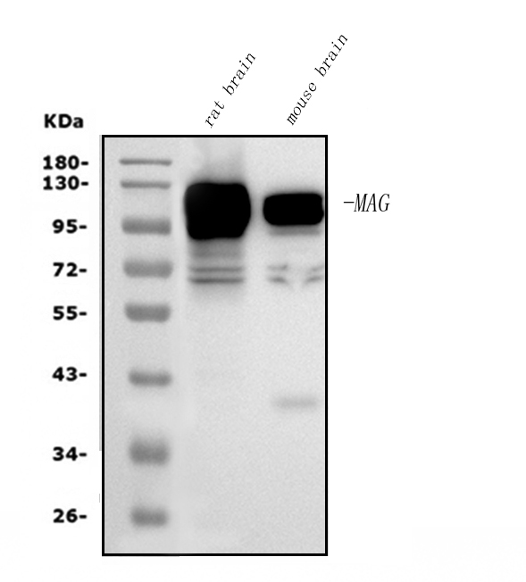
Western blot analysis of MAG using anti-MAG antibody (M03019).
Electrophoresis was performed on a 5-20% SDS-PAGE gel at 70V (Stacking gel) / 90V (Resolving gel) for 2-3 hours. The sample well of each lane was loaded with 50ug of sample under reducing conditions.
Lane 1: rat brain tissue lysates,
Lane 2: mouse brain tissue lysates.
After Electrophoresis, proteins were transferred to a Nitrocellulose membrane at 150mA for 50-90 minutes. Blocked the membrane with 5% Non-fat Milk/ TBS for 1.5 hour at RT. The membrane was incubated with mouse anti-MAG antigen affinity purified monoclonal antibody (Catalog # M03019) at 0.25 μg/mL overnight at 4°C, then washed with TBS-0.1%Tween 3 times with 5 minutes each and probed with a goat anti-mouse IgG-HRP secondary antibody at a dilution of 1:10000 for 1.5 hour at RT. The signal is developed using an Enhanced Chemiluminescent detection (ECL) kit (Catalog # EK1001) with Tanon 5200 system. A specific band was detected for MAG at approximately 100KD. The expected band size for MAG is at 100KD.

Click image to see more details
IHC analysis of MAG using anti-MAG antibody (M03019).
MAG was detected in paraffin-embedded section of mouse brain tissue. Heat mediated antigen retrieval was performed in EDTA buffer (pH8.0, epitope retrieval solution). The tissue section was blocked with 10% goat serum. The tissue section was then incubated with 2μg/ml mouse anti-MAG Antibody (M03019) overnight at 4°C. Biotinylated goat anti-mouse IgG was used as secondary antibody and incubated for 30 minutes at 37°C. The tissue section was developed using Strepavidin-Biotin-Complex (SABC) (Catalog # SA1021) with DAB as the chromogen.

Click image to see more details
IHC analysis of MAG using anti-MAG antibody (M03019).
MAG was detected in paraffin-embedded section of rat brain tissue. Heat mediated antigen retrieval was performed in EDTA buffer (pH8.0, epitope retrieval solution). The tissue section was blocked with 10% goat serum. The tissue section was then incubated with 2μg/ml mouse anti-MAG Antibody (M03019) overnight at 4°C. Biotinylated goat anti-mouse IgG was used as secondary antibody and incubated for 30 minutes at 37°C. The tissue section was developed using Strepavidin-Biotin-Complex (SABC) (Catalog # SA1021) with DAB as the chromogen.

Click image to see more details
Flow Cytometry analysis of U87 cells using anti-MAG antibody (M03019).
Overlay histogram showing U87 cells stained with M03019 (Blue line). The cells were fixed with 4% paraformaldehyde and blocked with 10% normal goat serum. And then incubated with mouse anti- MAG Antibody (M03019, 1μg/1x106 cells) for 30 min at 20°C. DyLight®488 conjugated goat anti-mouse IgG (BA1126, 5-10μg/1x106 cells) was used as secondary antibody for 30 minutes at 20°C. Isotype control antibody (Green line) was mouse IgG (1μg/1x106) used under the same conditions. Unlabelled sample without incubation with primary antibody and secondary antibody (Red line) was used as a blank control.

Click image to see more details
IF analysis of GFAP and MAG using anti-GFAP antibody and anti-MAG antibody (M03019).
GFAP and MAG was detected in a paraffin-embedded section of rat cerebellum tissue. Heat mediated antigen retrieval was performed in EDTA buffer (pH 8.0, epitope retrieval solution). The tissue section was blocked with 10% goat serum. The tissue section was then incubated with 5 μg/mL rabbit anti-GFAP antibody and mouse anti-MAG antibody (M03019) overnight at 4°C. DyLight®594 Conjugated Goat Anti-Rabbit IgG (BA1142), DyLight®488 Conjugated Goat Anti-Mouse IgG (BA1126) was used as secondary antibody at 1:100 dilution and incubated for 30 minutes at 37°C. The section was counterstained with DAPI. Visualize using a fluorescence microscope and filter sets appropriate for the label used.
Specific Publications For Anti-MAG Picoband® Antibody (monoclonal, 2G11) (M03019)
Loading publications
Recommended Resources
Here are featured tools and databases that you might find useful.
- Boster's Pathways Library
- Protein Databases
- Bioscience Research Protocol Resources
- Data Processing & Analysis Software
- Photo Editing Software
- Scientific Literature Resources
- Research Paper Management Tools
- Molecular Biology Software
- Primer Design Tools
- Bioinformatics Tools
- Phylogenetic Tree Analysis
Customer Reviews
Have you used Anti-MAG Picoband® Antibody (monoclonal, 2G11)?
Share your experimental results or join a short interview to earn up to $1,000 in product credits or other rewards.
0 Reviews For Anti-MAG Picoband® Antibody (monoclonal, 2G11)
Customer Q&As
Have a question?
Find answers in Q&As, reviews.
Can't find your answer?
Submit your question
