Click image to see more details
-

-
-
-
-
+8
Product Info Summary
| SKU: | PB9148 |
|---|---|
| Size: | 100 μg/vial |
| Reactive Species: | Human, Mouse, Rat |
| Host: | Rabbit |
| Application: | Flow Cytometry, IF, IHC, IHC-F, ICC, WB, ELISA (Cap) |
Customers Who Bought This Also Bought

Product info
Product Name
Anti-MyD88 Antibody Picoband®
SKU/Catalog Number
PB9148
Size
100 μg/vial
Form
Lyophilized
Description
Boster Bio Anti-MyD88 Antibody Picoband® catalog # PB9148. Tested in ELISA, Flow Cytometry, IF, IHC, IHC-F, ICC, WB applications. This antibody reacts with Human, Mouse, Rat. The brand Picoband indicates this is a premium antibody that guarantees superior quality, high affinity, and strong signals with minimal background in Western blot applications. Only our best-performing antibodies are designated as Picoband, ensuring unmatched performance.
Storage & Handling
Store at -20˚C for one year from date of receipt. After reconstitution, at 4˚C for one month. It can also be aliquotted and stored frozen at -20˚C for six months. Avoid repeated freeze-thaw cycles.
Cite This Product
Anti-MyD88 Antibody Picoband® (Boster Biological Technology, Pleasanton CA, USA, Catalog # PB9148)
Host
Rabbit
Contents
Each vial contains antibody formulated with stabilizing components, 0.9 mg NaCl, 0.2 mg Na2HPO4, and 0.05 mg NaN3.
*This antibody is supplied in a stabilized formulation.
Compatibility with conjugation reactions depends on the chemistry of the conjugation method used.
For conjugation methods that are not compatible with the stabilizing components present in this formulation, a carrier-free antibody format is required.
Clonality
Polyclonal
Isotype
Rabbit IgG
Immunogen
E.coli-derived human MyD88 recombinant protein (Position: A44-F264). Human MyD88 shares 84% and 83% amino acid (aa) sequences identity with mouse and rat MyD88, respectively.
Cross-reactivity
No cross-reactivity with other proteins
Reactive Species
PB9148 is reactive to PTGS2 in Human, Mouse, Rat
Observed Molecular Weight
36 kDa
Calculated molecular weight
69.0 kDa
Background of PTGS2
MYD88 (MYELOID DIFFERENTIATION PRIMARY RESPONSE GENE 88), is a protein that, in humans, is encoded by the MYD88 gene. MyD88 is a key downstream adapter for most Toll-like receptors (TLRs) and interleukin-1 receptors (IL1Rs). And it is mapped on 3p22.2. MYD88 encodes a cytosolic adapter protein that plays a central role in the innate and adaptive immune response. This protein functions as an essential signal transducer in the interleukin-1 and Toll-like receptor signaling pathways. Overexpression of MYD88 caused an increase in the level of transcription from the interleukin-8 promoter. The C-terminal domain of MYD88 has significant sequence similarity to the cytoplasmic domain of IL1RAP. Inhibiting the IL1R-MYD88 pathway in vivo could block the damage from acute inflammation that occurs in response to sterile cell death, and do so in a way that might not compromise tissue repair or host defense against pathogens.
Antibody Validation
Boster validates all antibodies on WB, IHC, ICC, Immunofluorescence, and ELISA with known positive control and negative samples to ensure specificity and high affinity, including thorough antibody incubations.
Application & Images
Applications
PB9148 is guaranteed for Flow Cytometry, IF, IHC, IHC-F, ICC, WB, ELISA (Cap) Boster Guarantee
Recommend Dilution
| Application | Dilution | Species |
|---|---|---|
| Western blot | 0.1-0.5μg/ml | |
| Immunohistochemistry (Paraffin-embedded Section) | 0.5-1μg/ml | |
| Immunohistochemistry (Frozen Section) | 0.5-1μg/ml | |
| Immunocytochemistry | 0.5-1μg/ml | |
| Immunofluorescence | 2μg/ml | |
| Flow Cytometry (Fixed) | 1-3μg/1x106 cells | |
| ELISA (Cap) | 1-5μg/ml |
Tested application
Suggested blocking solution with 5% non-fat milk or BSA; (*)Recommended protein loading: 20-40 µg per lane
Use TE buffer pH 9.0 for antigen retrieval; (*) citrate buffer pH 6.0 is an alternative.
Validation Images & Assay Conditions
Click image to see more details
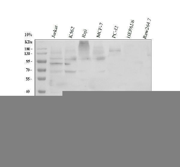
Western blot analysis of MyD88 using anti-MyD88 antibody (PB9148).
Electrophoresis was performed on a 10% SDS-PAGE gel at 80V (Stacking gel) / 120V (Resolving gel) for 2 hours. The sample well of each lane was loaded with 30 ug of sample under reducing conditions.
Lane 1: human Jurkat whole cell lysates,
Lane 2: human K562 whole cell lysates,
Lane 3: human Raji whole cell lysates,
Lane 4: human MCF-7 whole cell lysates,
Lane 5: rat PC-12 whole cell lysates,
Lane 6: mouse HEPA1-6 whole cell lysates,
Lane 7: mouse Raw264.7 whole cell lysates.
After electrophoresis, proteins were transferred to a nitrocellulose membrane at 150 mA for 50-90 minutes. Blocked the membrane with 5% non-fat milk/TBS for 1.5 hour at RT. The membrane was incubated with rabbit anti-MyD88 antigen affinity purified polyclonal antibody (PB9148) at 0.5 μg/mL overnight at 4°C, then washed with TBS-0.1%Tween 3 times with 5 minutes each and probed with a goat anti-rabbit IgG-HRP secondary antibody (Catalog # BA1054) at a dilution of 1:5000 for 1.5 hour at RT. The signal is developed using an ECL Plus Western Blotting Substrate (Catalog # AR1196-200) with Tanon 5200 system. A specific band was detected for MyD88 at approximately 36 kDa. The expected band size for MyD88 is at 33 kDa.

Click image to see more details
IF analysis of MYD88 using anti-MYD88 antibody (PB9148)
MYD88 was detected in paraffin-embedded section of human mammary cancar tissues. Heat mediated antigen retrieval was performed in citrate buffer (pH6, epitope retrieval solution ) for 20 mins. The tissue section was blocked with 10% goat serum. The tissue section was then incubated with 1μg/mL rabbit anti-MYD88 Antibody (PB9148) overnight at 4°C. Cy3 Conjugated Goat Anti-Rabbit IgG (BA1032) was used as secondary antibody at 1:100 dilution and incubated for 30 minutes at 37°C. The section was counterstained with DAPI. Visualize using a fluorescence microscope and filter sets appropriate for the label used.

Click image to see more details
IHC analysis of MYD88 using anti-MYD88 antibody (PB9148).
MYD88 was detected in paraffin-embedded section of rat lung tissue. Heat mediated antigen retrieval was performed in citrate buffer (pH6, epitope retrieval solution) for 20 mins. The tissue section was blocked with 10% goat serum. The tissue section was then incubated with 1μg/ml rabbit anti-MYD88 Antibody (PB9148) overnight at 4°C. Biotinylated goat anti-rabbit IgG was used as secondary antibody and incubated for 30 minutes at 37°C. The tissue section was developed using Strepavidin-Biotin-Complex (SABC)(Catalog # SA1022) with DAB as the chromogen.

Click image to see more details
IHC analysis of MYD88 using anti-MYD88 antibody (PB9148).
MYD88 was detected in paraffin-embedded section of human intestinal cancer tissue. Heat mediated antigen retrieval was performed in citrate buffer (pH6, epitope retrieval solution) for 20 mins. The tissue section was blocked with 10% goat serum. The tissue section was then incubated with 1μg/ml rabbit anti-MYD88 Antibody (PB9148) overnight at 4°C. Biotinylated goat anti-rabbit IgG was used as secondary antibody and incubated for 30 minutes at 37°C. The tissue section was developed using Strepavidin-Biotin-Complex (SABC)(Catalog # SA1022) with DAB as the chromogen.

Click image to see more details
IHC analysis of MYD88 using anti-MYD88 antibody (PB9148).
MYD88 was detected in paraffin-embedded section of mouse spleen tissue. Heat mediated antigen retrieval was performed in citrate buffer (pH6, epitope retrieval solution) for 20 mins. The tissue section was blocked with 10% goat serum. The tissue section was then incubated with 1μg/ml rabbit anti-MYD88 Antibody (PB9148) overnight at 4°C. Biotinylated goat anti-rabbit IgG was used as secondary antibody and incubated for 30 minutes at 37°C. The tissue section was developed using Strepavidin-Biotin-Complex (SABC)(Catalog # SA1022) with DAB as the chromogen.

Click image to see more details
IF analysis of MYD88 using anti-MYD88 antibody (PB9148)
MYD88 was detected in paraffin-embedded section of human tonsil tissues. Heat mediated antigen retrieval was performed in citrate buffer (pH6, epitope retrieval solution ) for 20 mins. The tissue section was blocked with 10% goat serum. The tissue section was then incubated with 1μg/mL rabbit anti-MYD88 Antibody (PB9148) overnight at 4°C. Cy3 Conjugated Goat Anti-Rabbit IgG (BA1032) was used as secondary antibody at 1:100 dilution and incubated for 30 minutes at 37°C. The section was counterstained with DAPI. Visualize using a fluorescence microscope and filter sets appropriate for the label used.

Click image to see more details
IF analysis of MYD88 using anti-MYD88 antibody (PB9148)
MYD88 was detected in paraffin-embedded section of human colon cancar tissues. Heat mediated antigen retrieval was performed in citrate buffer (pH6, epitope retrieval solution ) for 20 mins. The tissue section was blocked with 10% goat serum. The tissue section was then incubated with 1μg/mL rabbit anti-MYD88 Antibody (PB9148) overnight at 4°C. Cy3 Conjugated Goat Anti-Rabbit IgG (BA1032) was used as secondary antibody at 1:100 dilution and incubated for 30 minutes at 37°C. The section was counterstained with DAPI. Visualize using a fluorescence microscope and filter sets appropriate for the label used.

Click image to see more details
IF analysis of MYD88 using anti-MYD88 antibody (PB9148)
MYD88 was detected in paraffin-embedded section of human mammary cancar tissues. Heat mediated antigen retrieval was performed in citrate buffer (pH6, epitope retrieval solution ) for 20 mins. The tissue section was blocked with 10% goat serum. The tissue section was then incubated with 1μg/mL rabbit anti-MYD88 Antibody (PB9148) overnight at 4°C. Cy3 Conjugated Goat Anti-Rabbit IgG (BA1032) was used as secondary antibody at 1:100 dilution and incubated for 30 minutes at 37°C. The section was counterstained with DAPI. Visualize using a fluorescence microscope and filter sets appropriate for the label used.

Click image to see more details
IF analysis of MYD88 using anti-MYD88 antibody (PB9148)
MYD88 was detected in paraffin-embedded section of human placenta tissues. Heat mediated antigen retrieval was performed in citrate buffer (pH6, epitope retrieval solution ) for 20 mins. The tissue section was blocked with 10% goat serum. The tissue section was then incubated with 1μg/mL rabbit anti-MYD88 Antibody (PB9148) overnight at 4°C. Cy3 Conjugated Goat Anti-Rabbit IgG (BA1032) was used as secondary antibody at 1:100 dilution and incubated for 30 minutes at 37°C. The section was counterstained with DAPI. Visualize using a fluorescence microscope and filter sets appropriate for the label used.

Click image to see more details
Flow Cytometry analysis of A549 cells using anti-MYD88 antibody (PB9148).
Overlay histogram showing A549 cells stained with PB9148 (Blue line). To facilitate intracellular staining, cells were fixed with 4% paraformaldehyde and permeabilized with permeabilization buffer. The cells were blocked with 10% normal goat serum. And then incubated with rabbit anti-MYD88 Antibody (PB9148, 1μg/1x106 cells) for 30 min at 20°C. DyLight®488 conjugated goat anti-rabbit IgG (BA1127, 5-10μg/1x106 cells) was used as secondary antibody for 30 minutes at 20°C. Isotype control antibody (Green line) was rabbit IgG (1μg/1x106) used under the same conditions. Unlabelled sample without incubation with primary antibody and secondary antibody (Red line) was used as a blank control.

Click image to see more details
MT regulated brain barrier function and TLR4/MyD88/NF-κB signaling pathway in sleep-deprived rats. (A) Western blot bands showing the protein expression levels of Iba1 and Aβ42 in the HP, respectively. (B, C) Relative protein expression level of Iba1 and Aβ42 in the HP, respectively. (D) Western blot brands showing the protein expression levels of ZO-1 and occludin in the HP, respectively. (E) Western blot brands showing the protein expression levels of TLR4, MyD88, IKB-α, p-IKB-α, NF-κB p65, and p-NF-κB p65 in the HP, respectively. (F, G) Relative protein expression level of ZO-1 and occludin in the HP, respectively. (H–K) Relative protein expression level of TLR4, MyD88, p-IKB-α/IKB-α, and p-NF-κB p65/NF-κB p65, respectively. (L–N) Relative mRNA expression of ZO-1, occluding, and TLR4 in the HP, respectively. The data are expressed as the means ± SEM. # p < 0.05, ## p < 0.01, ### p < 0.001 vs. Control group; * p < 0.05, ** p < 0.01, *** p < 0.001 vs. Model group.
Index in PubMed under a CC BY license. PMID: 39101143
Specific Publications For Anti-MyD88 Antibody Picoband® (PB9148)
Loading publications
Recommended Resources
Here are featured tools and databases that you might find useful.
- Boster's Pathways Library
- Protein Databases
- Bioscience Research Protocol Resources
- Data Processing & Analysis Software
- Photo Editing Software
- Scientific Literature Resources
- Research Paper Management Tools
- Molecular Biology Software
- Primer Design Tools
- Bioinformatics Tools
- Phylogenetic Tree Analysis
Customer Reviews
Have you used Anti-MyD88 Antibody Picoband®?
Share your experimental results or join a short interview to earn up to $1,000 in product credits or other rewards.
1 Reviews For Anti-MyD88 Antibody Picoband®
The antibody shows high efficiency and specificity in Western blot detection of hemolymph cell proteins from Pacific oyster, with only slight nonspecific bands.
Excellent
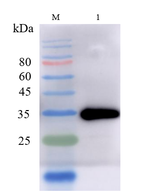
| SKU | PB9148 |
|---|---|
| Application | Western Blot |
| Sample | hemolymph of the Pacific oyster |
| Sample Processing Description | Add 10 µL of hemolymph cells to each lane and directly lyse in 1× Laemmli sample buffer. After adding β-mercaptoethanol, load the samples onto SDS-PAGE. |
| Primary Incubation | The membrane was incubated with the MyD88 primary antibody (1:1000) overnight at 4 °C. |
| Secondary Antibody | HRP-conjugated Goat Anti-Rabbit IgG Secondary Antibody |
| Secondary Incubation | Incubate at room temperature for 1 hour |
| Other Reagents used | Milk |
| Detection | Signal was developed using ECL substrate on a ChemiDoc MP system. |
| Results Summary | The antibody shows high efficiency and specificity in Western blot detection of hemolymph cell proteins from Pacific oyster, with only slight nonspecific bands. |
Yiqing Wang, Student at Dalian Ocean University
Verified customer
Submitted 2025-09-23
Customer Q&As
Have a question?
Find answers in Q&As, reviews.
Can't find your answer?
Submit your question
5 Customer Q&As for Anti-MyD88 Antibody Picoband®
Question
We have seen staining in rat pancreas. Are there any suggestions? Is anti-MyD88 antibody supposed to stain pancreas positively?
Verified Customer
Verified customer
Asked: 2020-05-04
Answer
From what I have seen in literature pancreas does express MYD88. From what I have seen in Uniprot.org, MYD88 is expressed in blood, dendritic cell, epidermal carcinoma, umbilical cord blood, pancreas, erythroleukemia, among other tissues. Regarding which tissues have MYD88 expression, here are a few articles citing expression in various tissues:
Dendritic cell, Pubmed ID: 8957090
Epidermal carcinoma, Pubmed ID: 9013863
Erythroleukemia, Pubmed ID: 23186163
Pancreas, Pubmed ID: 15489334
Umbilical cord blood, Pubmed ID: 14702039
Boster Scientific Support
Answered: 2020-05-04
Question
We need using your anti-MyD88 antibody for cellular response to lead ion studies. Has this antibody been tested with western blotting on cardiac muscle tissue? We would like to see some validation images before ordering.
Verified Customer
Verified customer
Asked: 2019-11-08
Answer
We appreciate your inquiry. This PB9148 anti-MyD88 antibody is tested on rat lung tissue, cardiac muscle tissue, tissue lysate, hela whole cell lysate, mcf whole cell lysate, hepg2 whole cell lysate, jurkat whole cell lysate, raji whole cell lysate, intestinal cancer tissue, mouse spleen tissue, a549 cells. It is guaranteed to work for ELISA, Flow Cytometry, IF, IHC-P, IHC-F, ICC, WB in human, mouse, rat. Our Boster guarantee will cover your intended experiment even if the sample type has not been be directly tested.
Boster Scientific Support
Answered: 2019-11-08
Question
My team were well pleased with the WB result of your anti-MyD88 antibody. However we have been able to see positive staining in erythroleukemia cytoplasm using this antibody. Is that expected? Could you tell me where is MYD88 supposed to be expressed?
Verified Customer
Verified customer
Asked: 2019-08-26
Answer
Based on literature, erythroleukemia does express MYD88. Generally MYD88 expresses in cytoplasm. Regarding which tissues have MYD88 expression, here are a few articles citing expression in various tissues:
Dendritic cell, Pubmed ID: 8957090
Epidermal carcinoma, Pubmed ID: 9013863
Erythroleukemia, Pubmed ID: 23186163
Pancreas, Pubmed ID: 15489334
Umbilical cord blood, Pubmed ID: 14702039
Boster Scientific Support
Answered: 2019-08-26
Question
We are currently using anti-MyD88 antibody PB9148 for mouse tissue, and we are content with the Flow Cytometry results. The species of reactivity given in the datasheet says human, mouse, rat. Is it possible that the antibody can work on dog tissues as well?
L. Taylor
Verified customer
Asked: 2016-03-04
Answer
The anti-MyD88 antibody (PB9148) has not been tested for cross reactivity specifically with dog tissues, though there is a good chance of cross reactivity. We have an innovator award program that if you test this antibody and show it works in dog you can get your next antibody for free. Please contact me if I can help you with anything.
Boster Scientific Support
Answered: 2016-03-04
Question
We have tried in the past anti-MyD88 antibody for WB on pancreas in a previous project. I am using rat, and We intend to use the antibody for ELISA next. We need examining pancreas as well as epidermal carcinoma in our next experiment. Could give a recommendation on which antibody would work the best for ELISA?
G. Kulkarni
Verified customer
Asked: 2015-06-30
Answer
I took a look at the website and datasheets of our anti-MyD88 antibody and it appears that PB9148 has been tested on rat in both WB and ELISA. Thus PB9148 should work for your application. Our Boster satisfaction guarantee will cover this product for ELISA in rat even if the specific tissue type has not been validated. We do have a comprehensive range of products for ELISA detection and you can check out our website bosterbio.com to find out more information about them.
Boster Scientific Support
Answered: 2015-06-30
