Click image to see more details
Product Info Summary
| SKU: | A00184 |
|---|---|
| Size: | 100 μg/vial |
| Reactive Species: | Human, Mouse, Rat |
| Host: | Rabbit |
| Application: | ELISA, Flow Cytometry, WB |
Customers Who Bought This Also Bought

Product info
Product Name
Anti-NCAM1 Antibody Picoband®
SKU/Catalog Number
A00184
Size
100 μg/vial
Form
Lyophilized
Description
Boster Bio Anti-NCAM1 Antibody Picoband® catalog # A00184. Tested in ELISA, Flow Cytometry, WB applications. This antibody reacts with Human, Mouse, Rat. The brand Picoband indicates this is a premium antibody that guarantees superior quality, high affinity, and strong signals with minimal background in Western blot applications. Only our best-performing antibodies are designated as Picoband, ensuring unmatched performance.
Storage & Handling
Store at -20˚C for one year from date of receipt. After reconstitution, at 4˚C for one month. It can also be aliquotted and stored frozen at -20˚C for six months. Avoid repeated freeze-thaw cycles.
Cite This Product
Anti-NCAM1 Antibody Picoband® (Boster Biological Technology, Pleasanton CA, USA, Catalog # A00184)
Host
Rabbit
Contents
Each vial contains 4mg Trehalose, 0.9mg NaCl, 0.2mg Na2HPO4, 0.01mg NaN3.
Clonality
Polyclonal
Isotype
Rabbit IgG
Immunogen
E.coli-derived human NCAM1 recombinant protein (Position: K152-Q690).
Cross-reactivity
No cross-reactivity with other proteins.
Reactive Species
A00184 is reactive to NCAM1 in Human, Mouse, Rat
Observed Molecular Weight
120-200 kDa
Calculated molecular weight
94.6 kDa
Background of NCAM1
NCAM is a membrane-bound glycoprotein that plays a role in cell-cell and cell-matrix adhesion through both its homophilic and heterophilic binding activity. The neural cell adhesion molecule appears on early embryonic cells and is important in the formation of cell collectives and their boundaries at sites of morphogenesis. Later in development it is found on various differentiated tissues and is a major CAM mediating adhesion among neurons and between neurons and muscle. NCAM gene is mapped to 11q23. The neural cell adhesion molecule (NCAM) can influence a number of diverse intercellular events, including junctional communication, the association of axons with pathways and targets, and signals that alter levels of neurotransmitter enzymes.
Antibody Validation
Boster validates all antibodies on WB, IHC, ICC, Immunofluorescence, and ELISA with known positive control and negative samples to ensure specificity and high affinity, including thorough antibody incubations.
Application & Images
Applications
A00184 is guaranteed for ELISA, Flow Cytometry, WB Boster Guarantee
Recommend Dilution
| Application | Dilution | Species |
|---|---|---|
| Western blot | 0.1-0.25μg/ml | Mouse, Rat |
| Flow Cytometry(Fixed) | 1-3 μg/1x106 cells | Human |
| ELISA | 0.1-0.5μg/ml | - |
Tested application
Suggested blocking solution with 5% non-fat milk or BSA; (*)Recommended protein loading: 20-40 µg per lane
Validation Images & Assay Conditions
Click image to see more details
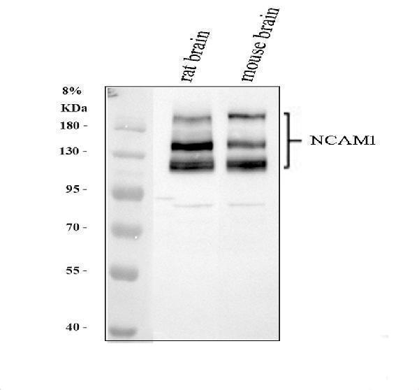
Western blot analysis of NCAM1 using anti-NCAM1 antibody (A00184).
Electrophoresis was performed on a 8% SDS-PAGE gel at 80V (Stacking gel) / 120V (Resolving gel) for 2 hours. The sample well of each lane was loaded with 30 ug of sample under reducing conditions.
Lane 1: rat brain tissue lysates,
Lane 2: mouse brain tissue lysates.
After electrophoresis, proteins were transferred to a nitrocellulose membrane at 150 mA for 50-90 minutes. Blocked the membrane with 5% non-fat milk/TBS for 1.5 hour at RT. The membrane was incubated with rabbit anti-NCAM1 antigen affinity purified polyclonal antibody (A00184) at 0.5 μg/mL overnight at 4°C, then washed with TBS-0.1%Tween 3 times with 5 minutes each and probed with a goat anti-rabbit IgG-HRP secondary antibody (Catalog # BA1054) at a dilution of 1:5000 for 1.5 hour at RT. The signal is developed using an ECL Plus Western Blotting Substrate (Catalog # AR1196-200) with Tanon 5200 system. A specific band was detected for NCAM1 at approximately 120-200 kDa. The expected band size for NCAM1 is at 95 kDa.

Click image to see more details
Flow Cytometry analysis of U937 cells using anti-NCAM1 antibody (A00184).
Overlay histogram showing U937 cells stained with A00184 (Blue line). To facilitate intracellular staining, cells were fixed with 4% paraformaldehyde and permeabilized with permeabilization buffer. The cells were blocked with 10% normal goat serum. And then incubated with rabbit anti-NCAM1 Antibody (A00184,1μg/1x106 cells) for 30 min at 20°C. DyLight®488 conjugated goat anti-rabbit IgG (BA1127, 5-10μg/1x106 cells) was used as secondary antibody for 30 minutes at 20°C. Isotype control antibody (Green line) was rabbit IgG (1μg/1x106) used under the same conditions. Unlabelled sample (Red line) was also used as a control.
Specific Publications For Anti-NCAM1 Antibody Picoband® (A00184)
Loading publications
Recommended Resources
Here are featured tools and databases that you might find useful.
- Boster's Pathways Library
- Protein Databases
- Bioscience Research Protocol Resources
- Data Processing & Analysis Software
- Photo Editing Software
- Scientific Literature Resources
- Research Paper Management Tools
- Molecular Biology Software
- Primer Design Tools
- Bioinformatics Tools
- Phylogenetic Tree Analysis
Customer Reviews
Have you used Anti-NCAM1 Antibody Picoband®?
Share your experimental results or join a short interview to earn up to $1,000 in product credits or other rewards.
0 Reviews For Anti-NCAM1 Antibody Picoband®
Customer Q&As
Have a question?
Find answers in Q&As, reviews.
Can't find your answer?
Submit your question
5 Customer Q&As for Anti-NCAM1 Antibody Picoband®
Question
We are currently using anti-NCAM1 antibody A00184 for rat tissue, and we are well pleased with the ELISA results. The species of reactivity given in the datasheet says human, mouse, rat. Is it true that the antibody can work on bovine tissues as well?
Verified Customer
Verified customer
Asked: 2020-04-02
Answer
The anti-NCAM1 antibody (A00184) has not been tested for cross reactivity specifically with bovine tissues, but there is a good chance of cross reactivity. We have an innovator award program that if you test this antibody and show it works in bovine you can get your next antibody for free. Please contact me if I can help you with anything.
Boster Scientific Support
Answered: 2020-04-02
Question
We want using your anti-NCAM1 antibody for ncam1 interactions studies. Has this antibody been tested with western blotting on mouse brain? We would like to see some validation images before ordering.
Verified Customer
Verified customer
Asked: 2019-05-21
Answer
We appreciate your inquiry. This A00184 anti-NCAM1 antibody is tested on rat brain tissue, mouse brain, u937 cells. It is guaranteed to work for ELISA, WB in human, mouse, rat. Our Boster guarantee will cover your intended experiment even if the sample type has not been be directly tested.
Boster Scientific Support
Answered: 2019-05-21
Question
My boss were satisfied with the WB result of your anti-NCAM1 antibody. However we have observed positive staining in neocortex isoform 1: cell membrane using this antibody. Is that expected? Could you tell me where is NCAM1 supposed to be expressed?
D. Bhatt
Verified customer
Asked: 2015-01-16
Answer
According to literature, neocortex does express NCAM1. Generally NCAM1 expresses in isoform 1: cell membrane. Regarding which tissues have NCAM1 expression, here are a few articles citing expression in various tissues:
Brain, Kidney, and Ovary, Pubmed ID: 15489334
Liver, Pubmed ID: 24275569
Plasma, Pubmed ID: 16335952
Skeletal muscle, Pubmed ID: 2887295, 3253057
Boster Scientific Support
Answered: 2015-01-16
Question
We have been able to see staining in mouse liver. Are there any suggestions? Is anti-NCAM1 antibody supposed to stain liver positively?
G. Carter
Verified customer
Asked: 2014-10-20
Answer
From literature liver does express NCAM1. From Uniprot.org, NCAM1 is expressed in neocortex, skeletal muscle, testis, brain, brain, kidney ovary, plasma, liver, among other tissues. Regarding which tissues have NCAM1 expression, here are a few articles citing expression in various tissues:
Brain, Kidney, and Ovary, Pubmed ID: 15489334
Liver, Pubmed ID: 24275569
Plasma, Pubmed ID: 16335952
Skeletal muscle, Pubmed ID: 2887295, 3253057
Boster Scientific Support
Answered: 2014-10-20
Question
We have tried in the past anti-NCAM1 antibody for WB on testis last year. I am using mouse, and I plan to use the antibody for ELISA next. I am interested in examining testis as well as brain in our next experiment. Could you please give me some suggestion on which antibody would work the best for ELISA?
G. Singh
Verified customer
Asked: 2014-09-22
Answer
I have checked the website and datasheets of our anti-NCAM1 antibody and it appears that A00184 has been validated on mouse in both WB and ELISA. Thus A00184 should work for your application. Our Boster satisfaction guarantee will cover this product for ELISA in mouse even if the specific tissue type has not been validated. We do have a comprehensive range of products for ELISA detection and you can check out our website bosterbio.com to find out more information about them.
Boster Scientific Support
Answered: 2014-09-22
