Click image to see more details
-

-
-
-
-
+3
Product Info Summary
| SKU: | A01246 |
|---|---|
| Size: | 0.1 mg |
| Reactive Species: | Human, Mouse, Rat |
| Host: | Rabbit |
| Application: | ELISA, IF, IHC-P, WB |
Customers Who Bought This Also Bought

Product info
Product Name
Anti-Occludin OCLN Antibody
SKU/Catalog Number
A01246
Size
0.1 mg
Form
Liquid
Description
Boster Bio Anti-Occludin OCLN Antibody (Catalog # A01246). Tested in ELISA, WB, IHC-P, IF applications. This antibody reacts with Human, Mouse, Rat.
Storage & Handling
Antibody can be stored at 4°C up to one year. Antibodies should not be exposed to prolonged high temperatures.
Cite This Product
Anti-Occludin OCLN Antibody (Boster Biological Technology, Pleasanton CA, USA, Catalog # A01246)
Host
Rabbit
Contents
OCLN Antibody is supplied in PBS containing 0.02% sodium azide.
Clonality
Polyclonal
Isotype
IgG
Immunogen
OCLN antibody was raised against a 15 amino acid synthetic peptide from near the carboxy terminus of human OCLN. The immunogen is located within the last 50 amino acids of OCLN.
Cross-reactivity
At least three isoforms of OCLN are known to exist.
Reactive Species
A01246 is reactive to OCLN in Human, Mouse, Rat
Observed Molecular Weight
68 kDa
Calculated molecular weight
59.1 kDa
Background of OCLN
Tight junctions act as a semi-permeable barrier to the transport of ions, solutes, and water and are considered to function as a fence that divides apical and basolateral domains of plasma membranes. Tight junctions coordinate a variety of signaling and trafficking molecules regulating cell differentiation, proliferation, and polarity and contain a number of junctional proteins including Occludin, Claudins, junctional adhesion molecules (JAMs), as well as multiple scaffold proteins. Occludin, the first identified component of tight junction strands, is thought function as a signal transmitter in multiple signaling pathways and can associate with multiple kinases and phosphatases such as PI3-kinase and protein phosphatases 1 and 2A. At least two isoforms of OCLN are known to exist.
Antibody Validation
Boster validates all antibodies on WB, IHC, ICC, Immunofluorescence, and ELISA with known positive control and negative samples to ensure specificity and high affinity, including thorough antibody incubations.
Application & Images
Applications
A01246 is guaranteed for ELISA, IF, IHC-P, WB Boster Guarantee
Recommend Dilution
OCLN antibody can be used for detection of OCLN by Western blot at 1 μg/mL. Antibody can also be used for immunohistochemistry starting at 2.5 μg/mL. For immunofluorescence start at 20 μg/mL.
Antibody validated: Western Blot in human samples; Immunohistochemistry in human and mouse samples and Immunofluorescence in human and mouse samples. All other applications and species not yet tested. Optimal dilutions for each application should be determined by the researcher.
Validation Images & Assay Conditions
Click image to see more details
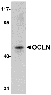
Western blot analysis of OCLN in human liver tissue lysate with OCLN antibody at 1 μg/mL.

Click image to see more details
Immunofluorescence of OCLN in Human Liver tissue with OCLN antibody at 20 μg/mL.

Click image to see more details
Immunohistochemistry of OCLN in human liver tissue with OCLN antibody at 2.5 μg/mL.

Click image to see more details
Immunofluorescence of OCLN in human liver tissue with OCLN antibody at 20 μg/ml.
Red: OCLN Antibody (A01246)
Blue: DAPI staining

Click image to see more details
Immunofluorescence of OCLN in mouse brain tissue with OCLN antibody at 20 μg/mL.
Red: OCLN Antibody (A01246)
Blue: DAPI staining

Click image to see more details
Immunohistochemistry of OCLN in mouse brain tissue with OCLN Antibody at 5 μg/mL.

Click image to see more details
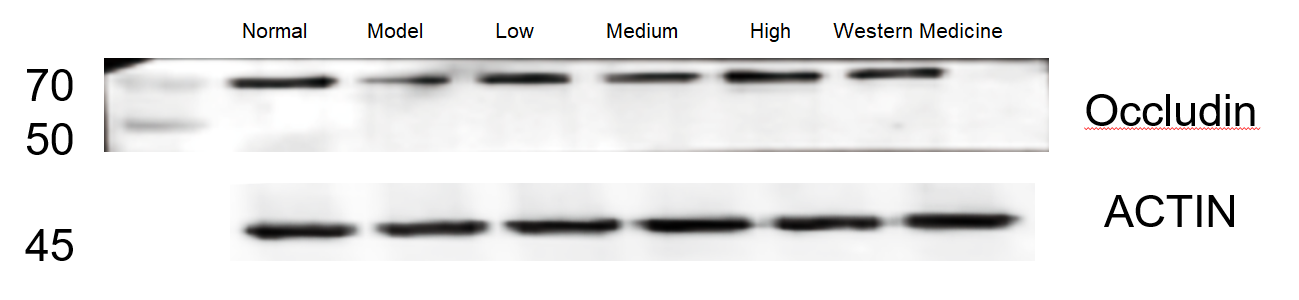
Western blot analysis of OCLN using anti-OCLN antibody (A01246).
Electrophoresis was performed on a 10% SDS-PAGE gel at 80V (Stacking gel) / 120V (Resolving gel) for 2 hours. The sample well of each lane was loaded with 30 ug of sample under reducing conditions.
Lane 1: Normal group-rat colon tissue,
Lane 2: Model group-colon tissue from model rats,
Lane 3: Low dose group-colon tissue from model rats,
Lane 4: Medium dose group-colon tissue from model rats,
Lane 5: High dose group-colon tissue from model rats,
Lane 6: Western medicine treated-colon tissue from model rats.
After electrophoresis, proteins were transferred to a nitrocellulose membrane at 150 mA for 50-90 minutes. Blocked the membrane with 5% non-fat milk/TBS for 1.5 hour at RT. The membrane was incubated with rabbit anti-OCLN antigen affinity purified monoclonal antibody (A01246) at 1:1000 overnight at 4°C, then washed with TBS-0.1%Tween 3 times with 5 minutes each and probed with a HRP Conjugated AffiniPure Goat Anti-rabbit IgG (H+L) at a dilution of 1:5000 for 1 hour at RT. The signal is developed using an ECL Plus Western Blotting Substrate (Catalog # AR1196-200) with ChemiDoc MP system. A specific band was detected for OCLN at approximately 68 kDa. The expected band size for OCLN is at 59.1 kDa.
Specific Publications For Anti-Occludin OCLN Antibody (A01246)
Loading publications
Recommended Resources
Here are featured tools and databases that you might find useful.
- Boster's Pathways Library
- Protein Databases
- Bioscience Research Protocol Resources
- Data Processing & Analysis Software
- Photo Editing Software
- Scientific Literature Resources
- Research Paper Management Tools
- Molecular Biology Software
- Primer Design Tools
- Bioinformatics Tools
- Phylogenetic Tree Analysis
Customer Reviews
Have you used Anti-Occludin OCLN Antibody?
Share your experimental results or join a short interview to earn up to $1,000 in product credits or other rewards.
1 Reviews For Anti-Occludin OCLN Antibody
In WB using Occludin antibody (Cat# A01246), clear bands showed decreased Occludin in the model group and best recovery in the high-dose herbal treatment group.
Excellent
| SKU | A01246 |
|---|---|
| Application | Western Blot |
| Sample | rat colon tissue |
| Sample Processing Description | RIPA lysis buffer containing protease inhibitor PMSF (100:1) was used to lyse samples for 10 min, followed by centrifugation at 12,000 rpm for 15 min; the supernatant was mixed with 5× loading buffer, boiled at 100 °C for 10 min, and loaded onto SDS-PAGE. |
| Other Reagents | 5% Non-fat milk |
| Primary Antibody | Occludin OCLN Antibody |
| Primary Incubation | 1:1000, overnight at 4 ℃ |
| Secondary Antibody | HRP Conjugated AffiniPure Goat Anti-rabbit IgG (H+L) |
| Secondary Incubation | 1:5000, 1 h in RT |
| Detection | Substrate: ECL substrate, Imaging system:ChemiDoc MP |
| Results Summary | The image shows WB results of Occludin and the loading control Actin in rat colon across different groups; Occludin expression was reduced in the model group, with the high-dose herbal treatment showing the best recovery and clear, well-defined target bands. |
Shiyu Zhang, Liaoning University of Traditional Chinese Medicine
Verified customer
Submitted 2026-02-26
Customer Q&As
Have a question?
Find answers in Q&As, reviews.
Can't find your answer?
Submit your question
